. 2025 Feb 18;25(1):285.
doi: 10.1186/s12885-025-13693-0.

Intraductal chemotherapy for triple-negative breast cancer: a pathway to minimally invasive clinical treatment

Affiliations

Intraductal chemotherapy for triple-negative breast cancer: a pathway to minimally invasive clinical treatment

Xinhong Wu et al. BMC Cancer. .

Abstract

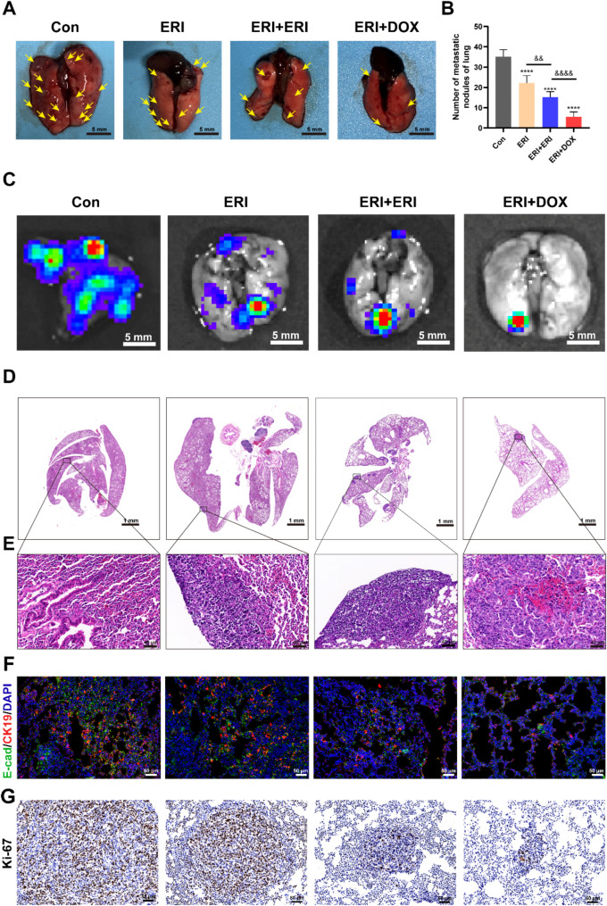
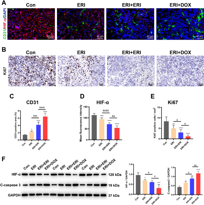
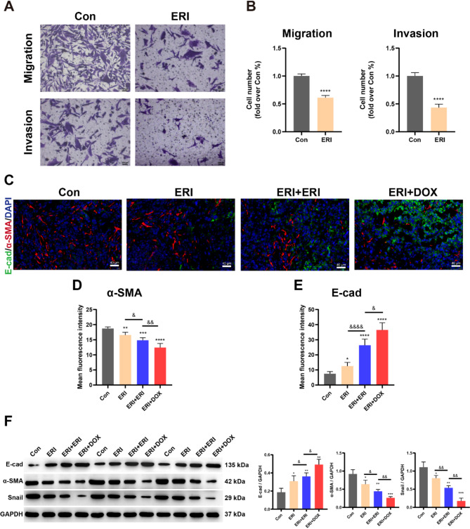
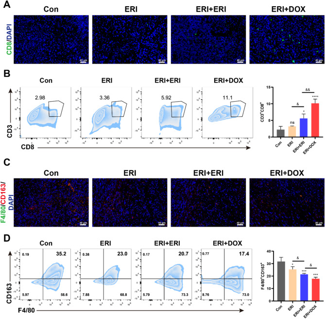
Triple-negative breast cancer (TNBC) is traditionally treated with systemic chemotherapy, often resulting in significant off-target toxicity. In this study, we assess the efficacy of intraductal chemotherapeutic delivery, aimed at reducing systemic side effects. Using an in situ TNBC model, created by intraductal injection of 4T1-luc cells, we identified day 3 post-tumor implantation as an optimal early intervention point. Echocardiographic analysis confirmed that intraductal administration of eribulin (ERI) or doxorubicin (DOX) did not cause cardiac dysfunction or apoptosis. Our results demonstrate that intraductal delivery of ERI and DOX significantly enhances anti-tumor and anti-metastatic effects. Mechanistically, ERI followed by DOX increased intratumoral perfusion, improved drug concentration, reversed epithelial-mesenchymal transition, and inhibited tumor cell invasion and metastasis. Additionally, this approach triggered immunogenic cell death and activated a systemic anti-tumor immune response. These findings underscore the potential of intraductal chemotherapy as a safe, highly effective approach, offering a preclinical foundation for minimally invasive TNBC therapies.

Keywords: Chemo-immunomodulation; Epithelial-mesenchymal transition; Intraductal therapy; Minimally invasive treatment; Triple-negative breast cancer.


Conflict of interest statement

Declarations. Ethics approval and consent to participate: The People’s Hospital of Wuhan University’s Animal Ethics Committee approved this round of animal testing (IACUC Issue No. 20200702), and all ethical guidelines were strictly followed. Consent for publication: Not applicable. Competing interests: The authors declare no competing interests.

Figures

Fig. 1
Fig. 1
Intraductal therapy. Intraductal injection of drugs kills tumor cells, causes cell immunogenic death, and activates anti-tumor immunity. Abbreviations, DC: dendritic cell; ECM: extracellular matrix; M1: M1 type macrophage; M2: M2 type macrophage; NK: natural killer cell; T: T lymphocyte.
Fig. 2
Fig. 2
I.duc injection of 4T1-luc cells to construct TNBC model in situ. A Light microscopic picture of day 3, 5, 7, and 9 breast tissue after inoculation. B HE stained microscopic image of the tumor. Scale bar = 40 μm.
Fig. 3
Fig. 3
Anti-tumor efficacy of i.duc intervention in 4T1-luc xenograft breast cancer model. A Schematic diagram of the experimental protocol for i.duc intervention. B Live imaging plots at day 7, 14, and 21 of mice implanted with 4T1-luc. C Tumor volume growth curves in various treatment groups. D, E Images of the tumor and data on its weight at day 27 of tumor growth. ns = no significance, *p < 0.05, **p < 0.01, ***p < 0.001, ****p < 0.0001; &&p < 0.01, &&&&p < 0.0001.
Fig. 4
Fig. 4
Efficacy of i.duc intervention on lung metastases. A, B Quantitative analysis of lung metastases in lung tissue at the study endpoint (day 27). Yellow arrows indicate lung metastases focuses. C Bioluminescence imaging plot of lung metastatic colonization. D, E HE staining of lungs for each group, with local magnification shown. Scale bar = 40 μm. F Immunofluorescence images showing E-cad (green) and CK19 (red) in lungs for each group, with DAPI (blue) indicating nuclei. Scale bar = 50 μm. G Immunohistochemical staining of Ki67 in tumors of each treatment group. Scale bar = 50 μm. ****p < 0.0001; &&p < 0.01, &&&&p < 0.0001.
Fig. 5
Fig. 5
Effect of i.duc intervention on tumor apoptosis. A, B Immunofluorescence of tunel (green) and C-caspase 3 (red) in tumor sections at the study endpoint (day 27). Scale = 40 μm. C, D Immunofluorescence of tunel and C-caspase 3 immunofluorescence statistics. ns = no significance, *p < 0.05, **p < 0.01, ****p < 0.0001; &p < 0.05, &&p < 0.01, &&&&p < 0.0001.
Fig. 6
Fig. 6
I.duc intervention impacts tumor blood supply and proliferation. A Immunofluorescence images showing CD31 (green) and HIF-α (red) in tumors of each treatment group, with DAPI (blue) indicating nuclei at the study endpoint (day 27). Scale bar = 40 μm. B Immunohistochemical staining of Ki67 in tumors of each treatment group. Scale bar = 40 μm. C–E Statistical analysis of CD31 and HIF-α immunofluorescence, and Ki67 immunohistochemistry. F Western blot analysis and corresponding quantification of the protein expression levels of C-caspase 3 and HIF-α in each group. *p < 0.05, **p < 0.01, ***p < 0.001, ****p < 0.0001; &p < 0.05, &&p < 0.01, &&&p < 0.001, &&&&p < 0.0001.
Fig. 7
Fig. 7
ERI-induced reversal of EMT in vitro and in vivo. A, B Representative images of migration and invasion assays of 4T1-luc cells following ERI treatment. Scale = 100 μm. C–E Representative images of immunofluorescence co-staining (C) of α-SMA (red) and E-cad (green) in each tumor group with quantification of α-SMA (D) and E-cad (E) at the study endpoint (day 27). Scale = 40 μm. F Western blot analysis and corresponding quantification of the protein expression levels of α-SMA, E-cad, and Snail in each group. *p < 0.05, **p < 0.01, ***p < 0.001, ****p < 0.0001; &p < 0.05, &&p < 0.01, &&&&p < 0.0001.
Fig. 8
Fig. 8
Mechanisms of chemo-immunotherapy via i.duc intervention. A, B Representative images of immunofluorescence staining and quantification of CD8+ cells in tumor tissue sections at the study endpoint (day 27). C, D Representative images of immunofluorescence staining and quantification of tumor-associated macrophages (F4/80+CD163+) in tumor tissue sections. Scale bar = 40 μm. ns = no significance; *p < 0.05, ***p < 0.001, ****p < 0.0001; &p < 0.05, &&p < 0.01.
Fig. 9
Fig. 9
Effects of i.duc and intravenous DOX on cardiac function. A Cardiac ultrasonogram after different treatment. B Changes in LVEF in different treatment groups. C, D Changes in %FS and LVIDs in different groups. E, F Quantitative analysis of cardiac tunel immunofluorescence in different groups. Scale bar = 40 μm. ns = no significance, *p < 0.05, **p < 0.01, ***p < 0.001, ****p < 0.0001; &p < 0.05, &&p < 0.01, &&&p < 0.001, &&&&p < 0.0001.

References

    1. Leon-Ferre RA, Goetz MP. Advances in systemic therapies for triple negative breast cancer. BMJ. 2023;381:e071674. - PubMed
    1. Bianchini G, Balko JM, Mayer IA, Sanders ME, Gianni L. Triple-negative breast cancer: challenges and opportunities of a heterogeneous disease. Nat Rev Clin Oncol. 2016;13(11):674–90. - PMC - PubMed
    1. Mohanraj B, Duan G, Peredo A, Kim M, Tu F, Lee D, Dodge GR, Mauck RL. Mechanically-Activated Microcapsules for ‘On-Demand’ Drug Delivery in Dynamically Loaded Musculoskeletal Tissues. Adv Funct Mater 2019, 29(15). - PMC - PubMed
    1. Wang G, Kumar A, Ding W, Korangath P, Bera T, Wei J, Pai P, Gabrielson K, Pastan I, Sukumar S. Intraductal administration of transferrin receptor-targeted immunotoxin clears ductal carcinoma in situ in mouse models of breast cancer-a preclinical study. Proc Natl Acad Sci U S A. 2022;119(24):e2200200119. - PMC - PubMed
    1. Stearns V, Mori T, Jacobs LK, Khouri NF, Gabrielson E, Yoshida T, Kominsky SL, Huso DL, Jeter S, Powers P, et al. Preclinical and clinical evaluation of intraductally administered agents in early breast cancer. Sci Transl Med. 2011;3(106):106ra108. - PMC - PubMed

MeSH terms

LinkOut - more resources