. 2025 Jan 2;36(1):102445.
doi: 10.1016/j.omtn.2024.102445. eCollection 2025 Mar 11.

MSCs act as biopatches for blood-retinal barrier preservation to enhance functional recovery after retinal I/R

Affiliations

MSCs act as biopatches for blood-retinal barrier preservation to enhance functional recovery after retinal I/R

Xiaoyue Wei et al. Mol Ther Nucleic Acids. .

Abstract

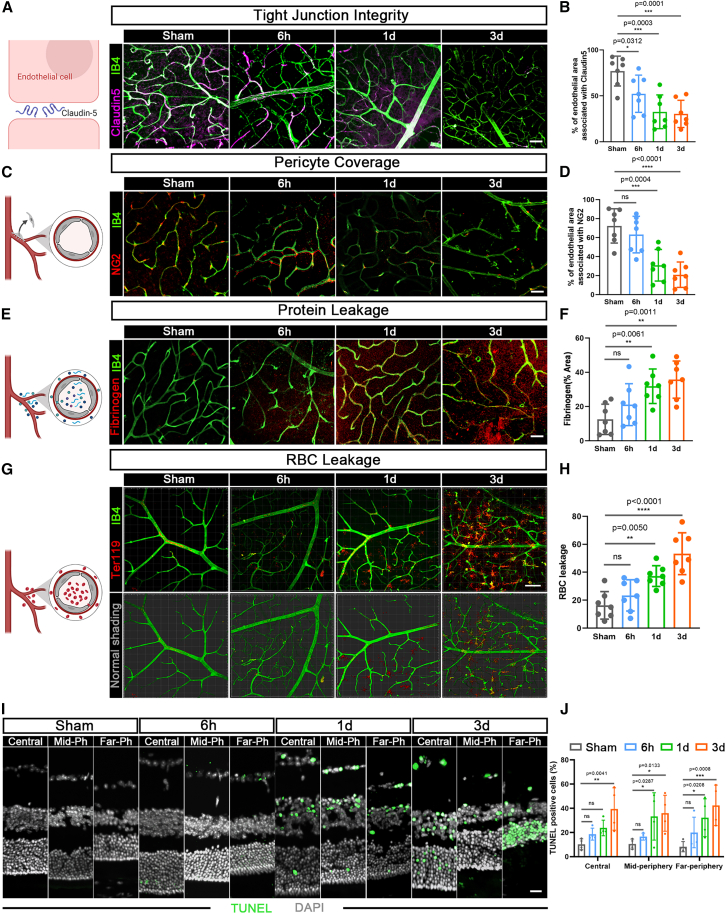
Retinal ischemia/reperfusion (I/R) is one of the most common pathologies of many vision-threatening diseases and is caused by blood-retinal barrier (BRB) breakdown and the resulting inflammatory infiltration. Targeting BRB is promising for retinal I/R treatment. Mesenchymal stromal cells (MSCs) are emerging as novel therapeutic strategies. Although intravitreal injection targets the retina, the restricted number of injected cells still requires the precise biodistribution of MSCs near the injury site. Here, we found that retinal I/R led to BRB breakdown, which induced protein and cell leakage from the circulation. Retinal cell death and diminished visual function were subsequently detected. Moreover, the expression of the chemokine CCL5 increased after retinal I/R, and CCL5 colocalized with the BRB. We then overexpressed CCR5 in human induced pluripotent stem cell-derived MSCs (iMSCs). In vivo, intravitreal-injected iMSCCCR5 preferentially migrated and directly integrated into the BRB, which preferably restored BRB integrity and eventually promoted retinal function recovery after retinal I/R. In summary, our work suggested that iMSCs act as biopatches for BRB preservation and that iMSC-based therapy is a promising therapeutic approach for retinal diseases related to I/R.

Keywords: MT: RNA/DNA Editing; blood-retinal barrier; cell therapy; mesenchymal stromal cells; retinal ischemia/reperfusion.


Conflict of interest statement

The authors declare that they have no competing interests.

Figures

None
Graphical abstract
Figure 1
Figure 1
BRB breakdown after retinal I/R (A, C, E, and G) Patterns (left) and representative confocal fluorescence images (right) of the distribution of Claudin5 (purple) associated with IB4+ blood vessels (green), NG2+ pericyte (red) coverage of IB4+ vessels (green) and protein (fibrinogen, red), and Ter119+ RBC (red) leakage in retinas from the sham and 6 h, 1 day, and 3 days after retinal I/R groups. Scale bar, 50 μm. (B, D, F, and H) Quantifications of (A), (C), (E), and (G) (n = 7 mice per group). (I) Representative confocal fluorescence images of TUNEL staining in the retinal center (Central), mid-periphery (Mid-Ph), and far periphery (Far-Ph) from the sham and 6 h, 1 day, and 3 days after retinal I/R groups. Scale bar, 20 μm. (J) Quantification of TUNEL-positive rates in retinas from the sham and 6 h, 1 day, and 3 days after retinal I/R groups (n = 4 mice per group). The data are expressed as the mean ± standard error of the mean (SEM). All statistical significance was calculated using two-way analysis of variance (ANOVA). ∗p < 0.05, ∗∗p < 0.01, ∗∗∗p < 0.001, and ∗∗∗∗p < 0.0001; ns, not significant.
Figure 2
Figure 2
CCL5 expression increased and colocalized with the BRB after retinal I/R (A) KEGG pathway enrichment analysis was performed for retinas from the sham and 1 day after retinal I/R groups to explore the possible signaling pathways that changed after retinal I/R (n = 3 mice per group). (B) RT-qPCR was performed to measure the expression of CCL2, CCL5, CCL7, CCL12, CXCL2, and CXCL12 in retinas from the sham and 1 day after retinal I/R groups (n = 6 mice per group). (C) RT-qPCR was performed to compare the expression of CCL5 in retinas from the sham and 6 h, 1 day, and 3 days after retinal I/R groups (n = 6 mice per group). (D and E) Flow cytometry analysis and quantification of the mean fluorescence intensity (MFI) of CCL5 in retinas from the sham and 1 day after retinal I/R groups (n = 4 mice per group). (F) Representative confocal fluorescence images of the distribution of CCL5 (red) around IB4+ blood vessels (green) in retinas from the sham and 1 day after retinal I/R groups. Scale bar, 50 μm. (G) Scheme (left) of the structure of the retina and the division of the retinal plexus into three layers, namely, the superficial, intermediate, and deep layers. Quantification (right) of the percentage of vessels colocalized with CCL5 in each layer. Analysis and statistics of colocalization were performed via ImageJ and a plug-in named coloc2. The data are expressed as the mean ± SEM. All statistical significance was calculated using Student’s t test or two-way ANOVA. ∗p < 0.05, ∗∗p < 0.01, and ∗∗∗∗p < 0.0001; ns, not significant.
Figure 3
Figure 3
Overexpression of CCR5 improved the migratory capacity of iMSCs in vitro and the vascular colonization capacity of iMSCs in vivo (A) Transwell migration assays were performed to compare the migratory capacity of iMSCtdTomato and iMSCCCR5 under the recruitment of hCCL5 and mCCL5, respectively. Scale bars, 100 μm. (B) Pattern (upward) and quantification of the transwell migration assay results. (C) Representative images of iMSCCCR5 (red) and iMSCtdTomato (red) retained in retinas at 4 and 7 dpt from the sham and retinal I/R groups. Scale bar, 100 μm. (D) Pattern (top) and quantification analyses of the numbers of iMSCCCR5 and iMSCtdTomato retained in retinas and their percentages on vessels at 4 and 7 dpt in the sham and retinal I/R groups (n = 6 mice per group). The data are expressed as the mean ± SEM. All statistical significance was calculated using one-way or two-way ANOVA. ∗∗p < 0.01 and ∗∗∗p < 0.001; ns, not significant.
Figure 4
Figure 4
iMSCCCR5 infusion reversed damage to the BRB caused by retinal I/R (A) Scheme of the evaluation of iMSC-based therapy on retinal I/R. (B–D and F) Representative confocal fluorescence images and quantification of the distribution of Claudin5 (purple) associated with IB4+ blood vessels (green), NG2+ pericyte (green) coverage of IB4+ blood vessels (gray), and Ter119+ RBC (red) leakage in retinas from the sham, PBS, iMSCtdTomato, and iMSCCCR5 groups (n = 7 mice per group). Scale bar, 50 μm. (E) Representative confocal fluorescence images of TUNEL staining and quantification of TUNEL-positive rates in the Central, Mid-Ph, and Far-Ph positions of retinas from the sham, PBS, iMSCtdTomato, and iMSCCCR5 groups (n = 4 mice per group). Scale bar, 20 μm. The data are expressed as the mean ± SEM. All statistical significance was calculated using one-way or two-way ANOVA. ∗p < 0.05, ∗∗p < 0.01, ∗∗∗p < 0.001, and ∗∗∗∗p < 0.0001; ns, not significant.
Figure 5
Figure 5
iMSCCCR5 infusion recovered damage to visual function caused by retinal I/R (A and B) ERG of amplitude and the time to peak of a- and b-waves among the sham, PBS, iMSCtdTomato, and iMSCCCR5 groups (n = 6 mice per group). The light intensity consisted of 4 increasing levels (dim to bright), including 0.01, 0.1, 1, and 3 cd s/m2. The data are expressed as the mean ± SEM. All statistical significance was calculated using one-way ANOVA. ∗p < 0.05, ∗∗p < 0.01, and ∗∗∗p < 0.001; ns, not significant.

References

    1. Lu C., Wang C., Xiao H., Chen M., Yang Z., Liang Z., Wang H., Liu Y., Yang Y., Wang Q. Ethyl pyruvate: A newly discovered compound against ischemia-reperfusion injury in multiple organs. Pharmacol. Res. 2021;171 doi: 10.1016/j.phrs.2021.105757. - DOI - PubMed
    1. Bresnick G.H., De Venecia G., Myers F.L., Harris J.A., Davis M.D. Retinal ischemia in diabetic retinopathy. Arch. Ophthalmol. 1975;93:1300–1310. doi: 10.1001/archopht.1975.01010020934002. - DOI - PubMed
    1. Tang L.H.C., Fung F.K.C., Lai A.K.W., Wong I.Y.H., Shih K.C., Lo A.C.Y. Autophagic Upregulation Is Cytoprotective in Ischemia/Reperfusion-Injured Retina and Retinal Progenitor Cells. Int. J. Mol. Sci. 2021;22 doi: 10.3390/ijms22168446. - DOI - PMC - PubMed
    1. Burton M.J., Ramke J., Marques A.P., Bourne R.R.A., Congdon N., Jones I., Ah Tong B.A.M., Arunga S., Bachani D., Bascaran C., et al. The Lancet Global Health Commission on Global Eye Health: vision beyond 2020. Lancet Global Health. 2021;9:e489–e551. doi: 10.1016/s2214-109x(20)30488-5. - DOI - PMC - PubMed
    1. Tan G.S.W., Chakravarthy U., Wong T.Y. Anti-VEGF Therapy or Vitrectomy Surgery for Vitreous Hemorrhage From Proliferative Diabetic Retinopathy. JAMA. 2020;324:2375–2377. doi: 10.1001/jama.2020.22829. - DOI - PubMed

LinkOut - more resources