. 2025 Jan 18;28(2):111830.
doi: 10.1016/j.isci.2025.111830. eCollection 2025 Feb 21.

Amniotic epithelial Cell microvesicles uptake inhibits PBMCs and Jurkat cells activation by inducing mitochondria-dependent apoptosis

Affiliations

Amniotic epithelial Cell microvesicles uptake inhibits PBMCs and Jurkat cells activation by inducing mitochondria-dependent apoptosis

Adrián Cerveró-Varona et al. iScience. .

Abstract

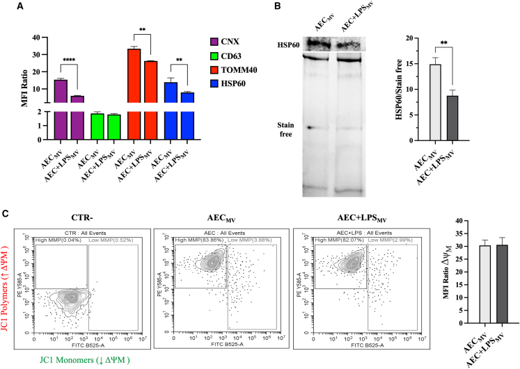
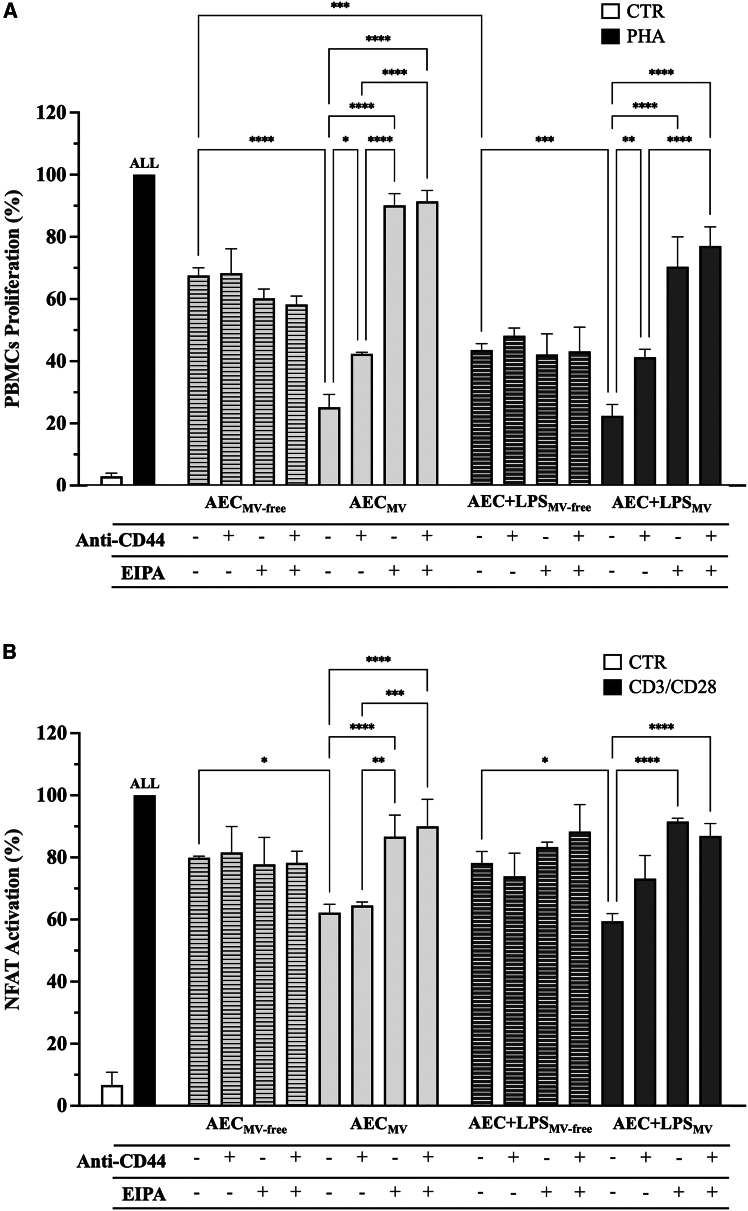
Amniotic epithelial cells (AECs) exhibit significant immunomodulatory and pro-regenerative properties, largely due to their intrinsic paracrine functions that are currently harnessed through the collection of their secretomes. While there is increasing evidence of the role of bioactive components freely secreted or carried by exosomes, the bioactive cargo of AEC microvesicles (MVs) and their crosstalk with the immune cells remains to be fully explored. We showed that under intrinsic conditions or in response to LPS, AEC-derived MV carries components such as lipid-mediated signaling molecules, ER, and mitochondria. They foster the intra/interspecific mitochondrial transfer into immune cells (PBMCs and Jurkat cells) in vitro and in vivo on the zebrafish larvae model of injury. The internalization of MV cargoes through macropinocytosis induces hyperpolarization of PBMC mitochondrial membranes and triggers MV-mediated apoptosis. This powerful immune suppressive mechanism triggered by AEC-MV cargo delivery paves the way for controlled and targeted cell-free therapeutic approaches.

Keywords: Cell biology; Functional aspects of cell biology; Immunology.


Conflict of interest statement

The authors declare no competing interests.

Figures

None
Graphical abstract
Figure 1
Figure 1
MV fraction is a constant component of AEC secretome (A–D) Exoid was used to characterize the particle size and concentration profiles of (A-B) AEC ± LPStot, (C-D) AEC ± LPSMV, and AEC± LPSMV-free. a-MEM and PBS were used as background. Data (mean) represent 3 independent sets of experiments (n = at least 3 biological replicates in each group per set; each biological replicate assayed in at least 3 technical replicates).
Figure 2
Figure 2
Lipidomic profile of MVs enrichment in lipid mediating signaling and ER/mitochondria is dependent of AEC activation status (A) Total lipid concentration in AEC ± LPStot, AEC± LPSMV-free, and AEC ± LPSMV fractions. (B) Bidimensional principal component analysis (PCA) of all fractions. (C) Hierarchical heatmap with Pearson’s coefficient of the lipidic fingerprint for each specific lipid in all fractions. (D) Spearman’s rank correlation analysis of the different lipid classes influenced by all CM fractions. (E and F) Lipid concentration for PE 38:5 and PE 40:5, respectively, in the AEC ± LPSMV fractions. (G) Cellular component classification AECtot, AECMV- free, and AECMV fractions treated ± LPS assessed with the Lipid Ontology web (LION/web) terms. (H) Function classification for the “lipid-mediated signaling” term of the lipid ontology web (LION/web). Data (mean ± SD) represent 3 independent sets of experiments (n = at least 3 biological replicates in each group per set; each biological replicate assayed in at least 3 technical replicates). ∗, ∗∗, ∗∗∗, and ∗∗∗∗ Statistically significant values between the different studied groups (p < 0.05, p < 0.01, p < 0.001, and p < 0.0001, respectively). One-way ANOVA and two tailored t-tests were employed for comparing normally distributed data.
Figure 3
Figure 3
Protein content-based MV fraction characterization confirmed the presence of functional mitochondria (A) Flow cytometric analysis of CNX, CD63, TOMM40, and HSP60, in AEC ± LPSMV. Data were presented as mean fluorescence intensity (MFI) ratio over negative CTR. (B) Representative WB images and relative densitometric analysis of HSP60 (normalized on total protein content via stain-free detection) in AEC ± LPSMV. (C) JC1 flow cytometric analysis to evaluate AEC ± LPSMV ΔΨM. Data were presented as MFI ratio over negative CTR. Data (mean ± SD) represent 3 independent sets of experiments (n = at least 3 biological replicates in each group per set; each biological replicate assayed in at least 3 technical replicates). ∗∗ and ∗∗∗∗ Statistically significant values between the different studied groups (p < 0.01 and p < 0.0001). One-way ANOVA and two tailored t-tests were employed for comparing normally distributed data.
Figure 4
Figure 4
Mitochondrial tracking demonstrated the AEC-derived MV cargo internalization in immune cells (A) Experimental design for B-D. (B) Representative confocal images of co-immunofluorescence staining of nuclei (DAPI), MitoTracker green (PBMCs’ mitochondria), and MitoTracker red (MV’ mitochondria) in PBMCs ± EIPA exposed to AEC ± LPSMV for 12h. (C) Flow cytometric investigation to confirm the mitochondria MV internalization (MitoTracker red) ± LPS in PBMCs and Jurkat ± CD44 and ± EIPA. Data are presented as % of cells positive to MitoTracker red. (D) qPCR analysis to corroborate the mitochondria MV internalization after 24h of exposition to AEC ± LPSMV by examining the ovine mtDNA copy number in Jurkat ± EIPA. (CTR: untreated Jurkat; CTR+: AECMV per se). Data (mean ± SD) represent 3 independent sets of experiments (n = at least 3 biological replicates in each group per set; each biological replicate assayed in at least 3 technical replicates). ALL, ∗, ∗∗, ∗∗∗, and ∗∗∗∗ Statistically significant values between the different studied groups (p < 0.0001, p < 0.05, p < 0.01, p < 0.001, and p < 0.0001, respectively). For the transfer of AECMV mitochondria into AEC see Figure S1. One-way ANOVA was employed for comparing normally distributed data. Scale bar, 20 μm.
Figure 5
Figure 5
Mitochondrial tracking demonstrated the iAEC-derived MV cargo internalization in immune cells (A) Experimental design for B-C. (B) Representative confocal images of co-immunofluorescence staining of nuclei (Hoescht 33342), cytoplasm (Calcein AM), and mitochondria-targeted turbo red florescence protein (TurboRFP-labeled mitochondria), in Jurkat ± EIPA exposed to iAECMV for 12h. (C) Flow cytometric investigation to confirm the mitochondria MV internalization (TurboRFP-labeled mitochondria) in Jurkat ± EIPA. Data are presented as % of cells positive to TurboRFP. Data (mean ± SD) represent 3 independent sets of experiments (n = at least 3 biological replicates in each group per set; each biological replicate assayed in at least 3 technical replicates). ∗∗∗ Statistically significant values between the different studied groups (p < 0.001). One-way ANOVA was employed for comparing normally distributed data. Scale bar, 5 μm.
Figure 6
Figure 6
Interspecific in vivo transfer of AEC-MV mitochondria cargo (A) Illustration describing the experimental plan followed for the treatment of 72 hpf zebrafish larvae with AEC ± LPSMV, and time points for the subsequent analyses. (B) Representative confocal microscopy images of Tg (mpx:GFP) larvae after 6 h post damage (caudal fin cut) and treatment with AEC ± LPSMV reportig the localization of ovine mitochondria MV (MitoTracker red), zebrafish neutrophils (green) and cell nuclei (blue) in the caudal fin. (C) Quantification by qPCR of the amount of ovine mtDNA copy number contained in the zebrafish larvae after 24 and 48 hpd. (CTR: untreated zebrafish larvae; CTR+: AECMV per se). Data (mean ± SD) represent 3 independent sets of experiments (n = at least 10 biological replicates in each group per set). All, and ∗ Statistically significant values between the different studied groups (p < 0.0001, and p < 0.05, respectively). One-way ANOVA was employed for comparing normally distributed data. Scale bar, 100 μm.
Figure 7
Figure 7
AECMV cargo transfer promoted an immunosuppressive response in PBMCs and Jurkat cells (A) PHA-stimulated PBMCs ± Anti-CD44 and ± EIPA were treated with the different CM (AEC ± LPSMV and AEC± LPSMV-free) for 48h and assessed for their proliferation. Data were normalized on PHA-stimulated PBMCs (100% of proliferation). (B) CD3/CD28-stimulated Jurkat reporter cells ± Anti-CD44 and ± EIPA were evaluated for the inhibition of NFAT activation after treatment with the different CM (±LPS) for 48h. Data were normalized on CD3/CD28 stimulated Jurkat (100% of NFAT activation). Data (mean ± SD) represent 3 independent sets of experiments (n = at least 3 biological replicates in each group per set; each biological replicate assayed in at least 3 technical replicates). ALL, ∗, ∗∗, ∗∗∗, and ∗∗∗∗ Statistically significant values between the different studied groups (p < 0.01, p < 0.05, p < 0.01, p < 0.001, and p < 0.0001, respectively). One-way ANOVA was employed for comparing normally distributed data.
Figure 8
Figure 8
Exposure of injured zebrafish larvae to AEC-derived MV stimulated early immune cell recruitment (A) Illustration depicting the experimental plan followed for the treatment of 72 hpf zebrafish larvae with AEC ± LPSMV and subsequent time lapse observation. (B) Exemplary images of Tg(lysz:dsRed2) larvae post tail fin resection, showcasing the localization of immune cells in the tail and their accumulation at the site of injury from 0.5 to 6 hpd. (C) Densitometric analysis of RFU, representing the amounts of immune cells detected in the total area and within the wounded area, normalized over CTR (black line). Data (mean ± SD) represent 3 independent sets of experiments (n = at least 3 biological replicates in each group per set). ∗, ∗∗ Statistically significant values between the different studied groups (p < 0.05, p < 0.01, respectively). One-way ANOVA was employed for comparing normally distributed data. Scale bar, 100 μm.
Figure 9
Figure 9
Induction of apoptosis in PBMCs via MV internalization (A and B) Flow cytometric analysis of Caspase 3/7 in PBMCs and Jurkat ± Anti-CD44 and ± EIPA exposed to AEC ± LPSMV for 48h. Data are presented as MFI ratio over negative CTR. (C–E and D–F) JC1 flow cytometric analysis to evaluate the ΔΨM of PBMCs and Jurkat ± EIPA exposed to AEC ± LPSMV for 12-72h. Data were presented as MFI ratio over negative CTR. Values were normalized on the basal ΔΨM of activated PBMCs. Data (mean ± SD) represent 3 independent sets of experiments (n = at least 3 biological replicates in each group per set; each biological replicate assayed in at least 3 technical replicates). (A) ALL, ∗, ∗∗, ∗∗∗, and ∗∗∗∗ Statistically significant values between the different studied groups (p < 0.0001, p < 0.05, p < 0.01, p < 0.001, and p < 0.0001, respectively). (B) ∗, Δ, and p < 0.05 between the groups studied (PHA, DMSO, AEC ± LPSMV, respectively). No apoptotic effect was recorded following AECMV mitochondria transfer into AEC (see Figure S1). One-way ANOVA was employed for comparing normally distributed data.

References

    1. Paolillo C., Londin E., Fortina P. Single-Cell Genomics. Clin. Chem. 2019;65:972–985. doi: 10.1373/clinchem.2017.283895. - DOI - PubMed
    1. Casado-Díaz A., Quesada-Gómez J.M., Dorado G. Extracellular Vesicles Derived From Mesenchymal Stem Cells (MSC) in Regenerative Medicine: Applications in Skin Wound Healing. Front. Bioeng. Biotechnol. 2020;8:146. doi: 10.3389/fbioe.2020.00146. - DOI - PMC - PubMed
    1. Robbins P.D., Morelli A.E. Regulation of immune responses by extracellular vesicles. Nat. Rev. Immunol. 2014;14:195–208. doi: 10.1038/nri3622. - DOI - PMC - PubMed
    1. Di Nicola M., Carlo-Stella C., Magni M., Milanesi M., Longoni P.D., Matteucci P., Grisanti S., Gianni A.M. Human bone marrow stromal cells suppress T-lymphocyte proliferation induced by cellular or nonspecific mitogenic stimuli. Blood. 2002;99:3838–3843. doi: 10.1182/blood.v99.10.3838. - DOI - PubMed
    1. Aggarwal S., Pittenger M.F. Human mesenchymal stem cells modulate allogeneic immune cell responses. Blood. 2005;105:1815–1822. doi: 10.1182/blood-2004-04-1559. - DOI - PubMed

LinkOut - more resources