

A ten-year evaluation of central nervous system cystic echinococcosis in a highly endemic area of Iran: Molecular epidemiology and clinicopathological characteristics
- PMID: 39968324
- PMCID: PMC11833634
- DOI: 10.1016/j.parepi.2025.e00414
A ten-year evaluation of central nervous system cystic echinococcosis in a highly endemic area of Iran: Molecular epidemiology and clinicopathological characteristics
Abstract
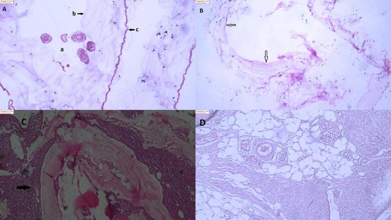
Cystic echinococcosis can involve various organs in humans with the brain and spine being particularly vulnerable. This research aimed to study clinicopathological features and molecular analysis of the central nervous system (CNS) echinococcosis cases in a central hospital for hydatid cyst surgery in northeastern Iran. CNS echinococcosis cases from surgically managed human CE cases at Ghaem hospital in northeastern Iran were analyzed from 2012 to 2022. Demographic and clinicopathological data were collected for CNS echinococcosis cases and formalin-fixed paraffin-embedded (FFPE) blocks were used for molecular analysis. The total prevalence of CNS echinococcosis cases was 1. 8 %. Most of the CE cases were reported in women (64. 7 %) and from rural areas (58. 8 %). The highest number of cases (41. 2 %) were aged ≤18 years, with majority being ranchers (47. 1 %). Thirteen cases (76.5 %) were found to have cysts in their brain, particularly in the supratentorial site. Headache was the most commonly reported sign in cases (9/13, 69.2 %). Infiltration of eosinophils, polymorphic inflammatory cells, and giant cells, gliosis, and foreign body granulomatous reaction, along with mild infiltration of mononuclear cells showing degeneration and necrotic foci in the brain infections. Spine infections included bone cartilage, ligaments, and hydatid cyst wall fragments. PCR analysis conducted on 17 samples revealed the presence of 13 isolates of E. granulosus sensu lato. Among these, 11 were classified within the E. granulosus sensu stricto (G1 and/or G3) complex, while 2 isolates were identified as belonging to the E. canadensis G6/G7. Cerebrospinal infection is a significant aspect of CE cases in northcentral Iran, with a higher prevalence among women and in rural areas. Children were the most affected age group, with the E. granulosus s.s. genotypes being the most common.
Keywords: Cerebral echinococcosis; Iran; PCR; Phylogenetic analysis; Spinal echinococcosis.
© 2025 The Author(s). Published by Elsevier Ltd on behalf of World Federation of Parasitologists.
Conflict of interest statement
The authors declare that they have no known competing financial interests or personal relationships that could have appeared to influence the work reported in this paper.
Figures


Similar articles
-
Application and evaluation of native antigen B from Echinococcus granulosus sensu stricto and E. canadensis alone or mixture for serodiagnosis of human G1-G3 and G6/G7 genotypes cystic echinococcosis sera, using ELISA and Western blotting.Parasitol Res. 2023 Sep;122(9):2227-2236. doi: 10.1007/s00436-023-07924-1. Epub 2023 Jul 13. Parasitol Res. 2023. PMID: 37438467
-
Histopathologic Alterations between Echinococcus granulosus sensu stricto and E. canadensis Genotypes of Human Cystic Echinococcosis Cysts in Shiraz, Iran.Iran J Parasitol. 2025 Jan-Mar;20(1):21-31. doi: 10.18502/ijpa.v20i1.18102. Iran J Parasitol. 2025. PMID: 40206365 Free PMC article.
-
Molecular characterization of human Echinococcus isolates and the first report of E. canadensis (G6/G7) and E. multilocularis from the Punjab Province of Pakistan using sequence analysis.BMC Infect Dis. 2020 Apr 3;20(1):262. doi: 10.1186/s12879-020-04989-6. BMC Infect Dis. 2020. PMID: 32245373 Free PMC article.
-
Echinococcus granulosus sensu lato genotypes infecting humans--review of current knowledge.Int J Parasitol. 2014 Jan;44(1):9-18. doi: 10.1016/j.ijpara.2013.08.008. Epub 2013 Nov 19. Int J Parasitol. 2014. PMID: 24269720 Review.
-
Species and genotypes belonging to Echinococcus granulosus sensu lato complex causing human cystic echinococcosis in Europe (2000-2021): a systematic review.Parasit Vectors. 2022 Mar 28;15(1):109. doi: 10.1186/s13071-022-05197-8. Parasit Vectors. 2022. PMID: 35346335 Free PMC article.
References
-
- Alvarez Rojas C.A., Romig T., Lightowlers M.W. Echinococcus granulosus sensu lato genotypes infecting humans--review of current knowledge. Int. J. Parasitol. 2014;44:9–18. - PubMed
-
- Ashraf M., Ahmed S., Ahmad S., Ahmad A. A large hydatid cyst in the brain of a 10-year child. J. Coll. Physicians Surg. Pak. 2022;32:538–540. - PubMed
-
- Bowles J., Blair D., McManus D.P. Genetic variants within the genus Echinococcus identified by mitochondrial DNA sequencing. Mol. Biochem. Parasitol. 1992;54:165–173. - PubMed
-
- Castillo S., Manterola C., Grande L., Rojas C. Infected hepatic echinococcosis. Clinical, therapeutic, and prognostic aspects. A systematic review. Ann. Hepatol. 2021;22 - PubMed
-
- Deplazes P., Rinaldi L., Alvarez Rojas C.A., Torgerson P.R., Harandi M.F., Romig T., Antolova D., Schurer J.M., Lahmar S., Cringoli G., Magambo J., Thompson R.C., Jenkins E.J. Global distribution of alveolar and cystic echinococcosis. Adv. Parasitol. 2017;95:315–493. - PubMed
LinkOut - more resources
Full Text Sources
Research Materials
Miscellaneous
