. 2025 Feb;292(2041):20242867.
doi: 10.1098/rspb.2024.2867. Epub 2025 Feb 19.

Phylogenomics of the rarest animals: a second species of Micrognathozoa identified by machine learning

Affiliations

Phylogenomics of the rarest animals: a second species of Micrognathozoa identified by machine learning

Shoyo Sato et al. Proc Biol Sci. 2025 Feb.

Abstract

The latest animal phylum to be discovered, Micrognathozoa, constitutes a rare group of limnic meiofauna. These microscopic 'jaw animals' are among the smallest metazoans yet possess highly complex jaw structures. The single species of Micrognathozoa, Limnognathia maerski Kristensen and Funch, 2000, was first described from Greenland, later reported from a remote Subantarctic island and more recently discovered in the Pyrenees on the European continent. Successful collections of these three known populations facilitated investigations of the intraphylum relationships and species limits within Limnognathia for the first time. Through detailed anatomical comparisons, we substantiate the lack of morphological differences between the three geographically disjunct populations. With transcriptomic data from single specimens, we conducted the first intraphylum phylogenetic analyses and extensively tested species hypotheses using standard approaches and novel machine learning methods. Analyses clearly delimited the Subantarctic population, here described as Limnognathia desmeti sp. nov., the second species of Micrognathozoa, but did not definitively split the Greenland and Pyrenees populations as separate species. Divergence dating analysis suggests the disjunct distribution of Micrognathozoa is not human mediated but the result of long-distance dispersal raising questions about their dispersal capabilities and potential undiscovered populations.

Keywords: Spiralia; confocal laser scanning microscopy; integrated taxonomy; meiobenthos; phylotranscriptomics; species delimitation.


Conflict of interest statement

We declare we have no competing interests.

Figures
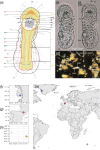
Distribution and habitus of Micrognathozoa.
Figure 1.
Distribution and habitus of Micrognathozoa. (a) Schematic of Micrognathozoa showing key morphological characteristics investigated in this study. (b) Ventral and (c) LM of Limnognathia desmeti sp. nov. (paratype, NHMD-001801226). (d) Dissecting scope image of Limnognathia desmeti sp. nov. among sampled substrate. (e) Map of the three populations of Micrognathozoa sampled for this study. Circles denote populations of Limnognathia maerski and diamonds denote Limnognathia desmeti sp. nov. (f) Isunngua Spring, Disko Island, Greenland. (g) Bassa Nera, Aigüestortes i Estany de Sant Maurici NP, Catalunya. (h) Pointe du Bougainville, Île de la Possession, Crozet Archipelago. Colours in the map correspond to the colours used in all other figures. abc, abdominal ciliophores; acf, anterior ciliated field; acp, adhesive ciliary pad; adpo, apical dorsal plate outline; ap, apicalia; apn, anterior protonephridia; ca, caudalia; ct, ciliary tufts; dj, dorsal jaw; do, dorsalia; dpo, dorsal plate outline; egg, egg; fi, fibularium; hc, head ciliophores; id, intermediate duct; la, lateralia; mc, mouth ciliation; mj, main jaw; np, neuropil; ov, oviduct; pcf, posterior ciliated field; pdvj, pseudodigit of the ventral jaw; pn, peripheral nerve; ppn, posterior protonephridia; tc, trunk ciliophores; vln, ventro-lateral nerve; vj, ventral jaw; vmn, ventro-median nerve.
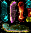
CLSM of Limnognathia. All images maximum intensity z-projection.
Figure 2.
CLSM of Limnognathia. All images maximum intensity z-projection. Anterior end up on (a)–(f), and left on (g). (a) Dorsal Limnognathia desmeti sp. nov. (paratype, NHMD-001801226) whole body showing epidermal plates (cyan = anti-serotonin-like immunostaining background signal (anti-5 HT)). (b) Left side Limnognathia desmeti sp. nov. (holotype, NHMD-001801220) whole body showing cilia and epidermal plates (yellow = acetylated α-tubulin-like immunoreactivity (α-tub-LIR), magenta = anti-5 HT background signal). (c) Ventral Limnognathia desmeti sp. nov. (holotype) whole body showing ciliated cells (glow = α-tub-LIR). (d) Dorsal anterior Limnognathia maerski Greenland, detail of apical plate (cyan = anti-synapsin1-like immunostaining background signal (ENZO Life Sciences, ADI-VAS-SV061-E)). (e) Lateral thoracic section Limnognathia desmeti sp. nov. (paratype, NHMD-001801223) detail of nephridial structures (glow = α-tub-LIR). (f) Ventro-posterior abdomen of Limnognathia desmeti sp. nov. (paratype, NHMD-001801223) detail of adhesive ciliary pad (yellow = α-tub-LIR). (g) Mid-sagittal body of Limnognathia desmeti sp. nov. (holotype) whole body showing ciliation and internal structures (yellow = α-tub-LIR, cyan = DAPI, magenta = anti-5 HT background signal). dob, dorsalia cell body; dp, dorsal plate; en, endodermis; gl, gut lumen; all other abbreviations are identical to figure 1. Cells of the apical plate are numbered 1−4.
Phylotranscriptomic and species delimitation analyses of Micrognathozoa and single nucleotide polymorphisms in 18S rRNA.
Figure 3.
Phylotranscriptomic and species delimitation analyses of Micrognathozoa and single nucleotide polymorphisms in 18S rRNA. (a) Summary of phylogenetic and species delimitation analysis. Topology obtained from the maximum likelihood and Bayesian analysis of the 50% occupancy matrix. All nodes received full support in both analyses (BS = 100%, PP = 1). Differences in topology among the four analyses (50 versus 70% occupancy and maximum likelihood versus Bayesian) are summarized by the stability plots. Colours correspond to figure 1fh. Results of species delimitation analyses are plotted to the right of the phylogeny. Blocks correspond to the number of recovered species. (b) Variation in 18S rRNA among Limnognathia. Fixed molecular diagnostic characters highlighted in green and other variable characters differing between the two species are highlighted in grey. Characters for Limnognathia desmeti sp. nov. are bolded.

References

    1. Kristensen RM, Funch P. 2000. Micrognathozoa: a new class with complicated jaws like those of Rotifera and Gnathostomulida. J. Morphol. 246, 1–49. (10.1002/1097-4687(200010)246:13.0.co;2-d) - DOI - PubMed
    1. De Smet WH. 2002. A new record of Limnognathia maerski Kristensen & Funch, 2000 (Micrognathozoa) from the subantarctic Crozet Islands, with redescription of the trophi. J. Zool. 258, 381–393. (10.1017/s095283690200153x) - DOI
    1. Sørensen MV. 2003. Further structures in the jaw apparatus of Limnognathia maerski (Micrognathozoa), with notes on the phylogeny of the Gnathifera. J. Morphol. 255, 131–145. (10.1002/jmor.10038) - DOI - PubMed
    1. Sørensen MV. 2000. An SEM study of the jaws of Haplognathia rosea and Rastrognathia macrostoma (Gnathostomulida), with a preliminary comparison with the rotiferan trophi. Acta Zool. 81, 200. (10.1046/j.1463-6395.2000.00032.x) - DOI
    1. Bekkouche N, Kristensen RM, Hejnol A, Sørensen MV, Worsaae K. 2014. Detailed reconstruction of the musculature in Limnognathia maerski (Micrognathozoa) and comparison with other Gnathifera. Front. Zool. 11, 71. (10.1186/s12983-014-0071-z) - DOI - PMC - PubMed

LinkOut - more resources