Randomized Controlled Trial
. 2025 May 2;46(17):1634-1644.
doi: 10.1093/eurheartj/ehaf037.

Non-invasive vagus nerve stimulation and exercise capacity in healthy volunteers: a randomized trial

Affiliations
Randomized Controlled Trial

Non-invasive vagus nerve stimulation and exercise capacity in healthy volunteers: a randomized trial

Gareth L Ackland et al. Eur Heart J. .

Abstract

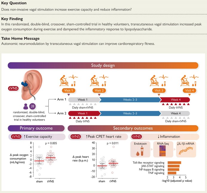
Background and aims: Vagal parasympathetic dysfunction is strongly associated with impaired exercise tolerance, indicating that coordinated autonomic control is essential for optimizing exercise performance. This study tested the hypothesis that autonomic neuromodulation by non-invasive transcutaneous vagus nerve stimulation (tVNS) can improve exercise capacity in humans.

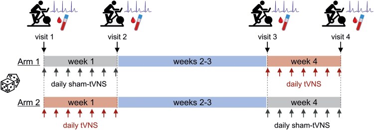
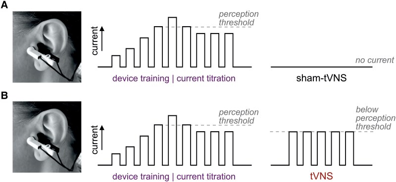
Methods: This single-centre, randomized, double-blind, sham-controlled, crossover trial in 28 healthy volunteers evaluated the effect of bilateral transcutaneous stimulation of vagal auricular innervation, applied for 30 min daily for 7 days, on measures of cardiorespiratory fitness (peak oxygen consumption (VO2peak)) during progressive exercise to exhaustion. Secondary endpoints included peak work rate, cardiorespiratory measures, and the whole blood inflammatory response to lipopolysaccharide ex vivo.

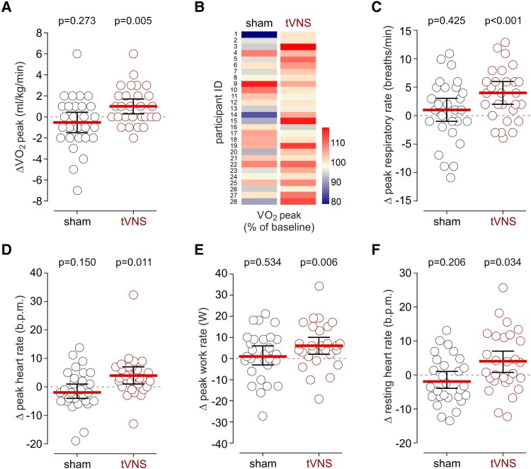
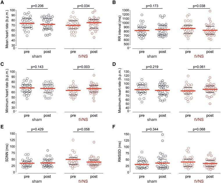
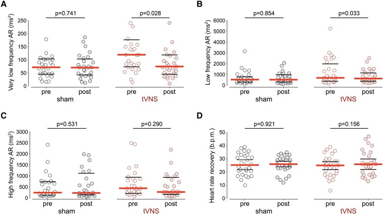
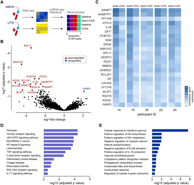
Results: tVNS applied for 30 min daily over 7 consecutive days increased VO2peak by 1.04 mL/kg/min (95% CI: .34-1.73; P = .005), compared with no change after sham stimulation (-0.54 mL/kg/min; 95% CI: -1.52 to .45). No carry-over effect was observed following the 2-week washout period. tVNS increased work rate (by 6 W; 95% CI: 2-10; P = .006), heart rate (by 4 bpm; 95% CI: 1-7; P = .011), and respiratory rate (by 4 breaths/min; 95% CI: 2-6; P < .001) at peak exercise. Analysis of the whole blood transcriptomic response to lipopolysaccharide in serial samples obtained from five participants showed that tVNS reduced the inflammatory response.

Conclusions: Non-invasive vagal stimulation improves measures of cardiorespiratory fitness and attenuates inflammation, offering an inexpensive, safe, and scalable approach to improve exercise capacity.

Keywords: Ageing; Autonomic nervous system; Exercise; Neuromodulation; Vagus nerve.


Figures

Structured Graphical Abstract
Methods, primary and secondary outcomes for randomized, double-blind, crossover, sham-controlled trial of non-invasive vagal stimulation in healthy volunteers. tVNS, transcutaneous vagus nerve stimulation; CPET, cardiopulmonary exercise test; RNAseq, RNA sequencing; IL-1β mRNA, interleukin-1 beta messenger RNA; JAK-STAT, Janus kinase signal transducer and activator of transcription; NF-kappa B, Nuclear factor kappa-light-chain-enhancer of activated B cells; TNF, tumour necrosis factor.
Figure 1
Figure 1
Study protocol. 28 volunteers (14 females) were randomized into two arms of the study to first receive either sham-tVNS or tVNS applied for 30 min daily for 7 consecutive days, followed by a 2-week washout period before receiving the alternative treatment. At each visit, after 10 min of ECG recording followed by orthostatic testing, participants undertook exhaustive cardiopulmonary exercise testing using cycle ergometry. Participants were issued individual sham-tVNS or tVNS device units and were trained and instructed to use the device for 30 min at a standardized time each day for 7 consecutive days. Volunteers were contacted by video call during each session to ensure correct electrode clip placement and correct device use, as well as to monitor for potential adverse effects. A 10 mL venous blood sample was obtained at each visit from the participants who consented to repeated venepuncture
Figure 2
Figure 2
Device settings. The individual sensitivities of the auricular tragi regions to electrical stimulation were determined during the first and third visits using a dedicated training device. During device training, participants were instructed to place the electrode clips on the left and right tragi. The current amplitude was gradually increased by the investigator conducting the training, starting from 0.1 mA, until the participant felt a tingling sensation. The current was then reduced to set the level of stimulation at ∼1.5 mA below this threshold. Once this threshold current was determined, participants were issued their personal device units, identical to the training device but with concealed controls. The stimulation current was set to 0 mA (A, sham-tVNS) or ∼1.5 mA below the individual perception threshold (B, tVNS), with 200 μs pulses generated at a frequency of 25 Hz
Figure 3
Figure 3
The effect of tVNS on measures of exercise capacity. (A) tVNS increased VO2peak by 1.04 mL/kg/min (95% CI: .34–1.73), compared to no change after sham stimulation (−0.54 mL/kg/min; 95% CI: −1.52 to .45). (B) Percentage changes in VO2peak after sham-tVNS or tVNS illustrated by heatmaps. tVNS increased VO2peak by 3.8% (95% CI: 1.5–6.1), compared with no change after sham-tVNS (mean difference: −1.3%, 95% CI: −4.26 to 1.56). (C) tVNS increased respiratory rate at peak exercise by 4 breaths/min (95% CI: 2–6), compared to no change after sham stimulation. (D) tVNS increased peak heart rate by 4 b.p.m. (95% CI: 1–7), compared to no change after sham stimulation. (E) tVNS increased power output at peak exercise by 6 W (95% CI: 2–10), compared to no change after sham stimulation. (F) tVNS increased resting heart rate measured in the supine position before CPET by 4 b.p.m. (95% CI: 1–7). Individual values and means (95% CI) are shown. P-values were determined by paired t-test comparisons between measurements taken at baseline and after sham-tVNS or tVNS treatment
Figure 4
Figure 4
The effect of tVNS on time-domain measures of heart rate variability. Mean heart rate (absolute values, A), RR interval (B), minimum heart rate (C), maximum heart rate (D), standard deviation of the RR intervals (SDNN, E), and the root mean square of successive differences between normal heartbeats (RMSSD, F) were derived from ECG recordings obtained from participants in the supine position before and after sham-tVNS or tVNS. Individual values, medians, and 25th–75th percentiles are shown. P-values were determined by paired t-test comparisons between measurements taken at baseline and after sham-tVNS or tVNS treatment
Figure 5
Figure 5
The effect of tVNS on frequency-domain measures of heart rate variability and heart rate recovery after exercise. Very-low frequency (<0.04 Hz, A), low frequency (0.04–0.15 Hz, B), and high frequency (0.15–0.4 Hz, C) bands of the HRV power spectrum were derived from the analysis of ECG recordings obtained from participants in the supine position before and after sham-tVNS or tVNS. (D) Heart rate recovery 1 min after the end of peak exercise, measured before and after sham-tVNS or tVNS treatment. Individual values, medians, and 25th–75th percentiles are shown. P-values were determined by paired t-test comparisons between measurements taken at baseline and after sham-tVNS or tVNS treatment
Figure 6
Figure 6
The effect of tVNS on the inflammatory response. (A) Whole blood samples were obtained from five volunteers who consented to repeated venepuncture. The samples were incubated with either sterile saline or lipopolysaccharide (LPS, 20 ng/mL). Bulk RNAseq was performed, followed by single-cell RNAseq referenced deconvolution. (B) Volcano plot illustrating differentially expressed genes in LPS-treated whole blood samples obtained from the participants that received sham-tVNS or tVNS. P-values were calculated using Benjamini–Hochberg adjustment (FDR < 0.05) for multiple testing. (C) Heatmap illustrating changes in the expression of 23 genes that were differentially affected by tVNS (false discovery rate <0.05; minimum fold-change ≥1.5). (D) Gene Ontology (KEGG) enrichment analysis for signaling pathways affected by tVNS. (E) Gene Ontology enrichment analysis for cellular processes affected by tVNS, derived from the genes differentially expressed in response to LPS stimulation

Similar articles

Cited by

References

    1. Kohl HW, Craig CL, Lambert EV, Inoue S, Alkandari JR, Leetongin G, et al. The pandemic of physical inactivity: global action for public health. Lancet Lond Engl 2012;380:294–305. 10.1016/S0140-6736(12)60898-8 - DOI - PubMed
    1. Valenzuela PL, Ruilope LM, Santos-Lozano A, Wilhelm M, Kränkel N, Fiuza-Luces C, et al. Exercise benefits in cardiovascular diseases: from mechanisms to clinical implementation. Eur Heart J 2023;44:1874–89. 10.1093/eurheartj/ehad170 - DOI - PubMed
    1. Mandsager K, Harb S, Cremer P, Phelan D, Nissen SE, Jaber W. Association of cardiorespiratory fitness with long-term mortality among adults undergoing exercise treadmill testing. JAMA Netw Open 2018;1:e183605. 10.1001/jamanetworkopen.2018.3605 - DOI - PMC - PubMed
    1. Blodgett JM, Ahmadi MN, Atkin AJ, Chastin S, Chan H-W, Suorsa K, et al. Device-measured physical activity and cardiometabolic health: the prospective physical activity, sitting, and sleep (ProPASS) consortium. Eur Heart J 2024;45:458–71. 10.1093/eurheartj/ehad717 - DOI - PMC - PubMed
    1. Bauman AE, Reis RS, Sallis JF, Wells JC, Loos RJ, Martin BW. Correlates of physical activity: why are some people physically active and others not? Lancet 2012;380:258–71. 10.1016/S0140-6736(12)60735-1 - DOI - PubMed

Publication types