. 2024 Dec 27;103(52):e41051.
doi: 10.1097/MD.0000000000041051.

Exploring phenotypic and genotypic diversity among methicillin-resistant, vancomycin-resistant, and sensitive Staphylococcus aureus

Affiliations

Exploring phenotypic and genotypic diversity among methicillin-resistant, vancomycin-resistant, and sensitive Staphylococcus aureus

Walid Bakeer et al. Medicine (Baltimore). .

Abstract

Background: Methicillin-Resistant Staphylococcus aureus (MRSA) is a global concern owing to the increasing prevalence of multidrug-resistant (MDR) strains. Vancomycin has been the primary treatment for MRSA; however, Vancomycin-resistant strains are being increasingly reported worldwide. Therefore, comparative studies are essential to support antimicrobial stewardship and improving clinical management. Ultimately, the findings from this study are expected to inform treatment strategies and guide public health interventions effectively.

Material and methods: This study investigated the prevalence, antimicrobial resistance, and virulence characteristics of Vancomycin-sensitive S. aureus (VSSA) and Vancomycin-resistant S. aureus (VRSA) within MRSA strains. By employing a combination of phenotypic methods, such as antimicrobial susceptibility testing, and genotypic techniques, including molecular typing and identification of virulence genes, we obtained comprehensive insights into VRSA and VSSA profiles.

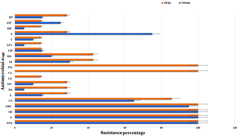
Results: Of 250 clinical samples, 62 (24.8%) were S. aureus and 27 (43.5%) were identified as MRSA. All MRSA isolates exhibited MDR patterns. Most MRSA strains were VSSA (20/27, 74.1%), while 7 (25.9%) were VRSA. The VRSA isolates showed more antimicrobial resistance than VSSA isolates; however, the VRSA isolates had less virulence than VSSA isolates. Linezolid was the most effective treatment, with a 3.7% resistance rate. A higher percentage of biofilm-producing MRSA (96.3%) was confirmed by both phenotypic and genotypic methods. All isolates, except one VRSA, showed multi-virulence patterns (harbored more than 3 virulence genes). High diversity and low clonality (D-value = 0.99) were found in both VSSA and VRSA. Based on our correlation findings, the emergence of vancomycin resistance could modify the association between antimicrobial resistance and virulence, potentially affecting the pathogenic profile of these strains. The study also revealed complex interactions among host factors (including age and gender), sample origin, antimicrobial resistance, biofilm production, and virulence genes.

Conclusion: This study highlights the alarming spread of MRSA and VRSA, which show significant resistance and virulence.


Conflict of interest statement

The authors have no conflicts of interest to disclose.

Figures

None
Graphical abstract
Figure 1.
Figure 1.
Antimicrobial resistance percentages of VSSA and VRSA isolates. AMC = amoxicillin/clavulanic acid, C = chloramphenicol, CIP = ciprofloxacin, CN = gentamicin, DA = clindamycin, DO = doxycycline, E = erythromycin, F = fusidic acid, FOX = cefoxitin, LEV = levofloxacin, LZ = linezolid, NIT = nitrofurantoin, OX = oxacillin, P = benzylpenicillin, PR = pristinomycin, RIF = rifampin, SXT = trimethoprim/sulfamethoxazole, TE = tetracycline, TEC = teicoplanin, VA = vancomycin, VRSA = Vancomycin-resistant S. aureus, VSSA= Vancomycin-sensitive S. aureus.
Figure 2.
Figure 2.
Percentages of virulence genes among VSSA and VRSA isolates. clfA = clumping factors A, eta = exfoliative toxins A, etb = exfoliative toxins B, hla = alpha hemolysin, hlb = beta hemolysin, hlg = gamma hemolysin, lukSF-PV = panton–valentin leukocydin, sea, seb, sec, sed, see, seg, seh, sei, and sej = staphylococcal enterotoxins A,B,C,D,E,G,H,I,J, tst = toxic shock syndrome toxin-1, VRSA = Vancomycin-resistant S. aureus, VSSA = Vancomycin-sensitive S. aureus.
Figure 3.
Figure 3.
Correlation analysis between biofilm formation and antimicrobial resistance profiles (A) and biofilm formation and virulence gene profiles (B) in MRSA isolates. (A) Correlation matrix showing the relationship between biofilm formation and antimicrobial resistance to various antibiotics. Each cell in the matrix represents the Pearson correlation coefficient (r) between two variables, with values ranging from −1 to +1. Positive correlations are shown in red, while negative correlations are in blue, with the intensity indicating the strength of the correlation. Antibiotics tested include rifampicin (RP), trimethoprim-sulfamethoxazole (SXT), rifampicin (RIF), fosfomycin (F), clindamycin (C), levofloxacin (LEV), ciprofloxacin (CIP), doxycycline (DO), tetracycline (TE), teicoplanin (TEI), vancomycin (VA), linezolid (LZ), nitrofurantoin (NIT), clindamycin (DA), erythromycin (E), and gentamicin (CN). (B) Correlation matrix displaying the association between biofilm formation and the presence of virulence genes in S. aureus. The virulence genes assessed include hla (α-hemolysin), lukED (leukocidin ED), hlg (γ-hemolysin), icaD and icaA (biofilm-related intercellular adhesion genes), tst (toxic shock syndrome toxin-1), see (enterotoxin E), and sed (enterotoxin D), As with panel A, red cells indicate positive correlations, and blue cells represent negative correlations, with color intensity reflecting correlation strength. MRSA = Methicillin-Resistant S. aureus.
Figure 4.
Figure 4.
Correlation between virulence genes and antimicrobial resistance among VSSA and VRSA Isolates. This figure shows correlation heatmaps illustrating the relationships between virulence genes and antimicrobial resistance among MRSA isolates: (A) Vancomycin-Susceptible S. aureus (VSSA) and (B) Vancomycin-Resistant S. aureus (VRSA). Positive correlations are represented in red, and negative correlations in blue, with darker shades indicating stronger correlations (range −1 to +1). These correlations highlight potential associations between virulence factors and resistance profiles in both VSSA and VRSA strains, which could suggest differing mechanisms of resistance and pathogenicity. AMC (amoxicillin-clavulanic acid), CN (gentamicin), E (erythromycin), DA (clindamycin), NIT (nitrofurantoin), TE (tetracycline), DO (doxycycline), CIP (ciprofloxacin), LEV (levofloxacin), C (chloramphenicol), F (fusidic acid), RIF (rifampicin), SXT (trimethoprim-sulfamethoxazole), RP (quinupristin-dalfopristin), icaA (intercellular adhesion A), icaD (intercellular adhesion D), tst (toxic shock syndrome toxin), see (enterotoxin E), sed (enterotoxin D), hla (alpha-hemolysin), hlg (gamma-hemolysin), lukED (Panton-Valentine leukocidin ED). MRSA = Methicillin-Resistant S. aureus.
Figure 5.
Figure 5.
Correlation of antimicrobial resistance, virulence genes, biofilm formation, patient demographics, and sample types among all detected MRSA isolates. This heatmap illustrates the correlations among antimicrobial resistance profiles, virulence genes, biofilm formation (MTP), demographic characteristics (gender, age groups), and sample types (urine, blood, wound, pus) in MRSA isolates. Positive correlations are displayed in red, and negative correlations in blue, with color intensity reflecting the strength of the correlation (range −1 to +1). These correlations provide insights into potential associations between resistance, virulence, patient factors, and sample origins, which may help in understanding risk factors and patterns of infection. AMC (amoxicillin-clavulanic acid), CN (gentamicin), E (erythromycin), DA (clindamycin), NIT (nitrofurantoin), TE (tetracycline), DO (doxycycline), CIP (ciprofloxacin), LEV (levofloxacin), C (chloramphenicol), F (fusidic acid), RIF (rifampicin), SXT (trimethoprim-sulfamethoxazole), RP (quinupristin-dalfopristin), CRO (ceftriaxone), VA (vancomycin), LZ (linezolid), hib, hla (alpha-hemolysin), lukED (Panton-Valentine leukocidin ED), hlg (gamma-hemolysin), icaA (intercellular adhesion A), icaD (intercellular adhesion D), tst (toxic shock syndrome toxin), see (enterotoxin E), sed (enterotoxin D), Biofilm Formation: MTP (Microtiter Plate Assay), Demographics: Gender (Female, Male), Age groups (Age (20/25), Age (25/30), etc.), Sample Types: Urine, Blood, Wound, Pus. MRSA = Methicillin-Resistant S. aureus.
Figure 6.
Figure 6.
Fan hierarchical clustering showing the heterogeneity and diversity of VSSA and VRSA strains. This circular dendrogram represents the hierarchical clustering of VRSA and VSSA strains. The strains are labeled by numerical values which represent the order of recovery around the circumference of the circle. Red color indicates the resistance pattern or the presence of this variable, while blue color indicates the sensitivity pattern or the absence of this variable. VRSA = Vancomycin-resistant S. aureus, VSSA= Vancomycin-sensitive S. aureus.

References

    1. Luzzago C, Locatelli C, Franco A, et al. . Clonal diversity virulence-associated genes and antimicrobial resistance profile of Staphylococcus aureus isolates from nasal cavities and soft tissue infections in wild ruminants in Italian Alps. Vet Microbiol. 2014;170:157–61. - PubMed
    1. Saleem N, Nawaz M, Ghafoor A, et al. . Phenotypic and molecular analysis of antibiotic resistance in lactobacilli of poultry origin from Lahore Pakistan. Pak Vet J. 2018;38:409.
    1. Shariati A, Dadashi M, Moghadam MT, van Belkum A, Yaslianifard S, Darban-Sarokhalil D. Global prevalence and distribution of vancomycin resistant vancomycin intermediate and heterogeneously vancomycin intermediate Staphylococcus aureus clinical isolates: a systematic review and meta-analysis. Sci Rep. 2020;10:12689. - PMC - PubMed
    1. El-Aziz NKA, El-Hamid MIA, Bendary MM, El-Azazy AA, Ammar AM. Existence of vancomycin resistance among methicillin-resistant Staphylococcus aureus recovered from animal and human sources in Egypt. Slovenian Vet Res. 2018;55:221–30.
    1. Shahkarami F, Rashki A, Rashki Ghalehnoo Z. Microbial susceptibility and plasmid profiles of methicillin-resistant Staphylococcus aureus and methicillin-susceptible S. aureus. Jundishapur J Microbiol. 2014;7:e16984. - PMC - PubMed

MeSH terms