

Synchronous pancreatic adenocarcinoma and duodenal mucosa‑associated lymphoid tissue lymphoma: A case report
- PMID: 39969306
- PMCID: PMC11688084
- DOI: 10.1097/MD.0000000000041173
Synchronous pancreatic adenocarcinoma and duodenal mucosa‑associated lymphoid tissue lymphoma: A case report
Abstract
Rationale: Duodenal mucosa-associated lymphoid tissue (MALT) lymphoma is a rare condition. Simultaneous presence of pancreatic ductal adenocarcinoma along with duodenal MALT lymphoma has not been documented in the scientific literature. We report an exceptionally rare case of synchronous duodenal MALT lymphoma and pancreatic ductal adenocarcinoma.
Patient concerns: A 75-year-old man was referred to our hospital with dyspepsia and weight loss.
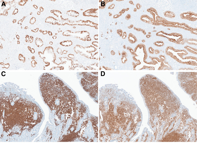
Diagnoses: Esophagogastroduodenoscopy was performed, revealing synchronous tumor comprising pancreatic ductal adenocarcinoma and MALT lymphoma of the duodenum.
Interventions: Given that the pancreatic carcinoma would be the primary determinant of prognosis, we prioritized treatment of the pancreatic carcinoma. Consequently, we performed a Whipple operation first. Post-operative pathologic examination revealed metastasis of pancreatic cancer to peri-pancreatic lymph nodes, whereas the MALT lymphoma was localized to the duodenum; therefore, only adjuvant chemotherapy for pancreatic cancer was performed.
Outcomes: To date, the patient has had no recurrence of either the pancreatic cancer or the MALT lymphoma.
Lessons: To the best of our knowledge, this is the first case to be reported. Awareness of this co-occurrence may help diagnosis and management of similar cases.
Copyright © 2024 the Author(s). Published by Wolters Kluwer Health, Inc.
Conflict of interest statement
The authors have no conflicts of interest to disclose.
Figures


Similar articles
-
Primary pulmonary extranodal marginal zone lymphoma/low grade B-cell lymphoma of MALT type combined with well-differentiated adenocarcinoma.Tumori. 2010 Jan-Feb;96(1):168-71. doi: 10.1177/030089161009600129. Tumori. 2010. PMID: 20437878
-
A case report of a collision tumor composed of pancreatic ductal adenocarcinoma and peri-pancreatic mucosa-associated lymphoid tissue lymphoma.World J Surg Oncol. 2023 Mar 28;21(1):110. doi: 10.1186/s12957-023-02981-3. World J Surg Oncol. 2023. PMID: 36973717 Free PMC article.
-
Synchronous duodenal mucosa-associated lymphoid tissue lymphoma and gastric cancer.Clin J Gastroenterol. 2021 Feb;14(1):109-114. doi: 10.1007/s12328-020-01241-1. Epub 2020 Sep 21. Clin J Gastroenterol. 2021. PMID: 32959165
-
Synchronous primary pulmonary adenocarcinoma and extranodal marginal zone lymphoma of mucosa-associated lymphoid tissue: A case report and literature review.Medicine (Baltimore). 2020 Jul 17;99(29):e20865. doi: 10.1097/MD.0000000000020865. Medicine (Baltimore). 2020. PMID: 32702827 Free PMC article. Review.
-
Synchronous Pancreatic Serous Cystic Neoplasm and Duodenal Neuroendocrine Tumor: Case Report and Review of the Literature.Int J Surg Pathol. 2018 Sep;26(6):551-557. doi: 10.1177/1066896918766245. Epub 2018 Apr 6. Int J Surg Pathol. 2018. PMID: 29623746 Review.
References
-
- Lepicard A, Lamarque D, Levy M, et al. . Duodenal mucosa-associated lymphoid tissue lymphoma: treatment with oral cyclophosphamide. Am J Gastroenterol. 2000;95:536–9. - PubMed
-
- Isaacson P, Wright DH. Malignant lymphoma of mucosa-associated lymphoid tissue. A distinctive type of B-cell lymphoma. Cancer. 1983;52:1410–6. - PubMed
-
- Fujishima F, Katsushima H, Fukuhara N, et al. . Incidence rate, subtype frequency, and occurrence site of malignant lymphoma in the gastrointestinal tract: population-based analysis in Miyagi, Japan. Tohoku J Exp Med. 2018;245:159–65. - PubMed
-
- Bertoni F, Coiffier B, Salles G, et al. . MALT lymphomas: pathogenesis can drive treatment. Oncology (Williston Park). 2011;25:1134–42, 1147. - PubMed
Publication types
MeSH terms
LinkOut - more resources
Full Text Sources
Medical
