. 2025 Mar;301(3):108325.
doi: 10.1016/j.jbc.2025.108325. Epub 2025 Feb 18.

Cdc42EP3-bound septin scaffolds promote actin polymerization

Affiliations

Cdc42EP3-bound septin scaffolds promote actin polymerization

Meagan R Tomasso et al. J Biol Chem. 2025 Mar.

Abstract

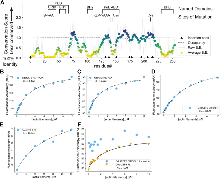
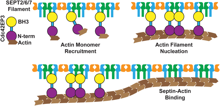
Septins are cytoskeletal filament-forming proteins that typically associate with membranes and perform critical functions in a variety of cellular processes. Septins often colocalize with actin and microtubule structures, yet our understanding of all the ways that septins contribute mechanistically to actin- and microtubule-based functions is incomplete. The Cdc42 effector protein Cdc42EP3 (also known as BORG2) promotes septin localization to actin structures in vivo, but little else is known about how Cdc42EP3 influences the interactions of septins and F-actin. Here, using purified components, we show that Cdc42EP3 binds directly to septins, actin filaments, and actin monomers. Moreover, septin-bound Cdc42EP3 accelerates actin filament polymerization. Thus, Cdc42EP3 is not merely a factor that crosslinks septins and F-actin, but one that promotes the formation of actin polymers along septin scaffolds.

Keywords: CDC42; Cdc42EP/BORG; G-actin; actin; bundling; cytoskeleton; filament nucleation; microfilaments; septin.


Conflict of interest statement

Conflicts of interest The authors declare that they have no conflicts of interest with the contents of this article.

Figures

Figure 1
Figure 1
Purification and biochemical characterization of Cdc42EP3. A, purified Cdc42EP3 resolved using SDS-PAGE, imaged using “Stain-Free” imaging (left) and Coomassie Brilliant Blue (CBB) stain (right). B, c(s) distributions of Cdc42EP3 at three different concentrations measured using SV-AUC. C, gel filtration chromatograms of Cdc42EP3, GMPPNP-loaded Cdc42 or both proteins. D, SDS-PAGE Stain-Free imaged gel of gel filtration fractions from (C). Letters in gel correspond to the lettered fractions in (C). E, gel filtration chromatograms of Cdc42EP3, GDP-loaded Cdc42 or both proteins. F, co-pelleting of Cdc42EP3 with SEPT2/6/7 filaments. Samples were centrifuged at 21,000g for 20 min, supernatant fraction was removed and pelleted fractions were resuspended in high salt buffer. Input (I), Supernatant (S) and Pelleted (P) fractions were analyzed using SDS-PAGE and imaged using Stain-Free imaging. G, band intensity quantification and two-tailed t test of the fraction of Cdc42EP3 that pelleted alone compared to with septin filaments, shown as swarm plot. ∗∗∗ means p < 0.001. The line represents the mean. H, fluorescence anisotropy assay measuring septin filament binding to Cdc42EP3.
Figure 2
Figure 2
Cdc42EP3 binds actin. A, centrifugation assay with actin filaments, Cdc42EP3, or both. Samples were centrifuged at 100,000g for 1 h and separated into supernatant and pelleted fractions. B, quantification of pelleted band intensity and two-tailed t test of the fraction of Cdc42EP3 that pelleted alone versus in the presence of actin filaments. ∗∗ means p < 0.01. Data are shown as a swarm plot, the line represents the mean. C, fluorescence anisotropy assay measuring actin filament (F-actin) binding to Cdc42EP3. Cdc42EP3 was maintained at a fixed 20 nM concentration while an increasing amount of actin filaments were titrated. D, Fluorescence anisotropy assay measuring Latrunculin A bound actin monomer binding to Cdc42EP3. Cdc42EP3 was maintained at a fixed 20 nM concentration while increasing amount of actin monomer was titrated in with 20 μM Latrunculin A. E, chromatogram of analytical gel filtration of Cdc42EP3, monomeric actin or a sample containing both, representative example from n = 3 repeats. F, two-tailed t test of elution volumes in (E). ∗∗ means p < 0.01. Data are shown as swarm plot and the line represents the mean. G, SDS-PAGE Stain-Free imaging of fractions corresponding to the gel filtration experiment in (E).
Figure 3
Figure 3
The N-terminal CRIB/BH1 region of Cdc42EP3 binds actin. A, conservation scores for residues in human Cdc42EP3, where conservation score is the Shannon Entropy. Values range from 0 to 3 with 0 being fully conserved (100% identity) and 3 the least conserved. The locations of named regions, endogenous cysteines and targeted mutations are indicated. Occupancy indicates the fraction of sequences with a residue present in a gapped alignment (1.0 means all sequences have a residue). Arrows indicate sites of insertions in non-human sequences. Fluorescence anisotropy assay measuring binding to actin filaments of TMR labeled (B) 20 nM KLP-AAA Cdc42EP3 mutant, (C) 20 nM IS-AA Cdc42EP3 mutant, (D) 20 nM isolated Cdc42EP3 CRIB/BH1 domains, or (E) 5 nM Cdd42EP3 (1-21) was maintained at fixed concentration while increasing amount of actin filaments were titrated in. TMR label introduced at C211 (B and C) or at the N-terminus (D and E). F, fluorescence anisotropy overlay of FL/WT Cdc42EP3 binding to actin filaments versus Cdc42EP3 binding to actin filaments when the CRIB and BH1 regions have been removed. Truncation did not produce a reliable F.A. change and was not fit. When fit, KD is indicated in the legend.
Figure 4
Figure 4
Septin polymers directly bind actin filaments. A, septins and actin were separately polymerized, mixed, and incubated on ice and centrifuged at 21,000g. B, quantification of band intensities of pelleted actin in the presence or absence of septins. C, septins and actin were mixed and co-polymerized while undergoing dialysis overnight, then centrifuged at 21,000g the following day. D, Pre-formed septin filaments were mixed and incubated with Latrunculin A bound actin monomers and spun down at 21,000g. E, quantification of band intensities of pelleted actin from D in the presence or absence of septins. F, pre-formed actin filaments were incubated with a non-polymerizable SEPT2/6/7 hexamer and then centrifuged at 100,000g. G, quantification of band intensities of pelleted septin hexamer in the presence or absence of actin filaments. For A, C, E, and G Input (I), Supernatant (S), and Pelleted (P) fractions were analyzed using SDS-PAGE and imaged using Stain-Free imaging. For B, E, and G, fractions of actin or septin pelleted are shown as swarm plots (horizontal lines show the mean). B, a one-way ANOVA followed by Tukey-HSD posthoc was performed with n = 3 for each group. ∗∗∗ indicates p < 0.001. E and G, A two-tailed t test was performed with n = 3 for each group. ∗ indicates p < 0.05 and ∗∗∗ indicate p < 0.001.
Figure 5
Figure 5
Co-polymerization and alignment of septins and actin in vitro. Representative fields of view for TIRF imaging of in vitro reconstituted mCherry labeled SEPT2/6/7 filaments, Alexa 488 labeled actin filaments, and Alexa 647 labeled Cdc42EP3 under hydrated conditions. Horizontal groups represent different channels and actin-septin merge composites for each sample. Reactions contain, as indicated, 2 μM actin (10% Alexa488-labeled, 2% biotinylated), 1 μM SEPT2-mCherry/SEPT6/SEPT7-StrepII tag filaments, and/or 700 nM Alexa647-Cdc42EP3. Fields of view were imaged for Alexa488-actin (green), for SEPT2-mCherry (magenta) or for Alexa647-Cdc42EP3. On the right, two-color merge with SEPT2 colored magenta and actin colored green. All panels are at the same scale and the scale bar at the lower right is 10 μm.
Figure 6
Figure 6
Cdc42EP3-septin complexes promote actin polymerization. A, 2 μM actin monomers (10% pyrene labeled) and 10 μM profilin were mixed with polymerization buffer and 500 nM Cdc42EP3 and/or 500 nM SEPT2/6/7 filaments, and actin polymerization was monitored by the increase in pyrene fluorescence intensity over time until polymerization was complete. Only every 15th data point is shown. B, the time to reach 50% complete polymerization (t50) was quantified for each sample. ∗ is p < 0.05 and ∗∗∗ is p < 0.001, using one-way ANOVA followed by Tukey post hoc test. Data shown as swarm plot, with the line representing the mean. C and D, 2 μM actin (10% Alexa488 labeled, 0.25% biotinylated) with 1 μM SEPT2/6/7 filaments (10% mCherry-SEPT2 labeled) form actin filament bundles comparable to those seen in Figure 5 in the absence (C) and presence (D) of 500 nM Alexa647-Cdc42EP3. E and F, images from early time points in TIRF actin assembly assays, showing actin filament assembly in the presence of septins or Cdc42EP3, as indicated at left. Elapsed time from mixing of septin and actin is shown at the lower left of each frame. C–F, scale bars are 50 μm. G, Filament counts over time for four different fields of view for the indicated samples. H, apparent nucleation rates (filament appearance within TIRF field in field of view). I, filament elongation rates from the same reactions. Each point is the average of at least 20 filaments in a field of view. H and I, individual data points are shown as swarm plots, with means shown as horizontal lines. Two-sample, two-tailed t test was used to determine significance, with ∗∗ as p < 0.01 and ∗∗∗ as p < 0.001.
Figure 7
Figure 7
Model for Cdc42EP3 promotion of actin assembly on septin scaffolds. Cartoon depiction of the mechanism by which Cdc42EP3 promotes actin assembly.

Similar articles

References

    1. Hartwell L.H. Genetic control of the cell division cycle in yeast. IV. Genes controlling bud emergence and cytokinesis. Exp. Cell Res. 1971;69:265–276. - PubMed
    1. Dolat L., Hunyara J.L., Bowen J.R., Karasmanis E.P., Elgawly M., Galkin V.E., et al. Septins promote stress fiber-mediated maturation of focal adhesions and renal epithelial motility. J. Cell Biol. 2014;207:225–235. - PMC - PubMed
    1. Kim S.K., Shindo A., Park T.J., Oh E.C., Ghosh S., Gray R.S., et al. Planar cell polarity acts through septins to control collective cell movement and ciliogenesis. Science. 2010;329:1337–1340. - PMC - PubMed
    1. Kim J., Cooper J.A. Septins regulate junctional integrity of endothelial monolayers. Mol. Biol. Cell. 2018;29:1693–1703. - PMC - PubMed
    1. Beber A., Taveneau C., Nania M., Tsai F.C., Di Cicco A., Bassereau P., et al. Membrane reshaping by micrometric curvature sensitive septin filaments. Nat. Commun. 2019;10:420. - PMC - PubMed

LinkOut - more resources