. 2025 Feb 19;16(1):1769.
doi: 10.1038/s41467-025-56844-4.

Ball-and-chain inactivation of a human large conductance calcium-activated potassium channel

Affiliations

Ball-and-chain inactivation of a human large conductance calcium-activated potassium channel

Shubhangi Agarwal et al. Nat Commun. .

Abstract

BK channels are large-conductance calcium (Ca2+)-activated potassium channels crucial for neuronal excitability, muscle contraction, and neurotransmitter release. The pore-forming (α) subunits co-assemble with auxiliary (β and γ) subunits that modulate their function. Previous studies demonstrated that the N-termini of β2-subunits can inactivate BK channels, but with no structural correlate. Here, we investigate BK β2-subunit inactivation using cryo-electron microscopy, electrophysiology and molecular dynamics simulations. We find that the β2 N-terminus occludes the pore only in the Ca2+-bound open state, via a ball-and-chain mechanism. The first three hydrophobic residues of β2 are crucial for occlusion, while the remainder of the N-terminus remains flexible. Neither the closed channel conformation obtained in the absence of Ca2+ nor an intermediate conformation found in the presence of Ca2+ show density for the N-terminus of the β2 subunit in their pore, likely due to narrower side access portals preventing their entry into the channel pore.


Conflict of interest statement

Competing interests: The authors declare no competing interests.

Figures

Fig. 1
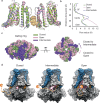
Fig. 1. N-terminus of β2 subunit inactivates hSlo1 channels by plugging the open pore.
a Representative excised inside-out patch clamp current traces in response to voltage steps from −100 mV to +140 mV with a holding potential of −80 mV, each with a duration of 500 msec, from hSlo1, hSlo1 + β2, and hSlo1 + β2N-β4 channels expressed in HEK293S GnTI- cells. The bath solution contained 10 μM free Ca2+. Prior to the depolarizing voltage steps, a 50 ms, −180 mV pulse was applied. b Plot of the ratio of steady state and maximal currents for hSlo1, hSlo1 + β2, and hSlo1 + β2N-β4 from currents as in a. Empty symbols with error bars are mean and standard errors for hSlo1 (n = 3), hSlo1 + β2 (n = 3), and hSlo1 + β2N-β4 (n = 5), where n is the number of independent patches. Filled symbols represent individual measurements. c Cryo-EM density map of hSlo1 in complex with β2N-β4 in the open state, class1, hSlo1 channel in gray, β4 in blue, β2 inactivating ball and chain in orange, Ca2+ bound to the RCK in yellow, Mg2+ bound to the RCK in pink and lipids in pale yellow. Models for the transmembrane regions for 2 opposing hSlo1 subunits (gating rings removed) in gray with the β2N-β4 subunit (β4 in blue and β2 in orange) to better display the ball density (orange) beneath the selectivity filter for class 1 (d), class 2 (e), class 3 (f). Black dashed line represents missing connecting regions (e.g. from residue 13 to 34 in df). Modeled residues of β2 N-terminus from 34 to 43 are in orange. Inset in d, shows the enlarged cryo-EM density of the ball with modeled residues F2-Y13. g Overlay of all densities hypothesized to belong to β2 from all three classes (df).
Fig. 2
Fig. 2. Flexible N-terminal β2 ball peptide makes non-unique hydrophobic interactions with the pore cavity.
a Cryo-EM density mesh (gray) of the ball peptide in conformation 1 (orange, same model as in Fig. 1d). b r.m.s.f. of alpha carbons of individual residues of the β2N-β4 subunit in the simulations against conformation 1 in a. The coordinates are taken at t = 400 ns in replica #1. The first three N-terminal residues (FIW) bind to the bottom of the selectivity filter (green), then each residue of the β2N-β4 is colored in scale by its r.m.s.f. value varying from 3 to 8 Å (bar). The inset shows the zoomed in view of the β2 ball beneath the selectivity filter. Two alternative conformations of the β2 ball (pink conformation 2 in c and blue conformation 3 in d) modeled in the density beneath selectivity filter (gray mesh) of Class 1 were used as starting models for additional simulations identical to those detailed in b, but with only the zoomed-in detail of the resulting ball peptide r.m.s.f. shown in boxes as the inset in b. e Surface representation of the ball peptide in conformation 1 and the channel pore area (gray) highlighting the ball-interacting residues from the pore lining helices. f Per-residue contact surface area between individual hSlo1 residues and FIW of β2 subunit is calculated using difference of solvent accessible surface area (SASA) of the hSlo1 and β2N-β4 subunit complex between the presence and absence of β-subunit. Bars are the averages of n = 3 independent replicas, and error bars are standard deviations. The residues where average contact surface area is less than 1 Å2 are excluded.
Fig. 3
Fig. 3. N-terminal chain domain of the inactivating β2 subunit binds to the gating ring and not the pore of the Ca2+-free BK channel.
a, b Divalent-free, closed state structure of hSlo1 + β2N-β4 in detergent (GDN). c, d Divalent-free, closed state structure of hSlo1 + β2N-β4 in MSP2N2 nanodiscs. Segmented cryo-EM density map of hSlo1 channel in gray, β4 in blue, β2 inactivating chain in orange (a, c). The transparent detergent micelle and nanodisc and orange β2 densities are superimposed at a higher threshold for visibility. Modeled hSlo1 channel monomer (gray), β4 in blue, β2 inactivating chain in orange, RCK region interacting with β2 inactivating chain in green (b, d). Insets of b and d highlight the residues of β2 in orange interacting with the RCK residues in green. Shown are overlays of the gating ring conformers of the Ca2+-free (e) and Ca2+-bound (f) channels indicated in the figure panels in surface representation, viewed from the extracellular side (Ca2+-free: PDB 6V35, 9CZJ, and 9CZK; Ca2+-bound: PDB 6V22, 9CZQ, and 9CZM). hSlo1 + β4 is in gray, hSlo1 + β2N-β4N- (detergent) is in pink, and hSlo1 + β2N-β4N (nanodisc) is in cyan.
Fig. 4
Fig. 4. Side portals as access points for the β2 ball into the BK channel pore.
a Overlay of the pore cavity structures (two opposing subunits shown for clarity) of closed (green), intermediate (purple), and open (pink) states to show the ball (orange, surface representation of conformation 1) may be able fit in all states, depending on its binding mode. b Pore radii vs. distance along the pore of closed, intermediate, and open states. c Overlay of gating rings of closed, intermediate, and open states, aligned on the TM domains highlighting the rotation relative to the TMs upon changing to intermediate or open states. Left, side view. Right, top view. d Views of the side portals (outlined in magenta) between the S6 helix and the gating ring in Ca2+-free closed, intermediate, and Ca2+-bound open BK channel structural models shown in surface representation. The β4 subunit is in blue and the visible regions of the N-terminus of β2 are in orange. The unresolved parts of the β2 chain and ball are rendered as dashed lines and orange sphere, respectively, only for the purposes of illustrating our hypothesis. The portal widens upon Ca2+-binding and pore opening due to the outward movements of both the gating ring and the S6 helix and allows entry of the ball in open but not in the closed or intermediate states. All structures are from detergent samples.
Fig. 5
Fig. 5. Key states in the process of BK channel inactivation by the β2 subunit.
a In the absence of Ca2+, the β2 chain binds to the resting gating ring and the channel is closed. Upon Ca2+-binding to the gating ring, both an intermediate (b) and an open (c) state form. b In the intermediate state, the gating ring (Ca2+ is bound to both RCK sites although the gating ring is still in the resting conformation) is slightly rotated with respect to the transmembrane domain, the S6 helix becomes partially unwound around the glycine hinge and slightly more dilated than in the closed state, and the β2 chain binds at a different location on the gating ring. c In the open state, the gating ring is activated and the S6 helices bend at the hinge glycine to open the pore wider. We did not observe an open, not inactivated state in our study. d When the pore is open, the β2 ball enters the pore to inactivate the channel and may also bind at an alternate site that does not (completely) occlude the pore. This state is drawn slightly transparent to convey its uncertainty. e The β2 ball binds to the pore and plugs the permeation pathway to inactivate the channel. The model shown highlights the main conformational states structurally identified here and it is not meant to account for all previously reported functional data.

References

    1. Pallotta, B. S., Magleby, K. L. & Barrett, J. N. Single channel recordings of Ca2+-activated K+ currents in rat muscle cell culture. Nature293, 471–474 (1981). - PubMed
    1. Latorre, R., Vergara, C. & Hidalgo, C. Reconstitution in planar lipid bilayers of a Ca2+-dependent K+ channel from transverse tubule membranes isolated from rabbit skeletal muscle. Proc. Natl Acad. Sci. USA79, 805–809 (1982). - PMC - PubMed
    1. Latorre, R., Oberhauser, A., Labarca, P. & Alvarez, O. Varieties of calcium-activated potassium channels. Annu. Rev. Physiol.51, 385–399 (1989). - PubMed
    1. Calderone, V. Large-conductance, ca(2+)-activated k(+) channels: function, pharmacology and drugs. Curr. Med. Chem.9, 1385–1395 (2002). - PubMed
    1. Kaczorowski, G. J., Knaus, H. G., Leonard, R. J., McManus, O. B. & Garcia, M. L. High-conductance calcium-activated potassium channels; structure, pharmacology, and function. J. Bioenerg. Biomembr.28, 255–267 (1996). - PubMed

MeSH terms

Substances

LinkOut - more resources