. 2025 Feb 19;16(1):1767.
doi: 10.1038/s41467-025-57014-2.

Benefits of equilibrium between microbiota- and host-derived ligands of the aryl hydrocarbon receptor after stroke in aged male mice

Affiliations

Benefits of equilibrium between microbiota- and host-derived ligands of the aryl hydrocarbon receptor after stroke in aged male mice

Pedram Peesh et al. Nat Commun. .

Abstract

Recent studies have highlighted the crucial role of microglia (MG) and their interactions with the gut microbiome in post-stroke neuroinflammation. The activation of immunoregulatory pathways, including the aryl hydrocarbon receptor (AHR) pathway, is influenced by a dynamic balance of ligands derived from both the host and microbiota. This study aimed to investigate the association between stroke-induced dysbiosis and the resultant imbalance in AHR ligand sources (loss of microbiota-derived [indole-based] and increase of host-derived [kynurenine-based]) after stroke. Microbiota-derived AHR ligands decreased in human plasma and remained low for days following an ischemic stroke highlighting the translational significance. Transient-middle-cerebral-artery-occlusion was performed in aged wild-type and germ-free male mice. MG-AHR expression and activity increased in both in vivo and ex vivo stroke models. Germ-free mice showed altered neuroinflammation and antigen presentation while aged mice showed reduced infarct volume and neurological deficits following treatment with microbiota-derived AHR ligands after stroke. Restoring a balanced pool of host- and microbiota-derived AHR ligands may be beneficial after stroke and may represent a therapeutic target.


Conflict of interest statement

Competing interests: The authors declare no competing interests.

Figures

Fig. 1
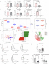
Fig. 1. Increased AHR expression in microglia (MG) and other Iba-1+ immune cells in postmortem brain samples of patients with stroke.
Activation of MG AHR after oxygen-glucose deprivation and reperfusion (OGD/R) using human cell lines, with decreased concentrations of indoles in human plasma samples after stroke. a, b Final sample selection was based on radiological and gross histological confirmation of ischemic stroke. c Representative images of double immunofluorescent labeling for AHR (green) and Iba-1 (pink) in brain sections. They show AHR expression in Iba-1+ cells and quantification of AHR + MG per unit area in the infarct (p < 0.0001) and peri-infarct regions (p < 0.0001) post-stroke (n = 29) relative to the cortical regions from controls (n = 10). Each dot represents a patient. Scale bar: 100 um. d Representative images of double immunofluorescent labeling for AHR (green) and Iba-1 (pink) in brain sections showing AHR expression in Iba-1+ cells in acute and chronic timepoint postmortem brain samples with quantification of AHR expression in Iba-1 positive cells. e RT-PCR demonstrating increased AHR and Cyp1ba expression levels after OGD/R performed on human MG cell line (HMC3) (n = 5/grp). f Plasma concentrations of indole-3-carboxaldehyde (p < 0.0001, exact value shown on figure) and indole-3-propionate (p < 0.0001, exact value shown on figure, pairwise test: Games-Howell, bars shows significant) are significantly decreased 24 h to 7 days after stroke in patients (n = 60) relative to healthy controls (n = 64). Unpaired two-tailed t-test (e) and Tukey’s two-way ANOVA with multiple comparisons (c, d) were used for statistical analyses. All data (with error bars) are presented as mean ± SEM.
Fig. 2
Fig. 2. Bioavailability of gut microbiota-dependent indole-based AHR ligands was disrupted after stroke.
a Metabolomics analysis of plasma samples from wild-type (WT) and germ-free (GF) mice. Host-derived metabolites (tryptophan [Trp] and Kynurenine [Kyn]) did not differ between WT and GF plasma samples. However, indole-3-carboxaldehyde and indole-3-propionate concentrations were significantly reduced in GF plasma (n = 5/grp). b Metabolomic analysis of brains revealed specific indole-based AHR ligands that depend on the presence of a microbiota. Indole-3-carboxaldehyde (p = 0.0122) and indole-3-propionate (p < 0.0001) were undetectable in GF brains relative to WT control brains, while Trp and Kyn concentrations did not differ (n = 5/grp). c1 Major bacterial populations involved in the regulation of AHR ligands and Trp metabolism (such as phylum Actinobacteria predominant genus being Bifidobacteria) were significantly (Mann–Whitney test: p < 0.001614, FDR-Adi.p < 0.003766; overall Median: 0.08179) reduced by aging and experimental stroke in aged WT mice. c2 No significant differences (Observed OTUs: p < 0.7 and Shannon: p < 0.54) in alpha diversity (measures within sample diversity) and operational taxonomic units (p = 0.7 or 0.5) between the groups were observed. d Principal component analysis plot of 16S data shows a significant (p = 0.002) clustering effect of stroke animals relative to the sham group. e The LDA score clearly shows changes in bacterial families in stroke versus sham animals. f Cladogram visualization of 16S data shows a clear pathological shift in bacterial diversity in stroke animals relative to healthy sham animals. g Metabolomics analysis of aged WT brain and plasma samples at multiple timepoints post-stroke (n = 5/grp). Kyn concentrations increased while concentrations of indole-3-carboxaldehyde and indole-3-propionate were significantly decreased in both brain and plasma (n = 5/grp). h Metabolomic analysis of aged WT plasma samples shows increased concentrations of Kyn at 3 days post-stroke and no significant difference between groups at 30 days post-stroke (n = 5/control, 6/treated). Concentrations of indole-3-carboxaldehyde and indole-3-propionate (p = 0.0182) were decreased at 3 days post-stroke (n = 5/control, 6/treated). Unpaired two-tailed t-tests were used for statistical analyses (a, b, h). All data (with error bars) are presented as mean ± SEM. All data points (each dot represents) presented are biological replicates.
Fig. 3
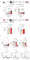
Fig. 3. Host- and microbiota-derived AHR ligands have opposing effects on MG survival after oxygen-glucose deprivation and reperfusion (OGD/R).
a Schematic showing sorted MG cells from naive aged WT mice treated with indole-based AHR ligands (indole-3-carboxaldehyde and indole-3-propionate) ex vivo. The cells received OGD/R to model stroke conditions. b MG AHR expression and MG survival post-OGD/R differ based on the AHR ligands present. AHR expression was increased after OGD/R and depleting tryptophan (Trp) from OGD/R media reversed this effect (n = 4/group for Vehicle, OGD, Trp Dep OGD (p < 0.0100), and Trp Dep Kyn OGD (p < 0.0001) and n = 8 for Trp Dep Indoles OGD (p < 0.0001) group). The addition of Kynurenine (Kyn) or indole-based AHR ligands (indole-3-carboxaldehyde and indole-3-propionate) led to an increase in AHR expression post-OGD/R. MG survival after OGD/R was significantly lower when Kyn was present relative to when Trp was depleted. Post-OGD/R MG survival increased when indole-based ligands were added (n = 4/group for Vehicle, OGD, Trp Dep OGD, and Trp Dep Kyn OGD and n = 8 for Trp Dep Indoles OGD group). c Schematic showing treatment of mice with indoles (indole-3-carboxaldehyde and indole-3-propionate) in vivo via oral gavage. MG were sorted from indole-treated mice and received OGD/R. d MG survival was increased post-OGD/R in the group treated with indoles relative to the vehicle group (n = 5/grp, p = 0.0249). e Kyn-mediated activation of AHR was detrimental, whereas indole-mediated activation of AHR did not worsen MG survival after OGD/R (n = 6/grp, p = 0.0256). f qPCR-based examination showed that the expression levels of IL-1b, IL−4, and IL-10 are significantly reduced in indole-treated relative to the Kyn-treated group (n = 6/grp, p < 0.0001 for all for all three comparisons). The schematic figures were created in BioRender. Unpaired two-tailed t-test (d) and Tukey’s two-way ANOVA with multiple comparisons (b, e, f) were used for statistical analyses. All data (with error bars) are presented as mean ± SEM. All data points presented are biological replicates.
Fig. 4
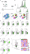
Fig. 4. Post-stroke treatment with microbiota-derived indole-based AHR ligands regulated MG-mediated neuroinflammation and antigen presentation molecules in GF mice.
a Schematics showing timeline of MCAO and indole treatment in GF mice. b Increased expression of MG AHR is associated with increased expression of antigen presenting and co-stimulatory molecules MHC-II and CD80. c Phenographs of non-MG and MG cells of homogenized brains of GF mice post-stroke from vehicle and indole-treated mice. d Surface expressions of AHR, CD11b, MHC-II, and CD80 by MG are significantly increased 24 h after stroke in GF mice treated with indoles at 3 h and 8 h after stroke relative to GF stroke mice receiving vehicle (n = 9/grp). e Surface expressions of MHC-II and CD80 by lymphocytes were significantly increased in mice treated with indoles (n = 9/grp). f Surface expression of MHC-II is significantly decreased and expression of CD80 is increased by monocytes in mice treated with indoles (n = 9/grp). g GF stroke mice treated with indoles had significantly lower reduction in body weight (normalized to pre-stroke bodyweight at 24 h after stroke, lower brain weight (normalized to bodyweight at 24 h post-stroke) and lower brain volume. Neurological deficit scores were not different at 24 h post-stroke in GF mice (n = 10/vehicle, 13/treated). h Expression of microglial Lamp1 (p = 0.0028), IL1B (p = 0.0078), and microglial phagocytosis (% red bead uptake) (p = 0.0256) were decreased in GF mice treated with indoles at 3- and 8-h post-stroke (n = 6/vehicle, 8/treated). Expression of IL10 was increased (p = 0.0825) following treatment with indoles (n = 6/vehicle, 8/treated). i, j Volcano plots and heatmap visualization of Nanostring mRNA expression analysis of homogenized brain tissues from the controls and indole-treated groups of GF stroke mice. The transcriptional profiles of AHR downstream target genes (Cyp1b1, Ifng, Ccl8, Il2, and Ccl20) in the indole-treated group (n = 8) were different. The figures were created in BioRender. Unpaired two-tailed t-test. All data (with error bars) are presented as mean ± SEM. All data points presented are biological replicates.
Fig. 5
Fig. 5. Post-stroke treatment with microbiota-derived indole-based AHR ligands (indole-3-carboxaldehyde and indole-3-propionate) reduces neurological deficits and infarct size in aged wild-type (WT) mice 24 h after stroke.
a Schematic showing the timeline of middle cerebral artery occlusion and indole treatment in aged WT mice. b Aged WT mice treated with indoles had a significant decrease in neurological deficit scores (p = 0.0150), smaller reduction in body weight (normalized to pre-stroke bodyweight, p = 0.0262) at 24 h after stroke, lower brain weight (normalized to bodyweight at 24 h post-stroke, p = 0.0018) and lower brain volume (n = 9/vehicle, 12/treated, p = 0.0003). Whiskers on box plot indicate minimum (2.00) and maximum (3.00). c Quantification of brain infarct volumes as analyzed by 2,3,5-triphenyl-tetrazolium staining in controls and treated mice (n = 9/vehicle, 12/treated). The total, cortical, and striatal infarct sizes suddenly increased in the stroke mice treated with indoles relative to the vehicle group (n = 9/vehicle, 12/treated). Unpaired two-tailed t-test. All data (with error bars) are presented as mean ± SEM. All data points presented are biological replicates. The figures were created in BioRender.
Fig. 6
Fig. 6. Schematic diagram representing the data.
The schematics highlights the stroke induced pathological changes of host- and indole-based ligands availability of AHR measured in the circulation and in the ischemic hemisphere of the brain. It emphasizes that the oral treatment with indoles attenuated inflammation in the microglial of the post-stroke brain leading to less severe stroke outcome via AHR dependent pathway. The scheme was created under the BioRender Agreement No: DP27GHBB1A).

Update of

References

    1. Virani, S. S. et al. Heart disease and stroke statistics—2020 update: a report from the American Heart Association. Circulation141, e139–e596 (2020). - PubMed
    1. Benjamin, E. J. et al. Heart disease and stroke statistics—2018 update: a report from the American Heart Association. Circulation137, e67–e492 (2018). - PubMed
    1. Powers, W. J. et al. Guidelines for the early management of patients with acute ischemic stroke: 2019 update to the 2018 guidelines for the early management of acute ischemic stroke: a guideline for healthcare professionals from the American Heart Association/American Stroke Association. Stroke50, e344–e418 (2019). - PubMed
    1. Rinaldo, L. et al. Racial and ethnic disparities in the utilization of thrombectomy for acute stroke. Stroke50, 2428–2432 (2019). - PubMed
    1. Murray, C. J. L. et al. Global, regional, and national disability-adjusted life years (DALYs) for 306 diseases and injuries and healthy life expectancy (HALE) for 188 countries, 1990–2013: quantifying the epidemiological transition. Lancet386, 2145–2191 (2015). - PMC - PubMed

LinkOut - more resources