. 2025 Feb 19;10(1):34.
doi: 10.1038/s41541-025-01089-x.

iDC-targeting PfCSP mRNA vaccine confers superior protection against Plasmodium compared to conventional mRNA

Affiliations

iDC-targeting PfCSP mRNA vaccine confers superior protection against Plasmodium compared to conventional mRNA

Sean Yanik et al. NPJ Vaccines. .

Abstract

Malaria resurgence in 2022 saw 249 million clinical cases and 608,000 deaths, mostly in children under five. The WHO-approved circumsporozoite protein (CSP)-targeting vaccines, RTS,S and R21, remain limited in availability. Strong humoral responses are crucial for sporozoite neutralization before hepatocyte infection, yet first-generation vaccines provide suboptimal protection, necessitating improved strategies. With the success of mRNA-LNP vaccines against COVID-19, there is interest in leveraging this approach to malaria. Here, we developed a novel chemokine fusion mRNA vaccine targeting immature dendritic cells (iDC) to enhance immunity against P. falciparum CSP (PfCSP). Mice immunized with MIP3α-CSP mRNA-LNP exhibited stronger CD4 + T cell responses and higher anti-NANP6 antibody titers than conventional CSP mRNA-LNP. Importantly, upon P. berghei PfCSP transgenic sporozoite challenge, MIP3α-CSP mRNA provided significantly greater protection from liver infection, strongly associated with multifunctional CD4 + T cells and anti-NANP6 titers. This study underscores iDC targeting as a promising strategy to enhance malaria vaccine efficacy.


Conflict of interest statement

Competing interests: D.W. is named on patents describing the use of nucleoside-modified mRNA - lipid nanoparticle vaccines. Y.T. is an employee of Acuitas Therapeutics that is involved in the development of mRNA-LNP therapeutics. D.W. and M.-G.A. are listed as inventors on patents covering LNP for nucleic acid therapeutic delivery for vaccines. Y.S., V.V., J.T.G., J.M., Y.L., E.G., Y. F-G., N.C., D.S., R.M, and P.S. have no competing interests.

Figures
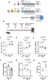
Fig. 1
Fig. 1. Antibody responses and protection from liver stage infection following standard 3-dose mRNA vaccination.
A Design of CSP and MIP3α-CSP constructs. Representations of protein sequences for full length CSP, the MIP3α -CSP construct, and the CSP construct used in the approved RTS,S vaccine are shown. Both CSP and MIP3α-CSP sequences contain the CSP C-terminal domain containing important T cell epitopes, CSP junctional region, and central repeat region of CSP with 1/4/38 copies of NPDP/NVDP/NANP respectively. For comparison, the RTS,S construct contains 0/0/19 copies of NPDP/NVDP/NANP respectively. In the MIP3α-CSP construct, the human MIP3α gene was fused to the N-terminus of 3D7 PfCSP gene via a 14 amino acid linker sequence. The tPA signal sequence is located at the N terminus of the MIP3α gene in this construct. B C57BL/6J mice (n = 5/grp) were immunized 3x at two-week intervals with 10ug MIP3α-CSP LNP-mRNA or CSP LNP-mRNA. Two weeks after the 3rd immunization, vaccinated and naïve mice were challenged with 3000 Pb PfCSP-luc intravenously delivered sporozoites. Forty-two h after infection, parasite liver loads, measured by luminescence, were captured. Full length recombinant CSP specific (C) and NANP6 peptide specific (D) antibody titers in mouse serum are shown. Endpoint titers (OD450 = 1) were used to quantify antibody titers for both groups. Data points are individual mice (n = 5) performed in duplicate, with horizontal lines representing mean values. Ordinary one-way ANOVAs with Tukey’s test for multiple comparisons were performed to compare differences between groups. E Log 10 titer ratio of anti-full length CSP antibodies to anti-NANP6 antibodies are shown, with horizontal lines representing mean values. An ordinary one-way ANOVA with Tukey’s test for multiple comparisons was performed to compare differences between groups. F NANP6 specific avidity indices are shown for antibodies from CSP and MIP3α-CSP groups. Dots represent avidity indices of individual mice. The avidity index was calculated using the following equation ((OD 1 titer in chaotropic agent)/(OD 1 titer in PBS)*100). A two-tailed unpaired t test was performed to determine p values. Bars represent mean ± SEM. G Luminescence values for each mouse in naïve, CSP and MIP3α-CSP groups were calculated in photons/sec, with horizontal lines representing group means. An ordinary one-way ANOVA with Tukey’s test for multiple comparisons was performed to compare differences between groups. H Percent inhibition of liver infection was calculated relative to naïve, sporozoite challenged mice. Data is shown for individual animals, with horizontal lines representing mean values. A two-tailed unpaired t test was used to compare groups.
Fig. 2
Fig. 2. T cell responses in mice following mRNA dose de-escalation and delayed boosting.
A C57BL/6J mice (n = 7/grp) were immunized 3x with 25 μg, 15 μg, and finally 5 μg of MIP3α-CSP LNP-mRNA or CSP LNP-mRNA. In this delayed dosing experiment, the 2nd and 3rd immunizations occurred 2 and 8 weeks after the first immunization respectively. Splenocytes were harvested from each mouse 42 h after challenge for usage in T cell stimulation assays. Cells were stained, run on flow cytometry, and gated for CD4+ (B) or CD8+ (C) T cells. Median fluorescence intensities of CD4 + IFNγ + (B) and CD8 + IFNγ + (C) T cells are shown (left). The percentage of IFN-γ+ cells among CD4+ cells (B) and CD8+ cells (C) are also shown (right). Horizontal lines indicate group means. Ordinary one-way ANOVAs with Tukey’s test for multiple comparisons were performed to compare differences between groups. D, E Cells were stained, run on flow cytometry, and gated for CD4 + T cells. Median fluorescence intensities of CD4 + TNFα + cells (D) and CD4 + IL2+ cells (E) are shown (left). The percentage of TNFα + (D) or IL2+ cells (E) among CD4+ cells are also shown (right). Horizontal lines indicate group means. Ordinary one-way ANOVAs with Tukey’s test for multiple comparisons were performed to compare differences between groups. F, G The percentage of CD4+ or CD8+ (G) T cells that were IFNγ, TNFα, and IL-2 triple positive (left axis) or IFNγ, TNFα double positive are shown (right axis). Horizontal lines indicate group means. Ordinary one-way ANOVAs with Tukey’s test for multiple comparisons were performed to compare differences between groups.
Fig. 3
Fig. 3. Antibody responses and protection from liver stage infection following mRNA dose de-escalation and delayed boosting.
A Full length recombinant CSP antibody titer in mice sera. B NANP6 peptide-specific antibody titer in mouse sera. Endpoint titers (OD450 = 1) were used to quantify antibody titers for both groups. Data points are individual mice (n = 7) performed in duplicate, with horizontal lines representing mean values. Two-tailed unpaired t tests were performed to compare differences between groups. C Log 10 titer ratio of anti-full length CSP antibodies to anti-NANP6 antibodies are shown, with horizontal lines representing mean values. A two-tailed unpaired t test was performed to compare differences between groups. Data is shown as mean ± SEM with individual points representing individual mice performed in duplicate. Full length recombinant CSP specific (D) and NANP6 peptide specific (E) avidity indices are shown for antibodies from CSP and MIP3α-CSP vaccinated mice. Dots represent avidity indices of individual mice performed in duplicate. The avidity index was calculated using the following equation ((OD 1 titer in chaotropic agent)/(OD 1 titer in PBS)*100). Two-tailed unpaired t tests were performed to determine p values. Bars represent mean ± SEM. F The IgG1/IgG2c ratio of CSPFL specific and NANP6 specific antibodies in CSP and MIP3α-CSP LNP-mRNA vaccinated groups are shown. P values were calculated using two-tailed unpaired t tests. Dots represent the ratio of antibody isotypes of individual mice performed in duplicate, and horizontal bars represent mean values of each group. G Luminescence values for each mouse in naïve, CSP and MIP3α-CSP groups were calculated in photons/sec, with horizontal lines representing group means. An ordinary one-way ANOVA with Tukey’s test for multiple comparisons was performed to compare differences between groups. H Percent inhibition of liver infection was calculated relative to naïve, sporozoite challenged mice. Data is again shown for individual animals, with horizontal lines representing mean values. A two-tailed unpaired t test was used to compare groups.
Fig. 4
Fig. 4. Immunological correlates of protection.
A Relationship between log10 NANP6 peptide antibody titers (y-axis) and % inhibition of liver stage parasitemia relative to naïve, sporozoite challenged mice (x-axis). Unfilled and filled symbols represent single mice from challenge studies 1 and 2 (standard regimen and delayed, dose de-escalation regimen) respectively. Squares represent mice from the MIP3α-CSP LNP-mRNA vaccinated group, circles represent mice from the CSP LNP-mRNA vaccinated group, and triangles represent mice from the rCSPFL vaccinated group. P values were calculated using Spearman’s ranked correlation coefficient. Relationship between inhibition of liver stage parasitemia (x-axis) and the percentage of IFNγ + cells among CD4+ cells (B), the percentage of IFNγ, TNFα, and IL-2 triple positive cells among CD4+ cells (C), and the percentage of IFNγ, TNFα, and IL-2 triple positive CD8+ cells (D). Circles represent mice from the CSP vaccinated group, and squares represent mice from the MIP3α-CSP vaccinated group. P values were calculated using Spearman’s ranked correlation coefficient.

Update of

References

    1. Organization WH. World Malaria Report 2022. https://www.who.int/teams/global-malaria-programme/reports/world-malaria....
    1. Carter, R. & Mendis, K. N. Evolutionary and historical aspects of the burden of malaria. Clin. Microbiol Rev.15, 564–594 (2002). - PMC - PubMed
    1. Schuerman, L. RTS,S malaria vaccine could provide major public health benefits. Lancet394, 735–736 (2019). - PubMed
    1. Datoo, M. S. et al. Safety and efficacy of malaria vaccine candidate R21/Matrix-M in African children: a multicentre, double-blind, randomised, phase 3 trial. Lancet403, 533–544 (2024). - PubMed
    1. Datoo, M. S. et al. Efficacy and immunogenicity of R21/Matrix-M vaccine against clinical malaria after 2 years’ follow-up in children in Burkina Faso: a phase 1/2b randomised controlled trial. Lancet Infect. Dis.22, 1728–1736 (2022). - PubMed

LinkOut - more resources