. 2025 Mar;639(8055):792-799.
doi: 10.1038/s41586-024-08574-8. Epub 2025 Feb 19.

SPO11 dimers are sufficient to catalyse DNA double-strand breaks in vitro

Affiliations

SPO11 dimers are sufficient to catalyse DNA double-strand breaks in vitro

Cédric Oger et al. Nature. 2025 Mar.

Abstract

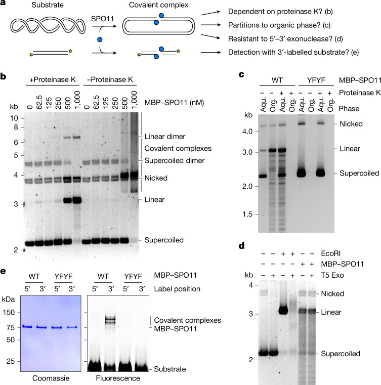
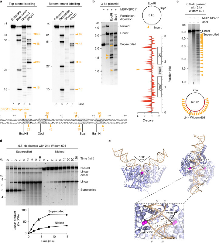
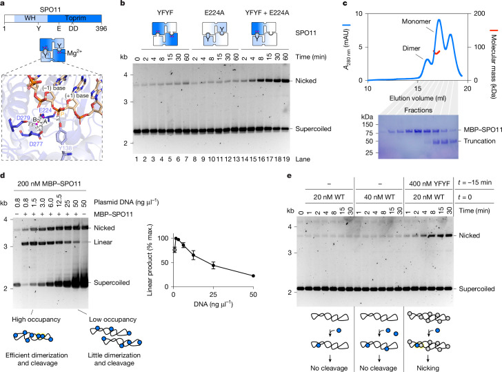
SPO11 initiates meiotic recombination through the induction of programmed DNA double-strand breaks (DSBs)1,2, but this catalytic activity has never been reconstituted in vitro3,4. Here, using Mus musculus SPO11, we report a biochemical system that recapitulates all the hallmarks of meiotic DSB formation. We show that SPO11 catalyses break formation in the absence of any partners and remains covalently attached to the 5' broken strands. We find that target site selection by SPO11 is influenced by the sequence, bendability and topology of the DNA substrate, and provide evidence that SPO11 can reseal single-strand DNA breaks. In addition, we show that SPO11 is monomeric in solution and that cleavage requires dimerization for the reconstitution of two hybrid active sites. SPO11 and its partner TOP6BL form a 1:1 complex that catalyses DNA cleavage with an activity similar to that of SPO11 alone. However, this complex binds DNA ends with higher affinity, suggesting a potential role after cleavage. We propose a model in which additional partners of SPO11 required for DSB formation in vivo assemble biomolecular condensates that recruit SPO11-TOP6BL, enabling dimerization and cleavage. Our work establishes SPO11 dimerization as the fundamental mechanism that controls the induction of meiotic DSBs.


Conflict of interest statement

Competing interests: The authors declare no competing interests.

Figures

Fig. 1
Fig. 1. In vitro reconstitution of SPO11-dependent DNA DSB formation.
a, Purification scheme of mouse SPO11 protein. b, SDS–PAGE analysis of ion exchange fractions of purified MBP–SPO11. c, Scheme of the in vitro DNA cleavage assay. Products are illustrated following deproteination of samples with proteinase K. d, Plasmid DNA cleavage analysis using fractions of SPO11 from b in the presence of divalent metal ions (Mg2+ and Mn2+). The band labelled by an asterisk corresponds to a plasmid dimer. e, Effect of active site-directed double mutation Y137F/Y138F (YFYF/) on the DNA cleavage activity of SPO11. WT, wild type. f, Requirement for divalent metal ions in DNA cleavage activity of SPO11. For gel source data, see Supplementary Fig. 1.
Fig. 2
Fig. 2. SPO11 is covalently bound to 5′ DNA ends.
a, Predictions tested in be. b, Agarose gel analysis of DNA cleavage products with or without proteinase K treatment before electrophoresis. All reactions were treated with SDS to eliminate non-covalent binding. c, Phenol–chloroform partitioning of DNA cleavage products with wild-type and mutant SPO11, with or without proteinase K treatment before phenol extraction. d, Analysis of the resistance of EcoRI- and SPO11-dependent cleavage products to the 5′–3′ exonuclease T5 Exo. e, SDS–PAGE analysis of covalent SPO11–DNA complexes from cleavage assays using wild-type or mutant SPO11 (YFYF) in the presence of 3′ or 5′ fluorescently labelled 80 bp substrates. For gel source data, see Supplementary Fig. 1. Aqu., aqueous phase; Org., organic phase.
Fig. 3
Fig. 3. Cleavage pattern and substrate specificity.
a, Sequencing gel analysis of DNA cleavage reactions using 5′ radioactively labelled 80-bp substrates. Lanes 2 and 6 were produced by digestion of the substrate using restriction enzymes indicated below the gel; lanes 3 and 7 were produced by partial digestion of the substrate with DNase I. SPO11 cleavage sites (lanes 4 and 8) are highlighted with orange arrowheads. Positions ±3 from the dyad axes are shown in bold. b, Agarose gel analysis of SPO11 cleavage sites on the standard plasmid substrate (pCCB959) using restriction digestion of SPO11 reaction products. Bottom right, cyclizability (C-score) of the plasmid substrate, as predicted by DNAcycP. The positions of the preferential cleavage site are indicated (arrowheads). c, Analysis of SPO11 cleavage sites with a plasmid substrate containing 24 copies of the Widom 601 sequence (pOC157). d, Effect of DNA topology on the rate of SPO11-dependent cleavage. Quantifications show the mean and range from two independent experiments. e, AlphaFold 3 model of SPO11 dimer bound to a 40-bp duplex DNA substrate. Mg2+ ions are shown in magenta. The nucleotides that would form the 5′ overhang are labelled +1 and +2. For gel source data, see Supplementary Fig. 1.
Fig. 4
Fig. 4. SPO11 cleavage requires dimerization.
a, Domain structure of SPO11 (top) and arrangement of the SPO11 dimer (middle). Bottom, zoomed-in view of the composite active site within the AlphaFold 3 model of a DNA-bound SPO11 dimer. Mg2+ ions are labelled A and B. Active site residues and scissile phosphate (P) are shown. b, Time-course analysis of SPO11 cleavage with mixtures of two catalytically inactive mutants. c, SEC–MALS analysis of MBP–SPO11 following amylose affinity purification. Blue traces represent absorbance measurements at 280 nm derived from size-exclusion chromatography; red traces represent molecular mass measurements across the peak. SDS–PAGE analyses of the corresponding fractions are shown. The leftmost peak probably corresponds to a dimer, although the molecular mass could not be determined; the rightmost peak corresponds to a truncated fragment. d, Titration of DNA at a constant concentration of SPO11; reactions were stopped after 15 min. Quantifications show the mean and range from two independent experiments. At the lowest concentration (open circle), the substrate is limiting so the amount of linear product is not representative of total break levels. e, Time-course analysis of SPO11 cleavage at the indicated concentrations of wild-type and catalytically inactive SPO11. With 20 nM wild-type SPO11, the increased nicking activity observed in the presence of the Y137F/Y138F (YFYF) double mutant cannot be explained by doubling the formation of cleavage-competent complexes, because no activity was detected with 40 nM wild-type SPO11. For gel source data, see Supplementary Fig. 1. mAU, milliabsorbance units; max., maximum; WH, winged helix.
Fig. 5
Fig. 5. The SPO11–TOP6BL complex.
a, Domain structure of SPO11 and TOP6BL. The C terminus of TOP6BL (grey) binds REC114 (ref. ). b, AlphaFold 3 model of the SPO11–TOP6BL heterotetramer bound to a 40-bp DNA substrate. The TOP6BL intrinsically disordered region (IDR) was omitted from the model. c, SEC–MALS analysis of SPO11–TOP6BL complexes tagged with MBP and His–Flag, respectively. Blue traces represent absorbance measurements at 280 nm from SEC; red traces represent molecular mass measurements across the peak. d, Plasmid (pOC157) cleavage with 45 nM SPO11 or SPO11–TOP6BL complexes with or without 0.01% NP-40. In lanes 3, 5, 7 and 9, proteinase K treatment was omitted before electrophoresis. Quantifications show individual data points, mean and range from two independent experiments. e, Gel shift analysis of the binding of SPO11 and SPO11–TOP6BL complexes to a 25-bp hairpin substrate with a two-nucleotide 5′ overhang. For gel source data, see Supplementary Fig. 1.
Extended Data Fig. 1
Extended Data Fig. 1. Impact of metal ions on SPO11 activity.
a, Kinetic analysis of SPO11 DNA cleavage in the presence of 5 mM Mg2+ or Mn2+. b, Phenol extraction of SPO11 cleavage reactions with EDTA, Mg2+, Ca2+ or Mn2+. Covalent complexes depend on the presence of metal ions, and both single-strand and double-strand cleavage products partition to the organic phase. For gel source data, see Supplementary Fig. 1.
Extended Data Fig. 2
Extended Data Fig. 2. Correlation between DNA bendability and SPO11 activity.
a, Agarose gel analysis of SPO11 DNA cleavage with linear substrates containing 0, 1, 3, or 6 copies of the Widom 601 sequence. The pUC19-derived substrates contain a single fluorophore (FAM) at one end, allowing the mapping of cleavage products. The position of the origin of replication (Ori), ampicillin resistance gene (AmpR) and the multiple cloning site (MCS) are indicated. Copies of the Widom 601 sequence are represented with pink arrows. b, Correlation between Widom 601 sequences, DNA bending, and SPO11 cleavage. The C-score is a bendability parameter predicted by DNAcycP. The insertion of Widom 601 sequences creates hotspots for SPO11, although the predicted bendability of the DNA sequence is not sufficient to account for the cleavage activity observed along the substrate. The cleavage hotspots produced by the Widom 601 sequence can also be due to a sequence preference of SPO11, irrespective of bendability. For gel source data, see Supplementary Fig. 1.
Extended Data Fig. 3
Extended Data Fig. 3. Relationship between DNA supercoiling, bending, and cleavage.
Time course analyses of SPO11 cleavage with (a) supercoiled and (b) linear plasmid DNA substrates without (pUC19) or with three copies of the Widom 601 sequence (pCCB1107). Quantification of the DNA substrate is shown under the gel. On the right, quantifications show the mean and range from two replicates. The two supercoiled substrates are consumed at the the same rate. In contrast, the linear substrate with Widom sequences is cleaved faster than the one without Widom sequences. This suggests that supercoiling accelerates cleavage by facilitating DNA bending. For gel source data, see Supplementary Fig. 1.
Extended Data Fig. 4
Extended Data Fig. 4. Structural modeling of SPO11 complexes and cleavage mechanism.
a, AlphaFold3 models colored by confidence score of SPO11 dimers and SPO11-TOP6BL complexes with or without 40 bp duplex DNA substrate. The C-terminus of TOP6BL was omitted from the model because it is predicted to be unstructured. b, Predicted alignment error plots. The structure and relative position (orange squares) of SPO11 monomers are predicted with lower confidence in the absence of DNA than in the presence of DNA, consistent with the monomeric stoichiometry of SPO11 and SPO11-TOP6BL complexes. In the absence of DNA, AlphaFold proposes an aberrant dimeric model of SPO11 throught interactions between WH domains (left). In the presence of DNA and TOP6BL, the relative position of SPO11 is predicted with much higher accuracy (compare orange squares on the rightmost PAE plot with the others). c, Sequence alignments of eukaryotic SPO11 and archaeal Top6A proteins. Blocks around active site residues are shown. Invariant amino acids are in orange, conservative substitutions are in light orange. Active site residues are indicated with an arrowhead. d, Cleavage assay with wild-type SPO11, double Y137F/Y138F (YFYF) and single Y137F and Y138F mutants. Conversion of the cleaved linear product into a smear in the absence of proteinase K indicates covalent attachment of SPO11 to the broken DNA ends. The low amount of linear and nicked products observed with the Y138F mutant is due to a non-specific nuclease contaminant in the protein preparation. e, Two-metal-ion reaction scheme, based on the mechanism proposed for type IIA topoisomerases. For gel source data, see Supplementary Fig. 1.
Extended Data Fig. 5
Extended Data Fig. 5. Optimization of the cleavage reaction.
a, Effect of the reaction temperature on SPO11 cleavage. b, Effect of the pH on SPO11 cleavage. The standard conditions chosen for all the reactions are 37 °C and pH 7.5. c, Titration of SPO11 protein in reactions that contained either 5 ng/µl (2.5 nM) or 25 ng/µl (12.5 nM) plasmid DNA. Quantifications show the mean and range from two independent experiments. Cleavage increased with protein concentration, but the total level of cleavage was higher in reactions that had lower DNA concentrations (compare linear product in lanes 5–7 with lanes 13–15). d, Titration of DNA in reactions that contained either 50 or 200 nM SPO11. Quantifications show the mean and range from two independent experiments. At 50 nM SPO11, cleavage was highest at the lowest DNA concentration tested (0.8 ng/µl, 0.4 nM). At 200 nM SPO11, a reduction of total DNA cleavage was observed at substrate concentrations above 12.5 ng/µl (6.2 nM). At lower concentrations (open circles), the substrate is limiting so the amount of linear product is not representative of total break levels. The reaction time in these experiments is 2 h. Note that in Fig. 4d, the inhibitory effect is observed at lower DNA concentrations because the reaction time was shorter (15 min). Longer reaction times provide more opportunity for dimerization and cleavage. For gel source data, see Supplementary Fig. 1.
Extended Data Fig. 6
Extended Data Fig. 6. Relationships between DNA binding and cleavage.
a,b,c, DNA cleavage requires higher SPO11 concentration than DNA binding. Comparison of the DNA-binding (a) and DNA cleavage (b) activities of SPO11. Reactions contained 5 ng/µl (2.5 nM) plasmid. Binding reactions were assembled for 30 min. Cleavage reactions were stopped after 2 h. (c) Quantification shows that the plasmid is bound efficiently at concentrations that do not support cleavage. d,e, SPO11 provides effective protection against DNase I treatment. Plasmid substrates (2.5 nM) were incubated with or without 600 nM catalytically inactive SPO11-Y137F/Y138F mutant, followed by two-minute treatment with the indicated concentration of DNase I. (e) Quantification of the supercoiled substrate remaining in panel d. Error bars are ranges from two independent experiments. f,g,h, DNA binding and cleavage by SPO11 are sensitive to salt. Effect of NaCl on (f) plasmid cleavage and (g) DNA binding. SPO11 concentration is 500 nM in panel f and 100 nM in panel g. The greater sensitivity of cleavage than DNA binding to salt could be explained by a mild reduction in DNA binding severely reducing the chances of dimerization on DNA (h). For gel source data, see Supplementary Fig. 1.
Extended Data Fig. 7
Extended Data Fig. 7. SPO11 can reseal single-strand DNA nicks.
a, Cleavage analysis at a constant SPO11 concentration in the presence of different ratios of wild-type and catalytically-inactive (Y137F/Y138F) mutants. The ladder that migrates between the supercoiled and nicked products is absent in reactions that contain only wild-type or inactive mutants, indicating that it is not due to a contaminating activity in one of the protein preparations. b, Kinetic analysis of cleavage products in reactions that contained mixtures of YFYF and E224A mutants. c, Phenol-chloroform partitioning of DNA cleavage products in reactions that contained mixtures of wild-type and mutant SPO11. Topoisomers partition to the organic phase as the covalent link with SPO11 has been released. d, Illustration of the plasmid relaxation activity observed with mixtures of wild type and inactive SPO11. Dissociation of the SPO11 dimer after single-strand nicking will lead to the swiveling of the DNA duplex around the intact phosphodiester bond (red arrows). Restauration of the SPO11 dimer then provides an opportunity for strand religation. Separation of the dimer interface could also be accompanied by the dissociation of the subunit not involved in catalysis from the DNA substrate, although this could would be expected to lead to full plasmid relaxation, which is not always observed. For gel source data, see Supplementary Fig. 1.
Extended Data Fig. 8
Extended Data Fig. 8. Comparison of the AlphaFold model of mouse SPO11-TOP6BL and the structure of Methanosarcina mazei Topo VI.
a, AlphaFold3 model of a 2:2 SPO11-TOP6BL heterotetramer. The structure was modeled with DNA, but the DNA was hidden to ease comparison. b, Crystal structure of Topo VI. SPO11 and Top6A are in blue. GHKL (green), helix two-turn helix (H2TH, orange), transducer (yellow), C-terminal domain (CTD, grey), intrinsically-disordered region (IDR, not shown).
Extended Data Fig. 9
Extended Data Fig. 9. Model of meiotic DSB formation in mice.
a, Proteins essential for DSB formation in mice. b, We propose that RMMI form condensates along the chromosome axis and recruit SPO11-TOP6BL complexes through an interaction between REC114 and the C-terminus of TOP6BL. The ensuing increase in local concentration of SPO11-TOP6BL complexes allows SPO11 dimerization and cleavage. c, Multiple dimers may assemble, leading to the formation of closely-spaced double DSBs,. d, Proposed thermodynamics of SPO11-TOP6BL cleavage reactions. Monomeric SPO11-TOP6BL complexes bind with high affinity to DNA. The assembly of an active pre-cleavage dimeric complex requires bending of the DNA substrate, which would correspond to a high-energy state. Cleavage is isoenergetic and therefore inherently reversible. Conformational transitions caused by or accompanied with the dissociation of SPO11 dimers stabilize SPO11-TOP6BL complexes on DNA ends, creating irreversible breaks.
Extended Data Fig. 10
Extended Data Fig. 10. AlphaFold models of SPO11 complexes from different species.
AlphaFold3 models of DNA-bound dimeric SPO11 core complexes from H. sapiens (Q9Y5K1, Q8N6T0), Z. mays (A0A804P805, A0A1D6GVU9), A. thaliana (Q9M4A2, Q9M4A1, Q5Q0E6), D. rerio (Q6P0S6, B3DIP6), D. melanogaster (Q7KPA5, Q9VS36), C. elegans (Q22236), S. cerevisiae (Spo11, Rec102 and Ski8 from SK1), S. pombe (P40384, P40385, Q09150), and S. macrospora (Q6WRU4, F7VPQ4 re-annotated, Q6URC5). SPO11 homologs are shown in light blue; the transducer domain of TOP6BL homologs are shown in yellow, the GHKL domain in green and C-terminal extension in light grey. Ski8 homologs present in fungal complexes are shown in grey. DNA is shown in orange. Mg2+ ions are in magenta. Despite extensive variability in the prediced structure and composition of cleavage complexes, in particular regarding TOP6BL homologs, all the models show SPO11 dimers engaged on a bent duplex DNA substrate, consistent with our interpretation that DNA bending is an important pre-requisite for cleavage.

References

    1. Keeney, S., Giroux, C. N. & Kleckner, N. Meiosis-specific DNA double-strand breaks are catalyzed by Spo11, a member of a widely conserved protein family. Cell88, 375–384 (1997). - PubMed
    1. Bergerat, A. et al. An atypical topoisomerase II from Archaea with implications for meiotic recombination. Nature386, 414–417 (1997). - PubMed
    1. de Massy, B. Initiation of meiotic recombination: how and where? Conservation and specificities among eukaryotes. Annu. Rev. Genet.47, 563–599 (2013). - PubMed
    1. Yadav, V. K. & Claeys Bouuaert, C. Mechanism and control of meiotic DNA double-strand break formation in S. cerevisiae. Front. Cell Dev. Biol.9, 642737 (2021). - PMC - PubMed
    1. Robert, T., Vrielynck, N., Mezard, C., de Massy, B. & Grelon, M. A new light on the meiotic DSB catalytic complex. Semin. Cell Dev. Biol.54, 165–176 (2016). - PubMed

LinkOut - more resources