. 2025 Mar;639(8056):1052-1059.
doi: 10.1038/s41586-025-08586-y. Epub 2025 Feb 19.

Clonal driver neoantigen loss under EGFR TKI and immune selection pressures

Affiliations

Clonal driver neoantigen loss under EGFR TKI and immune selection pressures

Maise Al Bakir et al. Nature. 2025 Mar.

Abstract

Neoantigen vaccines are under investigation for various cancers, including epidermal growth factor receptor (EGFR)-driven lung cancers1,2. We tracked the phylogenetic history of an EGFR mutant lung cancer treated with erlotinib, osimertinib, radiotherapy and a personalized neopeptide vaccine (NPV) targeting ten somatic mutations, including EGFR exon 19 deletion (ex19del). The ex19del mutation was clonal, but is likely to have appeared after a whole-genome doubling (WGD) event. Following osimertinib and NPV treatment, loss of the ex19del mutation was identified in a progressing small-cell-transformed liver metastasis. Circulating tumour DNA analyses tracking 467 somatic variants revealed the presence of this EGFR wild-type clone before vaccination and its expansion during osimertinib/NPV therapy. Despite systemic T cell reactivity to the vaccine-targeted ex19del neoantigen, the NPV failed to halt disease progression. The liver metastasis lost vaccine-targeted neoantigens through chromosomal instability and exhibited a hostile microenvironment, characterized by limited immune infiltration, low CXCL9 and elevated M2 macrophage levels. Neoantigens arising post-WGD were more likely to be absent in the progressing liver metastasis than those occurring pre-WGD, suggesting that prioritizing pre-WGD neoantigens may improve vaccine design. Data from the TRACERx 421 cohort3 provide evidence that pre-WGD mutations better represent clonal variants, and owing to their presence at multiple copy numbers, are less likely to be lost in metastatic transition. These data highlight the power of phylogenetic disease tracking and functional T cell profiling to understand mechanisms of immune escape during combination therapies.


Conflict of interest statement

Competing interests: M.A.B. has consulted for Achilles Therapeutics. J.L.R. reports speaker fees from Boehringer Ingelheim and GlaxoSmithKline and consults for Achilles Therapeutics Ltd. J.L.R. and has filed patents for cancer early detection (PCT/EP2023/076521 and PCT/EP2023/076511) using systemic TCR-seq data, on which S.G. is a co-inventor. A.T.G. and P.K. are former or current employees of Invitae or ArcherDx and report stock ownership. D.A.M. reports speaker fees from AstraZeneca, Eli Lilly and Takeda; consultancy fees from AstraZeneca, Thermo Fisher Scientific, Takeda, Amgen, Janssen, MIM Software, Bristol Myers Squibb (BMS) and Eli Lilly; and has received educational support from Takeda and Amgen. C.A. has received speaking honoraria or expenses from Novartis, Roche, AstraZeneca and BMS; has patents issued to detect tumour recurrence (PCT/GB2017/053289) and for methods for lung cancer detection (PCT/US2017/028013) and is co-inventor to a patent application to determine methods and systems for tumour monitoring (PCT/EP2022/077987); and is a current employee of AstraZeneca. C.T.H. has received speaker fees from AstraZeneca. K.L. has a patent on indel burden and CPI response pending and outside of the submitted work, speaker fees from Roche tissue diagnostics, research funding from CRUK TDL/Ono/LifeArc alliance and Genesis Therapeutics, and consulting roles with Monopteros Therapeutics and Kynos Therapeutics, all outside of the submitted work. M.D.F. acknowledges grant support from CRUK, AstraZeneca, Boehringer Ingelheim, MSD and Merck; is an advisory board member for Transgene; and has consulted for Achilles, Amgen, AstraZeneca, Bayer, Boxer, Bristol Myers Squibb, Celgene, EQRx, Guardant Health, Immutep, Ixogen, Janssen, Merck, MSD, Nanobiotix, Novartis, Oxford VacMedix, Pharmamar, Pfizer, Roche, Takeda and UltraHuman. T.A. reports employment and stock options with Ellipses Pharma, and is an advisory board member for Engitix, iOnctura, Labgenius and Further Group. S.R.H. is the co-inventor of the patents WO2015185067 and WO2015188839 for the barcoded MHC technology which is licenced to Immudex and co-inventor of the licensed patent for Combinatorial encoding of MHC multimers (EP2088/009356), licensee: Sanquin, NL. S.A.Q. is a co-founder, stockholder and Chief Scientific Officer of Achilles Therapeutics. S.V. is a co-inventor to a patent of methods for detecting molecules in a sample (patent no. 10,578,620). N.M. has stock options in and has consulted for Achilles Therapeutics and holds a European patent in determining HLA LOH (PCT/GB2018/052004), and is a co-inventor to a patent to identifying responders to cancer treatment (PCT/GB2018/051912). C.S. acknowledges grant support from AstraZeneca, Boehringer Ingelheim, Bristol Myers Squibb, Pfizer, Roche-Ventana, Invitae (previously ArcherDx Inc.—collaboration in minimal residual disease sequencing technologies) and Ono Pharmaceutical. He is an AstraZeneca Advisory Board member and Chief Investigator for the AZ MeRmaiD 1 and 2 clinical trials and is also Co-Chief Investigator of the NHS Galleri trial funded by GRAIL and a paid member of GRAIL’s Scientific Advisory Board. He receives consultant fees from Achilles Therapeutics (also SAB member), Bicycle Therapeutics (also a SAB member), Genentech, Medicxi, Roche Innovation Centre–Shanghai, Metabomed (until July 2022) and the Sarah Cannon Research Institute; had stock options in Apogen Biotechnologies and GRAIL until June 2021 and at present has stock options in Epic Bioscience and Bicycle Therapeutics, and has stock options and is co-founder of Achilles Therapeutics. C.S. holds patents relating to assay technology to detect tumour recurrence (PCT/GB2017/053289), targeting neoantigens (PCT/EP2016/059401), identifying patent response to immune checkpoint blockade (PCT/EP2016/071471), determining HLA LOH (PCT/GB2018/052004), predicting survival rates of patients with cancer (PCT/GB2020/050221), identifying patients who respond to cancer treatment (PCT/GB2018/051912), US patent relating to detecting tumour mutations (PCT/US2017/28013), methods for lung cancer detection (US20190106751A1) and both a European and US patent related to identifying insertion/deletion mutation targets (PCT/GB2018/051892), and is co-inventor to a patent application to determine methods and systems for tumour monitoring (PCT/EP2022/077987).

Figures

Fig. 1
Fig. 1. Patient pathway overview, cancer phylogenetics and ctDNA analyses.
a, Overview of the patient pathway annotated with samples acquired and analyses performed. b, Phylogenetic tree of the disease. Genes in black represent the NPV-targeted mutations; grey genes represent the variants that could not be included in the vaccine owing to solubility; red represents copy number gains and blue represents losses; and genes in green are putative driver genes. The asterisks indicate the neopeptides that resulted in a GZMB response. The clonality and the timing of the mutations relative to WGD are also annotated. c, ctDNA mean mutant allele frequency of phylogenetic clusters. AF, allele frequency; WT, wild type; SABR, stereotactic ablative body radiotherapy; −, not available/performed; +, collected/performed; EGFRamp, EGFR amplification. a, Credit: J. Brock.
Fig. 2
Fig. 2. T cell reactivity to personalized neoantigen vaccine epitopes.
a, Representative images from GZMB Fluorospot recall assay testing vaccine and non-vaccine neopeptides in PBMCs at 40 months (post-vaccine). b, Quantification of GZMB release by Fluorospot (40 months); bars represent the mean ± s.e.m. of triplicate cultures; *pAdj < 0.05, ****pAdj < 0.0001, one-way analysis of variance corrected for multiple testing by Benjamini–Hochberg. c, Fold change in the number of specific CDR3 beta chain sequences detected for EGFR and viral (CEF) peptides by MANAFEST in PBMCs sampled over time points shown (months, x axis), calculated relative to pre-vaccine (month 30); fold change set to 1 for no detectable T790M clones at month 30. d, The proportion of the repertoire in each sample that is occupied by specific CDR3s (shown as bar segments) reactive to peptides indicated at various time points as determined by MANAFEST. Connecting waves indicate clonotype sharing between samples. e, CIBERSORTx scores for the TRACERx 421 cohort and both RUL and SCLC-transformed liver metastasis (TRACERx 421, number of tumour regions, n = 954 regions from 347 patients), exploring the abundance of stromal (CD10+ and CD31+), immune (CD45+) and epithelial/cancer cells (EPCAM+). PHA, phytohaemagglutinin; FC, fold change; CK, cytokine; Mo, month; LUAD, lung adenocarcinoma; LUSC, lung squamous cell carcinoma.
Fig. 3
Fig. 3. TRACERx 421 cohort analyses.
Clonality of all pre-WGD mutations from tumour regions with evidence of WGD. Each column represents a single region from the TRACERx 421 cohort. The median proportion of pre-WGD mutations that were also clonal at a region level is 99.4% (IQR = 96.6–1). Regions in which pre-WGD mutations have a high proportion of subclonal mutations are enriched for tumours with subclonal WGD events and tumours that have a higher number of sequenced regions.
Extended Data Fig. 1
Extended Data Fig. 1. Whole exome sequencing results.
a, Binary heatmap of mutations detected on whole exome sequencing. Each row represents a sample, and each column is a unique somatic mutation. Grey bars indicate absence of a mutation, and dark red indicates the presence of a mutation in that sample. The five samples are all clonally related as indicated by the overlap of detected somatic mutations. b, Phased somatic copy number aberration profiles of the whole exome sequenced samples as well as the most recent common ancestor (MRCA): diagnostic primary lung biopsy (0 months), supraclavicular LN (7 months), RUL metastasis (19 months), SCLC transformed liver metastasis (38 months), mediastinal mass (45 months) and MRCA. c, RB1 LogR across all WES samples. This demonstrates evidence of deep loss of RB1 in the supraclavicular LN, RUL metastasis, SCLC transformed liver metastasis and mediastinal mass. RUL = right upper lobe, LN = lymph node, SCLC = small cell lung cancer.
Extended Data Fig. 2
Extended Data Fig. 2. Phased somatic copy number aberration profiles of chromosome 7 encompassing EGFR, and associated FISH assessments.
The mutant EGFR ex19del is represented by the magenta asterisk. Black triangles represent somatic mutations found on chromosome 7. a, Diagnostic primary lung biopsy (0 months). b, Supraclavicular LN (7 months). c, RUL metastasis (19 months). d, SCLC transformed liver metastasis (38 months). e, Mediastinal mass (45 months).
Extended Data Fig. 3
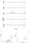
Extended Data Fig. 3. ctDNA analyses.
a, Low pass whole genome sequencing to assess for ctDNA content and explore gains and losses. b, ctDNA Mutant allele frequencies (AF) at sampled timepoints. The colours reflect the phylogenetic tree clusters. c, Zoomed in view of the ctDNA mutant allele frequencies, focussing on AF less than 0.015. Timepoints represent primary lung biopsy (0 months), RUL metastasis (19 months), RUL progression post osimertinib (30 months), SCLC transformed liver metastasis (38 months) & Mediastinal mass (45 months) RUL = right upper lobe, SCLC = small cell lung cancer.
Extended Data Fig. 4
Extended Data Fig. 4. T cell reactivity analysis and TCRseq tissue profiling.
a, Workflow for immunoreactivity assays. b, IFN-gamma Fluorospot at month 40; bars represent the mean +/− SEM of triplicate cultures; *P < 0.0001, one-way ANOVA. c, MANAFEST results showing significant clones in the conditions/time points indicated. d, MANAFEST parameters were adjusted over a sliding scale of expansion level for CDR3B sequences to be included in analysis (template threshold). Data show that post vaccine samples (months 40, 45) consistently harboured increased EGFR peptide reactivity relative to pre-vaccine timepoints (Month 30), irrespective of the template threshold used. In all analyses, clones enriched versus the cytokine alone sample and present at significantly higher levels in one condition vs all other conditions (OR > 10 and FDR < 0.01) by Fisher’s exact test, are classified as specific. e, TCRseq libraries of biopsies from 19 months (right upper lobe) and 38 months (liver metastasis), showing all significant CDR3s from MANAFEST assays. CEF = CMV, EBV, Flu viral pools of peptides. Illustration in a was created using BioRender (https://biorender.com).
Extended Data Fig. 5
Extended Data Fig. 5. TCRseq clustering of EGFR neopeptide stimulated MANAFEST cultures.
To identify groups of TCRs that shared similar sequence structure to the clones that were significantly expanded in PBMC samples from the MANAFEST assay, we clustered together the top 3000 CDR3B sequences from each timepoint (months 30, 40, 45) from each condition (Cytokine, CEF, ex19del, T790M), using the Gliph2 clustering algorithm. a, Gliph2 network plot, coloured by ‘total.score’ metric, herein referred to as cluster importance score. More significant clusters are located towards the middle of the plot. b, Overview of clusters which contain expanded sequences, as defined by the MANA webtool. As all samples were clustered together, the proportion of each condition’s contribution to the cluster is displayed in the bar. Cluster size refers to the number of unique TCR sequences within each cluster. We observed that clusters containing an expanded sequence determined by the MANA webtool were composed of sequences predominantly of the same condition. c, Clusters were segregated into conditions using a proportion cut-off of 50%. All clusters from Gliph2 output which had a sequence count above the threshold are displayed. Significance determined by two-tailed Wilcoxon test. There was no difference in cluster importance scores between the ex19del culture and the positive control (CEF), whilst the T790M condition showed the highest cluster importance score. Taken together, these data suggest that the expanded clones in their respective cultures were driven by peptide-specific stimulation, as we see more significant clustering in the conditions treated with peptide compared to cytokine alone. The box plots represent the upper and lower quartiles (box limits), the median (centre line) and the vertical bars span the 5th to 95th percentiles.
Extended Data Fig. 6
Extended Data Fig. 6. Copy number profiles for post-WGD lost variants.
Copy number changes observed at the genomic positions for the post-genome doubling variants FANCF p.A353G, STK38 p.P429S and PRPF39 p.Q318K in the: a, lung primary; b, supraclavicular LN; c, RUL lung; d, SCLC transformed liver metastasis; e, mediastinal mass. The dotted lines represent the genomic positions of the mutations. RUL = right upper lobe, SCLC = small cell lung cancer.
Extended Data Fig. 7
Extended Data Fig. 7. Bioinformatics immune analyses.
HLA copy number analyses for the various tissue-sequenced timepoints. There is no evidence of loss of heterozygosity in HLA- A, B or C for the: a, diagnostic primary lung biopsy (0 months); b, supraclavicular LN (7 months); c, RUL metastasis (19 months); d, SCLC transformed liver metastasis (38 months); e, mediastinal mass (45 months). TPM distribution for all expressed genes from RNA at the sequenced time points at: f, 19 months (RUL metastasis) g, 38 months (SCLC liver metastasis). Expression of EGFR and HLA-A, B & C are highlighted in black, red, blue and grey respectively. h, Danaher signature scores. Results demonstrate a reduction in CD45+ cells as well as many other immune cell types in the SCLC liver-transformed metastasis (post osimertinib and vaccine; 38 months) compared to the RUL metastasis (post erlotinib; 19 months). i, CIBERSORTx cell abundance scores. Similar to the Danaher scores, these results support that CD45+ cells (immune cells) are less abundant in the SCLC transformed liver metastasis compared to the RUL metastasis. TPM, transcripts per million.
Extended Data Fig. 8
Extended Data Fig. 8. TRACERx 421 immune and genomic data.
a, Comparison of histological tumour infiltrating lymphocyte (TIL) scores of the RUL and liver metastasis with primary tumours from the TRACERx 421 cohort (invasive adenocarcinoma n = 232, Squamous cell carcinoma n = 131, Other histology n = 46). b, Comparison of TIL scores with EGFR-mutant NSCLC cases within the TRACERx 421 cohort (n = 28). c, Using CIBERSORTx, the proportion of immune cells in all regions of the EGFR-mutant TRACERx 421 cohort with RNASeq data was calculated using the LM22 signature. The right upper lobe (RUL) and liver metastasis from this case are represented with dotted lines (blue and red, respectively). A solid black line represents the median of the EGFR-mutant TRACERx 421 tumours (n = 23 EGFR-mutant cases, with 54 primary tumour samples, 4 of which are metastatic). d, Illusion of clonality from single region biopsies; variants can appear to be clonal when they are in fact subclonal at the tumour level. e, Mutation heatmap for biopsies shown in panel. Whole-genome doubling (WGD) is usually an early clonal event in NSCLC, therefore, pre-WGD are likely to also be clonal. f, Mutations occurring post-WGD can be lost more easily than pre-WGD mutations through chromosomal instability as they occur on 1 chromosome. g, Mutations occurring pre-WGD, were more likely to be found in every metastasis sequenced than post-WGD mutations (96.7% versus 25.3%). h, Summary of clonal lung cancer driver gene SNV or DNV mutations timed relative to WGD. This only has genes with at least five mutations in the TRACERx 421 cohort. Top panel represents the number of mutations; lower panel represents the proportions of the variants in that gene. Dark red represents post-WGD, grey represents unclear timing & dark blue represents pre-WGD. WGD = whole genome doubling; RUL = Right upper lobe. Illustrations in df were created using BioRender (https://biorender.com).

References

    1. Li, F. et al. Neoantigen vaccination induces clinical and immunologic responses in non-small cell lung cancer patients harboring EGFR mutations. J. Immunother. Cancer9, e002531 (2021). - PMC - PubMed
    1. Lin, J. et al. An EGFR L858R mutation identified in 1862 Chinese NSCLC patients can be a promising neoantigen vaccine therapeutic strategy. Front. Immunol.13, 1022598 (2022). - PMC - PubMed
    1. Frankell, A. M. et al. The evolution of lung cancer and impact of subclonal selection in TRACERx. Nature616, 525–533 (2023). - PMC - PubMed
    1. Westover, D., Zugazagoitia, J., Cho, B. C., Lovly, C. M. & Paz-Ares, L. Mechanisms of acquired resistance to first- and second-generation EGFR tyrosine kinase inhibitors. Ann. Oncol.29, i10–i19 (2018). - PMC - PubMed
    1. Nahar, R. et al. Elucidating the genomic architecture of Asian EGFR-mutant lung adenocarcinoma through multi-region exome sequencing. Nat. Commun.9, 216 (2018). - PMC - PubMed

MeSH terms