. 2025 Feb 19;23(1):101.
doi: 10.1186/s12964-025-02091-5.

Cullin-RING ligase BioE3 reveals molecular-glue-induced neosubstrates and rewiring of the endogenous Cereblon ubiquitome

Affiliations

Cullin-RING ligase BioE3 reveals molecular-glue-induced neosubstrates and rewiring of the endogenous Cereblon ubiquitome

Laura Merino-Cacho et al. Cell Commun Signal. .

Abstract

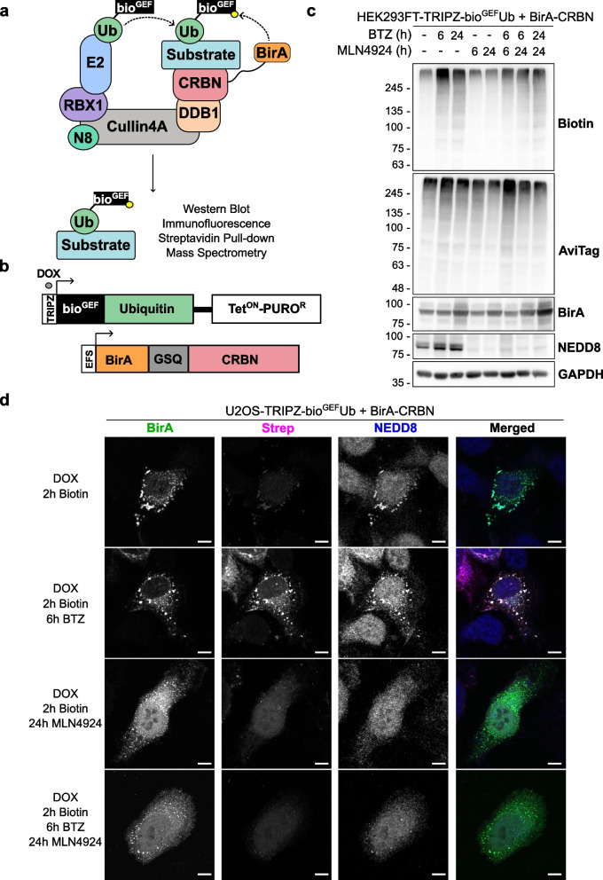
Background: The specificity of the ubiquitination process is mediated by the E3 ligases. Discriminating genuine substrates of E3s from mere interacting proteins is one of the major challenges in the field. We previously developed BioE3, a biotin-based approach that uses BirA-E3 fusions together with ubiquitin fused to a low-affinity AviTag to obtain a site-specific and proximity-dependent biotinylation of the substrates. We proved the suitability of BioE3 to identify targets of RING and HECT-type E3 ligases.

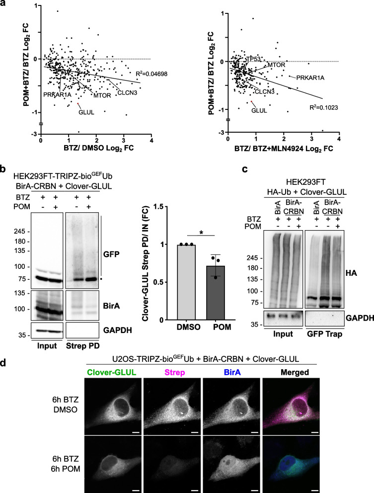
Methods: BioE3 experiments were performed in HEK293FT and U2OS stable cell lines expressing TRIPZ-bioGEFUb transiently transfected with BirA-cereblon (CRBN). Cells were seeded using biotin-free media, followed later by a short-biotin pulse. We evaluated the applicability of the BioE3 system to CRBN and molecular glues by Western blot and confocal microscopy, blocking the proteasome with bortezomib, inhibiting NEDDylation with MLN4924 and treating the cells with pomalidomide. For the identification of endogenous substrates and neosubstrates we analyzed the eluates of streptavidin pull-downs of BioE3 experiments by LC-MS/MS. Analysis of targets for which ubiquitination changes significantly upon treatment was done using two-sided Student's t-test. Orthogonal validations were performed by histidine pull-down, GFP-trap and computational modelling.

Results: Here we demonstrate that BioE3 is suitable for the multi-protein complex Cullin-RING E3s ligases (CRLs), the most utilized E3-type for targeted protein degradation (TPD) strategies. Using CRBN as proof of concept, one of the substrate receptors of CRL4 E3 ligase, we identified both endogenous substrates and novel neosubstrates upon pomalidomide treatment, including CSDE1 which contains a G-loop motif potentially involved in the binding to CRBN in presence of pomalidomide. Importantly, we observed a major rearrangement of the endogenous ubiquitination landscape upon treatment with this molecular glue.

Conclusions: The ability of BioE3 to detect and compare both substrates and neosubstrates, as well as how substrates change in response to treatments, will facilitate both on-target and off-target identifications and offer a broader characterization and validation of TPD compounds, like molecular glues and PROTACs.

Keywords: E3 ligases; Immunomodulatory drugs; Molecular Glue; Targeted Protein Degradation; Ubiquitin.


Conflict of interest statement

Declarations. Ethics approval and consent to participate: Not applicable. Consent for publication: Not applicable. Competing interests: C.G. is co-founder and Drug Discovery Scientific Advisor of Oniria Therapeutics. The other authors declare that they have no competing interests.

Figures

Fig. 1
Fig. 1
BioE3 labels CRL-dependent ubiquitinated substrates of CRBN. a, b Schematic representation of the BioE3 strategy adapted to the substrate receptor CRBN (a) and the constructs used in this work (b). TRIPZ, all-in-one inducible lentiviral vector; bioGEF, low affinity AviTag (see text); DOX, doxycycline; TetON, tetracycline inducible promoter; PUROR, puromycin resistant cassette; EFS, elongation factor 1α short promoter. c Western blot of BioE3 experiment performed on HEK293FT stable cell line expressing TRIPZ-bioGEFUb and transfected with EFS-BirA-CRBN. Indicated samples were treated with 100 nM bortezomib (BTZ) for 24 h, 200 nM BTZ for 6 h and/or 1 µM MLN4924 for 6 or 24 h. Molecular weight markers are shown to the left of the blots in kDa, antibodies used are indicated to the right. AviTag antibodies highlight all the ubiquitinated proteins, while biotin shows those ubiquitinated by CRBN. d Confocal microscopy images of BioE3 experiment performed on U2OS stable cell line expressing TRIPZ-bioGEFUb transfected with BirA-CRBN. Indicated samples were treated with 200 nM BTZ for 6 h and/or 1 µM MLN4924 for 24 h. Biotinylated material is stained with fluorescent streptavidin (Strep, magenta) and BirA (green) and NEDD8 (blue) with specific antibodies. Scale bar: 8 µm. All BioE3 experiments were performed by pre-incubating the cells in dialyzed FBS-containing media prior to transfections, doxycycline (DOX) induction at 1 µg/ml for 24 h and biotin supplementation at 50 µM for 2 h
Fig. 2
Fig. 2
Identification of SALL4 as a neosubstrate of CRBN upon pomalidomide treatment. a Confocal microscopy of BioE3 experiment performed in U2OS stable cell line stably expressing TRIPZ-bioGEFUb. Cells were transfected with EFS-BirA-CRBNWT or EFS-BirA-CRBNW386A (IMiD-binding deficient mutant). Indicated samples were treated with 10 µM pomalidomide (POM) and/or with 200 nM bortezomib (BTZ) for 6 h. Biotinylated material is stained with fluorescent streptavidin (Strep, magenta) and BirA (green) with specific antibodies. b, c BioE3 experiment performed in HEK293FT-TRIPZ-bioGEFUb (b) or U2OS-TRIPZ-bioGEFUb (c) stable cell lines transiently transfected with CMV-SALL4-YFP and EFS-BirA-CRBNWT or EFS-BirA-CRBNW386A and treated with 10 µM POM and/or 200 nM BTZ for 6 h. b Western blot validation of SALL4 as a neosubstrate upon pomalidomide treatment. The dot represents the possible monoubiquitinated protein, whereas the bar represents the polyUb-modified SALL4-YFP. Strep PD: streptavidin pull-down. Molecular weight markers are shown to the left of the blots in kDa, antibodies used are indicated to the right. Quantification graph of the ubiquitinated GFP-SALL4 level in the GFP panel is shown under the Western blots; *: p-value < 0.05. c) Biotinylated material is stained with fluorescent streptavidin (Strep, magenta) and BirA (blue) with a specific antibody. SALL4-YFP can be found in green. Scale bar: 8 µm. Yellow dotted lines indicate the nuclei. Insets show the amplification of the area indicated by a white dotted square in each panel. All BioE3 experiments were performed by pre-incubating the cells in dialyzed FBS-containing media prior to transfections, doxycycline (DOX) induction at 1 µg/ml for 24 h and biotin supplementation at 50 µM for 2 h
Fig. 3
Fig. 3
BioE3 identifies endogenous substrates of CRBN. a Western blot of BioE3 experiment performed on HEK293FT stable cell line expressing TRIPZ-bioGEFUb and transfected with EFS-BirA-CRBN. Indicated samples were treated with 200 nM bortezomib (BTZ) for 6 h, 1 µM MLN4924 for 24 h and 5 µM pomalidomide (POM) for 6 h. b, c Volcano plots of LC–MS/MS analysis comparing streptavidin pull-downs of BioE3 experiments showed in (a). Proteins significantly enriched (Log2 Fold Change (FC) BTZ/DMSO (b) or BTZ + MLN4924 (c) > 0 and p-value < 0.05) were considered as CRBN targets. Statistical analyses were done using two-sided Student’s t-test. d Venn diagram showing the endogenous targets of CRBN identified by BioE3 in (b) and (c). e Western blot of NEDDylated proteins from samples described in (a). Strep PD: streptavidin pull-down. Molecular weight markers are shown to the left of the blot in kDa, antibodies used are indicated to the right. All BioE3 experiments were performed by pre-incubating the cells in dialyzed FBS-containing media prior to transfections, doxycycline (DOX) induction at 1 µg/ml for 24 h and biotin supplementation at 50 µM for 2 h
Fig. 4
Fig. 4
BioE3 identifies neosubstrates of CRBN upon pomalidomide treatment. a Volcano plot of LC–MS/MS analysis comparing streptavidin pull-downs of BioE3 experiments showed in Fig. 3. Proteins significantly enriched (Log2 Fold Change (FC) POM + BTZ/BTZ > 0 and p-value < 0.05) were considered as CRBN neosubstrates. Statistical analysis was done using two-sided Student’s t-test. b Western blot of a BioE3 experiment performed in HEK293FT-TRIPZ-bioGEFUb stable cell line transiently transfected with EFS-BirA-CRBNWT or EFS-BirA-CRBNW386A and treated with 10 µM pomalidomide (POM) and/or 200 nM bortezomib (BTZ) for 6 h. We validated endogenous CSDE1 as a neosubstrate of CRBN upon pomalidomide treatment. All BioE3 experiments were performed by pre-incubating the cells in dialyzed FBS-containing media prior to transfections, doxycycline (DOX) induction at 1 µg/ml for 24 h and biotin supplementation at 50 µM for 2 h. Quantification graph of the ubiquitinated endogenous CSDE1 level is shown under the Western blots; *: p-value < 0.05. c Histidine pull-down confirming CSDE1 as a neosubstrate of CRBN. HEK293FT cells were transiently transfected with BirA-CRBN and His-Ub and treated with 200 nM BTZ for 6 h and with pomalidomide at the indicated concentrations and time points. Molecular weight markers are shown to the left of the blots in kDa, antibodies used are indicated to the right. Dots in (b) and (c) indicate an unidentified band that may correspond to a partially degraded form of CSDE1 or an isoform. d Comparison between the potential mean force (PMF) profiles for the interaction of W400 of CRBN with the backbone carbonyl group of T168 (top panel) and T727 (bottom panel) of CSDE1 in the presence of POM (orange line) and without ligand in the thalidomide-binding domain (blue line). The solid lines represent the PMF calculated using the Jarzynski equality for 100 independent replicas and the shaded areas represent the standard deviation obtained by bootstrapping
Fig. 5
Fig. 5
The ubiquitination of endogenous substrates of CRBN decreases upon pomalidomide treatment. a Comparison of the fold changes (FC) of the ubiquitination levels of the putative endogenous substrates showing pomalidomide (POM) treatment targets in the Y axis versus not treated in X axis, with or without MLN4924 treatments as in Fig. 3b and c. b, c BioE3 experiment performed in HEK293FT-TRIPZ-bioGEFUb (b) or U2OS-TRIPZ-bioGEFUb (c) stable cell lines transiently transfected with EFS-Clover3-GLUL and EFS-BirA-CRBN and treated with 10 µM POM and/or 200 nM bortezomib (BTZ) for 6 h. b Western blot validation by BioE3 of GLUL as a substrate of CRBN. The dot represents the unmodified protein, whereas the bar the Ub modified. Strep PD: streptavidin pull-down. Molecular weight markers are shown to the left of the blots in kDa, antibodies used are indicated to the right. Quantification graph of the ubiquitinated Clover-GLUL levels is shown to the right of the Western blots. c GFP-trap pull-down confirming the reduction in the ubiquitination by CRBN upon pomalidomide treatment. HEK293FT cells were transiently transfected with BirA-CRBN, Clover3-GLUL and HA-Ub and treated with 200 nM BTZ and with 10 µM pomalidomide for 6 h as indicated. d Biotinylated material is stained with fluorescent streptavidin (Strep, magenta) and BirA (blue) with a specific antibody. Clover3-GLUL can be found in green. Scale bar: 8 µm. All BioE3 experiments were performed by pre-incubating the cells in dialyzed FBS-containing media prior to transfections, doxycycline (DOX) induction at 1 µg/ml for 24 h and biotin supplementation at 50 µM for 2 h

References

    1. Dikic I, Schulman BA. An expanded lexicon for the ubiquitin code. Nat Rev Mol Cell Biol. 2023;24(4):273–87. - PMC - PubMed
    1. Cappadocia L, Lima CD. Ubiquitin-like protein conjugation: structures, chemistry, and mechanism. Chem Rev. 2018;118(3):889–918. - PMC - PubMed
    1. Lange SM, Armstrong LA, Kulathu Y. Deubiquitinases: From mechanisms to their inhibition by small molecules. Mol Cell. 2022;82(1):15–29. - PubMed
    1. Zheng N, Shabek N. Ubiquitin ligases: structure, function, and regulation. Annu Rev Biochem. 2017;86:129–57. - PubMed
    1. Morreale FE, Walden H. Types of ubiquitin ligases. Cell. 2016;165(1):248–e1. - PubMed

MeSH terms

LinkOut - more resources