. 2025 Feb 19;17(1):47.
doi: 10.1186/s13195-025-01695-w.

Cornuside alleviates cognitive impairments induced by Aβ1-42 through attenuating NLRP3-mediated neurotoxicity by promoting mitophagy

Affiliations

Cornuside alleviates cognitive impairments induced by Aβ1-42 through attenuating NLRP3-mediated neurotoxicity by promoting mitophagy

Fulin Zhou et al. Alzheimers Res Ther. .

Erratum in

Abstract

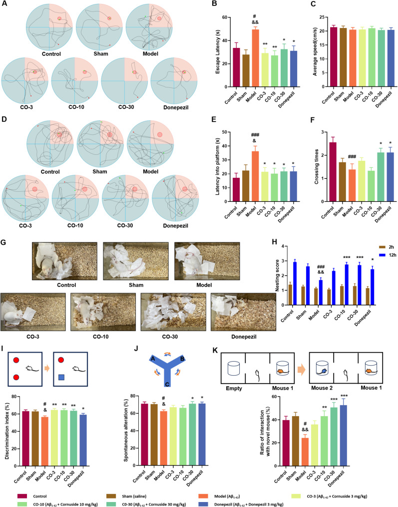
Alzheimer's disease (AD) is a progressive neurodegenerative disorder in which mitochondrial dysfunction and neuroinflammation play crucial roles in its progression. Our previous studies found that cornuside from Cornus officinalis Sieb.Et Zucc is an anti-AD candidate, however, its underlying mechanism remains unknown. In the present study, AD mice were established by intracerebroventricular injection of Aβ1-42 and treated with cornuside (3, 10, 30 mg/kg) for 2 weeks. Cornuside significantly ameliorated behavioral deficits, protected synaptic plasticity and relieved neuronal damage in Aβ1-42 induced mice. Importantly, cornuside decreased NLRP3 inflammasome activation, characterized by decreased levels of NLRP3, ASC, Caspase-1, GSDMD, and IL-1β. Furthermore, cornuside promoted mitophagy accompanied by decreasing SQSTM1/p62 and promoting LC3B-I transforming into LC3B-II, via Pink1/Parkin signaling instead of FUNDC1 or BNIP3 pathways. In order to investigate the relationship between NLRP3 inflammasome and mitophagy in the neuroprotective mechanism of cornuside, we established an in-vitro model in BV2 cells exposed to LPS and Aβ1-42. And cornuside inhibited NLRP3 inflammasome activation and subsequent cytokine release, also protected neurons from damaging factors in microenvironment of conditional culture. Cornuside improved mitochondrial function by promoting oxidative phosphorylation and glycolysis, decreasing the production of ROS and mitochondrial membrane potential depolarization. Besides, mitophagy was also facilitated with increased colocalization of MitoTracker with LC3B and Parkin, and Pink1/Parkin, FUNDC1 and BNIP3 pathways were all involved in the mechanism of cornuside. By blocking the formation of autophagosomes by 3-MA, the protective effects on mitochondria, the inhibition on NLRP3 inflammasome as well as neuronal protection in conditional culture were eliminated. There is reason to believe that the promotion of mitophagy plays a key role in the NLRP3 inhibition of cornuside. In conclusion, cornuside re-establishes the mitophagy flux which eliminates damaged mitochondria and recovers mitochondrial function, both of them are in favor of inhibiting NLRP3 inflammasome activation, then alleviating neuronal and synaptic damage, and finally improving cognitive function.

Keywords: Alzheimer’s disease; Cornuside; Mitophagy; NLRP3; Neuroprotection.


Conflict of interest statement

Declarations. Ethics approval and consent to participate: All animal procedures were approved by the Animal Care and Use Committee of China-Japan Friendship Hospital. Consent for publication: Not applicable. Competing interests: The authors declare no competing interests.

Figures

Fig. 1
Fig. 1
The effect of cornuside on the learning and memory of Aβ1−42 intracerebroventricular injected mice in the behavioral tests. Data were expressed as the mean ± SEM (n = 13–15). A. Exploring routes during the navigation trial. B, C. Escape latency and average speed in the navigation trial. D. Exploring routes in the spatial probe trial. E, F. Latency to the platform and crossing times in the spatial probe trial. G, H. Representative nests built by mice, scored at 2 and 12 h in the nest building test. I. Discrimination index in the novel object recognition test. J. Spontaneous alternation rate in the Y-maze. K. Ratio of interaction with a novel mouse in the novel object recognition test. #p < 0.05 and ###p < 0.001 vs. control group; &p < 0.05 and &&p < 0.01 vs. sham group; *p < 0.05, **p < 0.01 and ***p < 0.001 vs. model group
Fig. 2
Fig. 2
The effect of cornuside on synapses of hippocampus and cortex in Aβ1−42 intracerebroventricular injected mice. Data were expressed as the mean ± SEM (n = 3–6). A. Protein levels of PSD95 in the hippocampus and cortex. B-D. Fold changes of PSD95 and representative immunofluorescence images (scale bar: 40 μm) in the hippocampus and cortex. E. Number of synapses in the hippocampus, with representative transmission electron microscopy (TEM) photomicrographs (scale bar: 2 μm). #p < 0.05, ###p < 0.001 vs. control group; *p < 0.05 and ***p < 0.001 vs. model group
Fig. 3
Fig. 3
The effect of cornuside on neuron in the hippocampus and cortex of Aβ1−42 intracerebroventricular injected mice. Data were expressed as the mean ± SEM (n = 3–6). A, C. Representative images of TUNEL staining for the hippocampus and cortex, with a scale bar of 100 μm. B, D. The percentage of apoptotic cells in these regions. E, F. Representative images of IHC staining (scale bar: 100 μm) in CA3 and cortex. G Protein levels of NeuN in the hippocampus and cortex. H. TEM photomicrographs showing neuronal structures from different treatment groups (scale bar: 2 μm). The orange arrow indicates the nucleolus; the yellow arrow indicates the nuclear membrane; the blue arrow indicates mitochondrial ultrastructure; and the green arrow indicates the endoplasmic reticulum. #p < 0.05, ###p < 0.001 vs. control group; *p < 0.05, **p < 0.01 and ***p < 0.001 vs. model group
Fig. 4
Fig. 4
The effect of cornuside on NLRP3 inflammasome in Aβ1−42 intracerebroventricular injected mice. Data are expressed as the mean ± SEM (n = 4–6). A, B. Representative images of NLRP3 and ASC in the hippocampus and cortex (scale bar: 40 μm). C-F. Fold changes of NLRP3 and ASC are displayed. G, H. Protein levels of NLRP3, GSDMD, ASC, Caspase-1, and IL-1β. #p < 0.05, ##p < 0.01, ###p < 0.001 vs. control group; *p < 0.05, **p < 0.01 and ***p < 0.001 vs. model group
Fig. 5
Fig. 5
The effect of cornuside on mitophagy of hippocampus and cortex in Aβ1−42 intracerebroventricular injected mice. Data are expressed as the mean ± SEM (n = 4–6). A, B. Representative images of LC3B in the hippocampus and cortex (scale bar: 40 μm). C-I. Protein levels of PINK1, SQSTM1/p62, Parkin, BNIP3, FUNDC1 and LC3B. #p < 0.05, ##p < 0.01 vs. control group, *p < 0.05, **p < 0.01 and ***p < 0.001 vs. model group
Fig. 6
Fig. 6
The effect of cornuside on LPS + Aβ1−42 induced NLRP3 inflammasome activation of BV2 cells and conditioned stimulation of HT22 cells. Data are expressed as the mean ± SEM (n = 3–6). A-D. Cytokine levels determined in the supernatant of cultured BV2 cells. E, G. Protein levels of NLRP3, ASC, Pro-caspase-1, Caspase-1, Pro-IL-1β, IL-1β and GSDMD-NT. F. The representative images of NLRP3 in BV2 cells (scale bar: 20 μm). H, I. Neuron survival rate and LDH levels in supernatant. J, K. Flow cytometric representation and statistical analysis of apoptosis in conditioned medium. #p < 0.05, ##p < 0.01, ###p < 0.001 vs. control group; *p < 0.05, **p < 0.01 and ***p < 0.001 vs. model group
Fig. 7
Fig. 7
The effect of cornuside on mitochondrial function in LPS + Aβ1−42 induced d BV2 cells. Data were expressed as the mean ± SEM (n = 4–6). A. JC-1 flow cytometry representation and statistics. B-D. Levels of ROS and mtROS measured by flow cytometry, and the representative pictures with scale bar of 40 μm. E, F. The OCR and ECAR were measured using a Seahorse XF24 Extracellular Flux Analyzer instrument. #p < 0.05, ##p < 0.01, ###p < 0.001 vs. control group; *p < 0.05, **p < 0.01 and ***p < 0.001 vs. model group
Fig. 8
Fig. 8
The effect of cornuside on mitophagy in LPS + Aβ1−42 induced d BV2 cells. Data are expressed as the mean ± SEM (n = 4–6). A, B. Protein levels of PINK1, Parkin, FUNDC1, BNIP3, SQSTM1/p62 and LC3B. C, D IF staining of MitoTracker and LC3B colocalization, with Pearson colocalization coefficient. E, F. IF staining of MitoTracker and Parkin colocalization, with Pearson colocalization coefficient. (Scale bar: 20 μm). #p < 0.05, ##p < 0.01, ###p < 0.001 vs. control group; *p < 0.05, **p < 0.01 and ***p < 0.001 vs. model group
Fig. 9
Fig. 9
The effect of 3-MA on mitophagy, anti-inflammatory and neuroprotective effects of cornuside. Data are expressed as the mean ± SEM (n = 4–6). A Protein levels of PINK1, SQSTM1/p62, Parkin, BNIP3, FUNDC1 and LC3B. B. The red/green ratio of JC-1 was measured by flow cytometry. C. mtROS levels were measured by flow cytometry. D-G. cytokines levels in the supernatant. H, I. Protein levels of NLRP3, Pro-caspase-1, GSDMD-NT, Pro-IL-1β, ASC, Caspase-1 and IL-1β. J. LDH leakage in supernatant. K, L. Flow cytometric representation and statistical analysis of apoptosis in conditioned medium. *p < 0.05, **p < 0.01 and ***p < 0.001
Fig. 10
Fig. 10
Proposed mechanism of cornuside alleviating cognitive disorder in Aβ1−42 intracerebroventricular injected mice. Cornuside promoted mitophagy via PINK1/Parkin, BNIP3 and FUNDC1 pathways, leading to the elimination of damaged mitochondria. Following the improvement of mitochondrial quality and function, ROS production was significantly inhibited, which decreased the activation of NLRP3 inflammasome. Both mitochondrial improvement and NLRP3 inhibition could protect the neuron and synapse from damage, indicating the importance of mitophagy in cornuside alleviating neuro-inflammation and neuro-degeneration in Aβ1−42 intracerebroventricular injected mice

References

    1. Lane CA, Hardy J, Schott JM. Alzheimer’s disease. Eur J Neurol. 2018;25:59–70. - PubMed
    1. Hardy J, Selkoe DJ. The amyloid hypothesis of Alzheimer’s disease: progress and problems on the road to therapeutics. Science. 2002;297:353–6. - PubMed
    1. Reddy PH, Manczak M, Yin X, Grady MC, Mitchell A, Tonk S, Kuruva CS, Bhatti JS, Kandimalla R, Vijayan M, et al. Protective effects of Indian Spice Curcumin against Amyloid-β in Alzheimer’s Disease. J Alzheimers Dis. 2018;61:843–66. - PMC - PubMed
    1. Oddo S, Caccamo A, Shepherd JD, Murphy MP, Golde TE, Kayed R, Metherate R, Mattson MP, Akbari Y, LaFerla FM. Triple-transgenic model of Alzheimer’s disease with plaques and tangles: intracellular abeta and synaptic dysfunction. Neuron. 2003;39:409–21. - PubMed
    1. Goel P, Chakrabarti S, Goel K, Bhutani K, Chopra T, Bali S. Neuronal cell death mechanisms in Alzheimer’s disease. An insight. Front Mol Neurosci. 2022;15:937133. - PMC - PubMed

MeSH terms

LinkOut - more resources