Review
. 2025 Feb 19;20(1):20.
doi: 10.1186/s13024-025-00811-6.

The integrated stress response in neurodegenerative diseases

Affiliations
Review

The integrated stress response in neurodegenerative diseases

Maria Astrid Bravo-Jimenez et al. Mol Neurodegener. .

Abstract

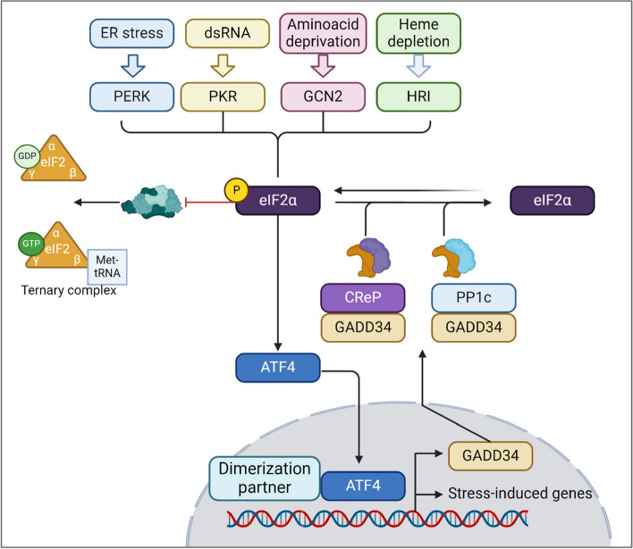
The integrated stress response (ISR) is a conserved network in eukaryotic cells that mediates adaptive responses to diverse stressors. The ISR pathway ensures cell survival and homeostasis by regulating protein synthesis in response to internal or external stresses. In recent years, the ISR has emerged as an important regulator of the central nervous system (CNS) development, homeostasis and pathology. Dysregulation of ISR signaling has been linked to several neurodegenerative diseases. Intriguingly, while acute ISR provide neuroprotection through the activation of cell survival mechanisms, prolonged ISR can promote neurodegeneration through protein misfolding, oxidative stress, and mitochondrial dysfunction. Understanding the molecular mechanisms and dynamics of the ISR in neurodegenerative diseases aids in the development of effective therapies. Here, we will provide a timely review on the cellular and molecular mechanisms of the ISR in neurodegenerative diseases. We will highlight the current knowledge on the dual role that ISR plays as a protective or disease worsening pathway and will discuss recent advances on the therapeutic approaches that have been developed to target ISR activity in neurodegenerative diseases.


Conflict of interest statement

Declarations. Competing interests: Authors declare no competing financial interests.

Figures

Fig. 1
Fig. 1
The integrated stress response signaling pathway. The ISR can be initiated upon sensing ER stress, dsRNA, amino acid deprivation, and heme depletion, which are recognized by PERK, PKR, GCN2, and HRI respectively. Once activated, the four kinases converge in the phosphorylation of eIF2α that then activates ATF4. ATF4 translocate to the nucleus in which promotes the translation of stress response genes. Evidence shows regulation of ISR through “negative feedback”. ATF4 induces the expression of GADD34, which promotes the dephosphorylation of eIF2α by recruiting CReP and PP1c, hence restoring protein synthesis. Figure was created in BioRender
Fig. 2
Fig. 2
Mitochondrial dysfunction and ISR: The activation of the integrated stress response (ISR) during mitochondrial dysfunction is triggered by various mechanisms. The fragmentation of mitochondrial DNA is among the leading causes of mitochondrial dysfunction, which is managed by an ISR sensor, protein kinase RNA-activated (PKR). Mitochondrial dysfunction during amino acid metabolism is managed by another class of ISR kinase- general control nonderepressible-2 (GCN2). The Tri-Carboxylic Acid (TCA) cycle is fed via the amino acid degradation during metabolic rewiring stage of mitochondrial stress, and the depletion of amino acids results in activation of GCN2. During this process, the equilibrium maintenance of reducing equivalents is maintained by the malate and aspartate shuttle. Upon induction of mitochondrial stress by generation of reactive oxygen species (ROS), a mitochondrial protease known as OMA1 regulates the mitochondrial stress dynamics by cleaving DAP3-binding cell death enhancer 1 (DELE1). This subsequently activates heme-regulated inhibitor (HRI) after translocating to the cytoplasm. The crosstalk between mitochondria and endoplasmic reticulum aids ER to sense the alterations in the levels of calcium (Ca2 +), ROS, and changes in energy productions; leading to activation of ER transmembrane protein- PKR-like ER kinase (PERK). Upon mitochondrial dysfunction and the ER-mitochondrial crosstalk, these protein kinases phosphorylate eIF2α and inhibit global protein translation to mitigate stress and restore the normal homeostasis. Until the stress is resolved, a translational shift due to upstream open reading frame (uORF) mediates selective translation of proteins such as ATF-4 and usurps the global translation to mitigate the stress. ATF-4 along with its other dimerization partners regulates amino acid synthesis genes, antioxidant pathways, chaperones and metabolism related genes to restore the homeostasis post stress. However, chronic stress results in activation of CHOP-mediated apoptotic cell death. Figure was created in BioRender
Fig. 3
Fig. 3
The integrated stress response in neurodegenerative disorders. The integrated stress response (ISR) can be initiated upon sensing ER stress. In Multiple Sclerosis (MS), IFNγ is shown to induce ER stress in myelinating oligodendrocytes (OLs) via the phosphorylation of PERK and eIF2α. The main downstream markers identified in lesions of human MS and the MS animal model EAE (experimental autoimmune encephalomyelitis) are CHOP and BiP. EAE studies show that the ISR can be either detrimental for developing oligodendrocytes or cytoprotective for neurons and mature oligodendrocytes. In Alzheimer’s Disease (AD), the formation of amyloid beta (Aβ) plaques and Tau protein aggregates can initiate ISR primarily through PERK and PKR. ISR markers such as p-PERK and p-eIF2α have been found mainly in the frontal cortex and hippocampus. Evidence implicates PERK activation as either neuroprotective by mitigating Aβ- and tau-induced neuronal death (neuron in green) or cytotoxic by promoting tau phosphorylation and aggregation that lead to neuronal damage (neuron in red). In Parkinson’s Disease (PD), α synuclein and Lewy bodies promote the initiation of the ISR via PERK and HRI phosphorylation. ISR markers are commonly found in the affected substantia nigra and dopaminergic neurons. The PERK-ATF4 signaling pathway is relevant in PD. The involvement of ATF4 in the transcription of Parkin (a pro-survival protein) indicates a cytoprotective role (neuron in green). However, excessive ATF4 activity may contribute to neuronal death by promoting the transcription of pro-apoptotic factors such as Trib3 and CHOP (neuron in red). In Amyotrophic Lateral Sclerosis (ALS) motor neurons in the spinal cord are primarily affected. The ISR is activated by protein aggregation, induced mainly by mutations in TDP-43, SOD1, and FUS. The main markers observed in ALS models and human ALS samples are p-eIF2α, ATF4, CHOP, and BiP. The ISR is mostly detrimental in ALS, and the activation of CHOP in ALS models is linked to apoptosis and neurodegeneration (neuron in red). Figure was created in BioRender

References

    1. Pakos-Zebrucka K, et al. The integrated stress response. EMBO Rep. 2016;17:1374–95. - PMC - PubMed
    1. Lin JH, Walter P, Yen TSB. Endoplasmic reticulum stress in disease pathogenesis. Ann Rev Pathol: Mech Dis. 2008;3:399–425. 10.1146/annurev.pathmechdis.3.121806.151434. - PMC - PubMed
    1. Balachandran S, et al. Essential role for the DsRNA-dependent protein kinase PKR in innate immunity to viral infection. Immunity. 2000;13:129–41. 10.1016/s1074-7613(00)00014-5. - PubMed
    1. Han AP, et al. Heme-regulated eIF2alpha kinase (HRI) is required for translational regulation and survival of erythroid precursors in iron deficiency. EMBO J. 2001;30:6909–18. - PMC - PubMed
    1. Deegan S, et al. A close connection between the PERK and IRE arms of the UPR and the transcriptional regulation of autophagy. Biochem Biophys Res Commun. 2015;456:305–11. - PubMed

MeSH terms

LinkOut - more resources