

Understanding the role of electrostatic force, van der Waals force, and osmotic pressure in retinal function and barrier integrity
- PMID: 39972495
- PMCID: PMC11837441
- DOI: 10.1186/s40942-025-00643-y
Understanding the role of electrostatic force, van der Waals force, and osmotic pressure in retinal function and barrier integrity
Abstract
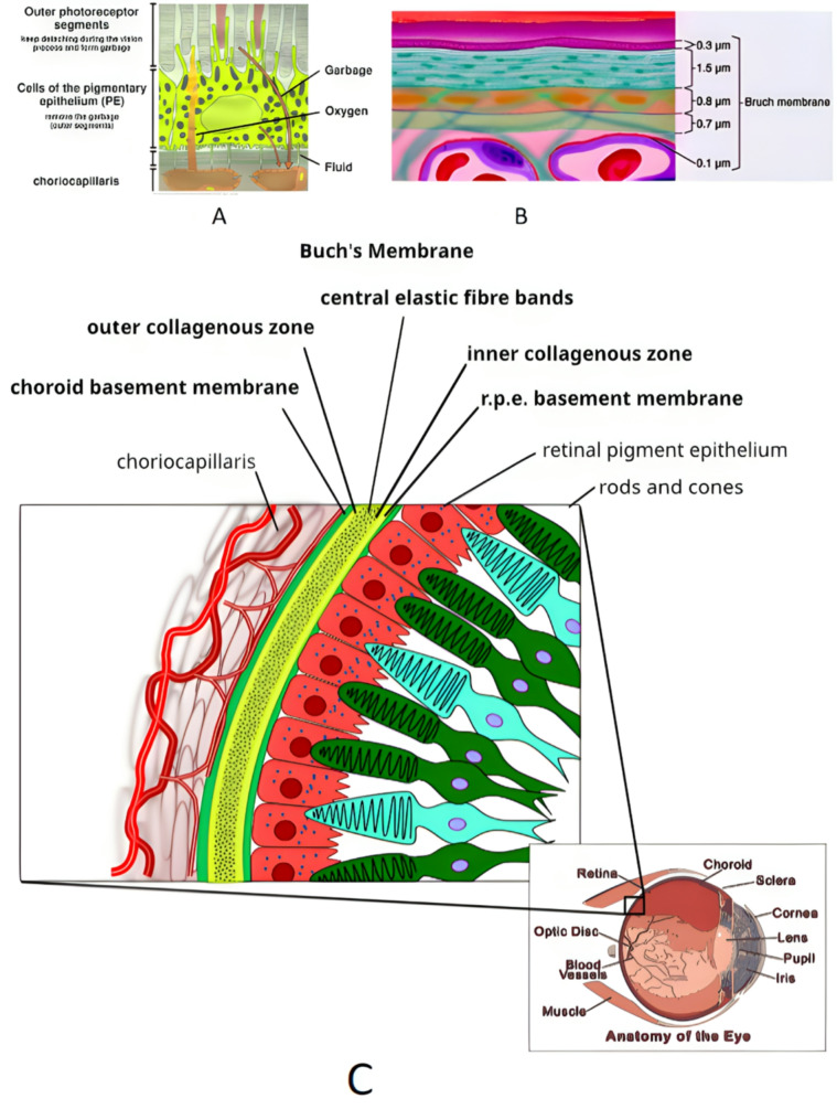
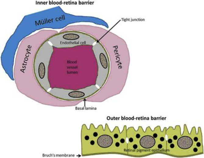
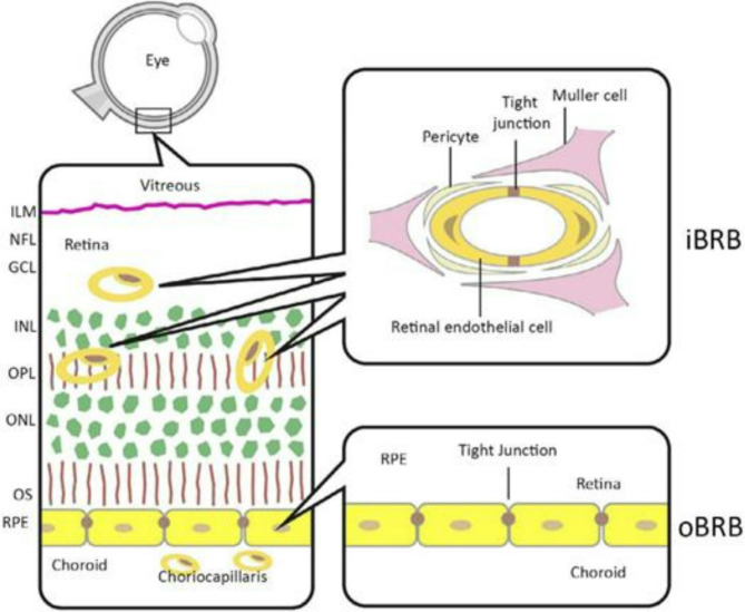
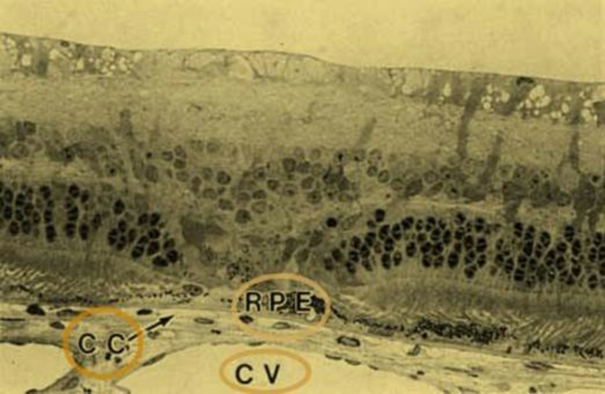
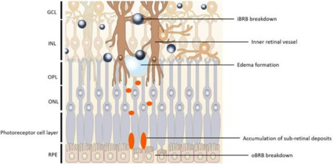
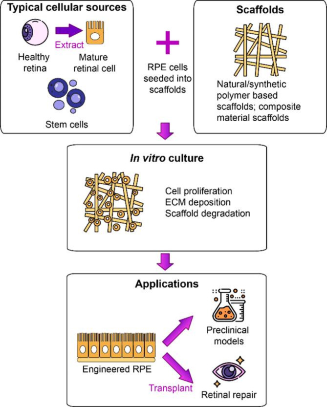
The retina's intricate interplay of forces and structures, with a focus on the retinal pigment epithelium (RPE) and photoreceptors, is essential for retinal health and function. Among these forces, electrostatic forces play a crucial role, working alongside van der Waals forces and oncotic pressure to maintain the retina's attachment to the RPE and ensure the integrity of the blood-retina barrier (BRB). The composition of the interphotoreceptor matrix (IPM), influenced by molecules like Retbindin secreted by rod photoreceptors, further modulates these forces, affecting processes like visual pigment regeneration and metabolite exchange. In the context of retinal tissue engineering and new technologies for support and cells-based treatments, electrostatic forces are harnessed to optimize nutrient supply to transplanted RPE cells by reducing pore size in electrospun polymer membranes. Scaffold-based strategies for retinal repair also utilize electrostatic, hydrophobic, van der Waals, and hydrogen bonding forces to enhance cell adhesion and growth, mimicking the basement membrane. Understanding the complex dynamics of these forces in retinal-RPE interactions holds promise for innovative treatments for retinal disorders, emphasizing the intricate balance between electrostatic forces, van der Waals forces, oncotic pressure, and more. These insights open exciting avenues for research and therapeutic interventions in ophthalmology. Additionally, van der Waals forces are explored in the context of cell adhesion, and their potential role in retinal health is discussed, particularly in relation to melanin's protective properties against blue light-induced damage. Tissue engineering approaches, both scaffold-free and scaffold-based, are discussed, highlighting the importance of physical surface treatments and adhesive forces in preserving engineered RPE tissue. Overall, this abstract provides a comprehensive overview of the multifaceted role of electrostatic and other forces in retinal biology and their implications for future research and clinical applications in ophthalmology.
Keywords: Electrostatic Force; Osmotic pressure; Outer segment photoreceptor; Retinal epithelium; Van Der Waals Force.
© 2025. The Author(s).
Conflict of interest statement
Declarations. Ethics approval and consent to participate: The article describes a review article. Therefore, no additional permission from our Ethics Committee was required. Competing interests: The authors declare no competing interests. Clinical trial number: Not applicable. Method of literature: Explore scientific articles and research papers on Minimally Invasive Glaucoma Surgery (MIGS) in Google Scholar and pubmed as a transformative approach to glaucoma management. Investigate MIGS techniques, classifications, indications, contraindications, devices, and their impact on patient outcomes, with a focus on precision, safety, and minimal invasiveness. Assess the comparative advantages of MIGS over traditional glaucoma treatments, including reduced complications and improved patient comfort. Examine clinical cases and studies showcasing the efficacy of MIGS in various types of glaucoma. Delve into the potential of MIGS to change the landscape of ophthalmology and enhance the quality of life for glaucoma patients.
Figures
Similar articles
-
Relative importance of electrostatic and van der Waals forces in particle adhesion to rough conducting surfaces.Phys Rev E. 2021 Apr;103(4-1):042906. doi: 10.1103/PhysRevE.103.042906. Phys Rev E. 2021. PMID: 34005883
-
Specific and nonspecific interaction forces between Escherichia coli and silicon nitride, determined by poisson statistical analysis.Langmuir. 2006 Aug 15;22(17):7296-301. doi: 10.1021/la0533415. Langmuir. 2006. PMID: 16893229
-
Electrospun membranes filtering 100 nm particles from air flow by means of the van der Waals and Coulomb forces.J Memb Sci. 2022 Feb 15;644:120138. doi: 10.1016/j.memsci.2021.120138. Epub 2021 Nov 27. J Memb Sci. 2022. PMID: 36567692 Free PMC article.
-
A review on data and predictions of water dielectric spectra for calculations of van der Waals surface forces.Adv Colloid Interface Sci. 2017 Dec;250:54-63. doi: 10.1016/j.cis.2017.10.004. Epub 2017 Oct 31. Adv Colloid Interface Sci. 2017. PMID: 29100682 Review.
-
Construction of a Universal Gel Model with Volume Phase Transition.Gels. 2020 Feb 27;6(1):7. doi: 10.3390/gels6010007. Gels. 2020. PMID: 32120904 Free PMC article. Review.
References
-
- Kaur C, Foulds W, Ling E. Blood–retinal barrier in hypoxic ischaemic conditions: basic concepts, clinical features and management. Prog Retin Eye Res. 2008;27(6):622–47. - PubMed
-
- Ishikawa M, Sawada Y, Yoshitomi T. Structure and function of the interphotoreceptor matrix surrounding retinal photoreceptor cells. Exp Eye Res. 2015;133:3–18. - PubMed
-
- Tan E, Sing S, Yeong W. Scaffolds for retinal repairs. Handbook of tissue Engineering Scaffolds: volume two. Elsevier; 2019. pp. 673–91.
-
- Levine L. Basic and clinical science course, Sect. 2: fundamentals and principles of ophthalmology. Basic Clin Sci Course. 2018;430:2018–9.
Publication types
LinkOut - more resources
Full Text Sources
