Case Reports
. 2025 Feb 19;30(4):102605.
doi: 10.1016/j.jaccas.2024.102605. Epub 2024 Nov 27.

Multimodality Imaging Findings in Cardiac Plasmacytoma

Affiliations
Case Reports

Multimodality Imaging Findings in Cardiac Plasmacytoma

Mohammed Adam Benharrats et al. JACC Case Rep. .

Abstract

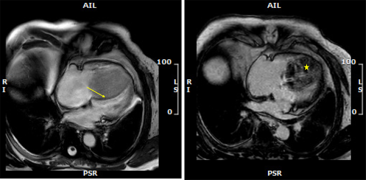
Extramedullary plasmacytoma involving the heart is a rare occurrence. We present the case of a 74-year-old man who received a diagnosis of cardiac plasmacytoma in the context of relapsed multiple myeloma. The tumor primarily presented as a solitary extramedullary plasmacytoma with extensive infiltration into the right ventricle, leading to mild symptoms such as dyspnea. There was no evidence of systemic involvement. A treatment with DRd (daratumumab, lenalidomide, and dexamethasone) was promptly initiated. This case demonstrates the effective use of multimodal imaging techniques to characterize cardiac masses and to determine the most appropriate management plan.

Keywords: cardiac magnetic resonance; computed tomography; echocardiography; heart; plasmacytoma; positron emission tomography-computed tomography.


Conflict of interest statement

Funding Support and Author Disclosures The authors have reported that they have no relationships relevant to the contents of this paper to disclose.

Figures

None
Graphical abstract
Figure 1
Figure 1
12-Lead Electrocardiogram Showing an Incomplete Right Bundle Branch Block and Negative T Waves in Leads V1-V3
Figure 2
Figure 2
Transthoracic Echocardiography (Left) Parasternal long-axis view. (Right) Apical view. A large invasive mass occupies nearly the entire right ventricle, involving the interventricular septum and the tricuspid valve.
Figure 3
Figure 3
Gated Cardiac Scan (Left) Contrast-enhanced imaging. (Upper right) Non–contrast-enhanced imaging. (Lower right) Delayed phase. The cardiac scan showed an irregular mass involving the right ventricle adjacent to the interventricular septum. The lesion displayed no fatty or calcified component, with a heterogeneous enhancement in the delayed phase.
Figure 4
Figure 4
Cardiac Magnetic Resonance Imaging (Left) Still frame of an axial SSFP cine sequence realized after injection of contrast material. A delineation between the interventricular septum and the mass attachment can be identified (yellow arrow). (Right) Late gadolinium enhancement sequence, showing heterogeneous gadolinium uptake of the mass (yellow star).
Figure 5
Figure 5
Cardiac Magnetic Resonance Imaging (Left) T1-weighted sequence showing a lightly hyperintense signal of the mass (yellow star). (Right) T2-STIR sequence showing a hyperintense signal of the mass (yellow star).
Figure 6
Figure 6
Positron Emission Tomography Scan Showing a Highly Hypermetabolic Mass
Figure 7
Figure 7
Coronary Angiography Showing Normal Epicardial Coronary Arteries A hypervascularized mass is seen. (Left) Right anterior oblique cranial projection. (Right) Left anterior oblique projection.
Figure 8
Figure 8
Histologic Findings (Upper left) Neoplastic plasma cells with positive staining for CD138 (×200). (Lower left) Focal sheets of neoplastic plasma cells (hematoxylin and eosin, ×400). (Right) Plasmacytoma, immunohistochemistry for kappa (upper right) and lambda (lower right) showing lambda light chain restriction (×200).
Visual Summary
Visual Summary
Imaging Modalities Used to Characterize the Intracardial Mass A 74-year-old man known to have multiple myeloma and extramedullary plasmacytoma in remission presented with worsening heart failure symptoms. A thorough investigation followed. A transthoracic echocardiogram (TTE) displayed a large mass in the right ventricle. Coronary angiography revealed substantial collateralization toward the mass. A cardiac scan confirmed its extension to the right ventricular outflow tract. Cardiac magnetic resonance (CMR) displayed an iso-to-hyperintense mass in T1, and T2 displayed a hyperintense mass. Fluordeoxyglucose–positron emission tomography (PET-FDG) demonstrated a highly hypermetabolic mass located in the right ventricle. A biopsy of the mass followed these imaging findings, and histologic examination of the biopsy specimen confirmed the diagnosis of cardiac plasmacytoma.

References

    1. Reynen K. Frequency of primary tumors of the heart. Am J Cardiol. 1996;77:107. - PubMed
    1. Tazelaar H.D., Locke T.J., McGregor C.G. Pathology of surgically excised primary cardiac tumors. Mayo Clin Proc. 1992;67:957–965. - PubMed
    1. Molina J.E., Edwards J.E., Ward H.B. Primary cardiac tumors: experience at the University of Minnesota. Thorac Cardiovasc Surg. 1990;38(suppl 2):183–191. - PubMed
    1. Mousavizadeh Ahmadabadi S.M., Banar S., Parvas E., Shahbazi N., Biranvand H. Uncommon manifestation of multiple myeloma: a case report of intracardiac plasmacytoma presenting with severe heart failure and bilateral pleural effusion. Int J Surg Case Rep. 2024;114 - PMC - PubMed
    1. Sequist L.V., Han J.Y., Ahn M.J., et al. Osimertinib plus savolitinib in patients with EGFR mutation-positive, MET-amplified, non-small-cell lung cancer after progression on EGFR tyrosine kinase inhibitors: interim results from a multicentre, open-label, phase 1b study. Lancet Oncol. 2020;21:373–386. - PubMed

Publication types

LinkOut - more resources