[Preprint]. 2025 Jan 24:2025.01.22.25320997.
doi: 10.1101/2025.01.22.25320997.

A novel lncRNA FAM151B-DT regulates autophagy and degradation of aggregation prone proteins

Affiliations

A novel lncRNA FAM151B-DT regulates autophagy and degradation of aggregation prone proteins

Arun Renganathan et al. medRxiv. .

Abstract

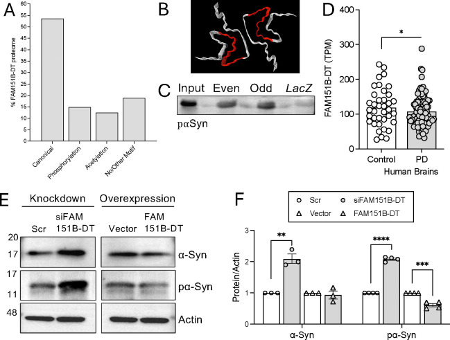
Neurodegenerative diseases share common features of protein aggregation along with other pleiotropic traits, including shifts in transcriptional patterns, neuroinflammation, disruptions in synaptic signaling, mitochondrial dysfunction, oxidative stress, and impaired clearance mechanisms like autophagy. However, key regulators of these pleotropic traits have yet to be identified. Here, we discovered a novel long non-coding RNA (lncRNA), FAM151B-DT, that is reduced in a stem cell model of frontotemporal dementia with tau inclusions (FTLD-tau) and in brains from FTLD-tau, progressive supranuclear palsy, Alzheimer's disease, and Parkinson's disease patients. We show that silencing FAM151B-DT in vitro is sufficient to enhance tau aggregation. To begin to understand the mechanism by which FAM151B-DT mediates tau aggregation and contributes to several neurodegenerative diseases, we deeply characterized this novel lncRNA and found that FAM151B-DT resides in the cytoplasm where it interacts with tau, α-synuclein, HSC70, and other proteins enriched in protein homeostasis. When silenced, FAM151B-DT blocks autophagy, leading to the accumulation of tau and α-synuclein. Importantly, we discovered that increasing FAM151B-DT expression is sufficient to promote autophagic flux, reduce phospho-tau and α-synuclein, and reduce tau aggregation. Overall, these findings pave the way for further exploration of FAM151B-DT as a promising molecular target for several neurodegenerative diseases.


Figures

Figure 1.
Figure 1.. FAM151B-DT expression is significantly reduced in MAPT mutant neurons and tauopathy brains.
A. Schematic representation of the discovery of lncRNAs that are differentially expressed (DE) in MAPT mutant iPSC-neurons compared with isogenic controls. FAM151B-DT was among the 15 common dysregulated lncRNAs in MAPT R406W, P301L, and IVS10+16 neurons . B. FAM151B-DT is significantly reduced in MAPT R406W, P301L, and IVS10+16 neurons compared with isogenic controls. Graph represents Log2FC. Data is representative of three biological replicates. C. FAM151B-DT is significantly reduced in human FTD with tau inclusions, AD, PSP brains compared with control brain tissues. Graph represents Log2FC. *, p≤0.05; ***, p<0.001; ****, p<0.0001.
Figure 2.
Figure 2.. FAM151B-DT expression regulates tau seeding.
A. Schematic representation of the FRET-based tau seeding assay. B. qPCR for FAM151B-DT reveals significant reduction of FAM151B-DT levels after siRNA treatment in tau biosensor cells. C. Silencing FAM151B-DT significantly increased integrated FRET density compared to control siRNA-treated tau biosensor cells. Quantification of the FRET signal, normalized to controls. Error bars represent SEM, n = 50,000 cells per experiment. D. qPCR for FAM151B-DT reveals significant increase in FAM151B-DT levels upon overexpression in tau biosensor cells. E. FAM151B-DT overexpression significantly decreased the integrated FRET density compared to ctrl-vector-treated tau-biosensor cells. Quantification of the FRET signal, normalized to controls. Error bars represent SEM, n = 50,000 cells per experiment. Data is representative of three biological replicates. Student’s t-test, *, p≤0.05; **, p<0.001; ****, p<0.0001.
Figure 3.
Figure 3.. Proteins interacting with FAM151B-DT.
A-B. Nuclear and cytosolic isolation of RNAs to define the localization of FAM151B-DT relative to well characterized lncRNAs (nuclear localized MALAT1 and cytosolic TUG1). A. iPSC-derived neurons. B. SH-SY5Y cells. C-D. Tau interacts with FAM151B-DT. C. Immunoprecipitation of tau (Tau5) in SH-SY5Y cells. D. RNA immunoprecipitation (RIP)-qPCR validation of the tau-bound RNA fraction shows robust FAM151B-DT expression. GAPDH included as a negative control. Data is representative of three biological replicates. Two-way ANOVA, ****, p<0.0001. E. Comprehensive identification of RNA binding proteins (ChIRP) workflow. Schematic representation includes the positions of even and odd probes along the entire FAM151B-DT RNA sequence. F. ChIRP-qPCR reveals enrichment of FAM151B-DT, compared to the housekeeping transcript GAPDH. One way ANOVA, ****, p<0.0001. G. ChIRP-mass spectrometry hits of proteins retrieved by even and odd oligos specific for FAM151B-DT compared with non-targeting LacZ control oligos. 215 total proteins were identified across the three samples using a protein probability ≥99%; ≥2 peptides mapped; and ≥95% peptide probability (coverage). H. GO enrichment analysis of the FAM151B-DT proteome (101 proteins) in SH-SY5Y cells. I. Schematic of string network analysis of FAM151B-DT proteome reveals multiple clusters (see Supplemental Figure 5). J. Visualizing cluster 1. Protein-protein interaction enrichment p-value = 1.0e−16. K. Top 10 most significant pathways in cluster 1 identified by KEGG-Pathway enrichment analysis.
Figure 4.
Figure 4.. FAM151B-DT and tau share interacting partners that are disrupted in MAPT mutant neurons and human brains.
A. Schematic illustrating workflow. The 101 FAM151B-DT interactors were compared with tau interactors defined in iPSC-neurons, human brains, and neurofibrillary tangles (NFT). The common set of interactors were then evaluated for differential expression at the RNA level in MAPT mutant iPSC-neurons and FTLD-tau brains. B. Upset plot for FAM151B-DT interactors that are also reported to interact with tau in human iPSC-derived neurons from MAPT WT, P301L, and V337M , soluble, total tau from human post-mortem brain tissues from AD patients , , , and NFT from AD patient brains . C. GO enrichment analysis of the FAM151B-DT interactome overlapped with tau and NFT interactomes (n=19). D. Heatmap representing differential expression of FAM151B-DT/tau shared interactome in MAPT mutant neurons and human brains from FTD with tau inclusions, PSP, and AD patients. *, p≤0.05. E. ChIRP-qPCR validation of FAM151B-DT pull-down using biotinylated DNA oligos. GAPDH was included as a negative control. Data are represented as mean ± SEM from three replicates. One-way ANOVA, ****, p< 0.0001. Right panel, ChIRP-immunoblot of FAM151B-DT pull-down fractions probed with HSC70 and tau (Tau5) antibodies. F. Immunoprecipitation of HSC70 in SH-SY5Y cells. Right panel, RIP-qPCR validation of HSC70-bound RNA fractions illustrates robust FAM151B-DT expression. GAPDH included as a negative control. Data are representative of three biological replicates. Two-way ANOVA, ** p < 0.01. G. Crystal structure HSC70 (PDB: 4FL9) with FAM151B-DT interaction sites annotated in red. H. Schematic of the FAM151B-DT-HSC70-tau complex.
Figure 5.
Figure 5.. FAM151B-DT modulates autophagy and tau.
SH-SY5Y cells were transiently transfected with scrambled (scr) or siFAM151B-DT siRNAs and control vector or FAM151B-DT overexpression plasmid. A. Representative immunoblots of SH-SY5Y cell lines detecting HSC70, lysosome (LAMP1, LAMP2A) and autophagy (LC3 and p62) markers, and tau (AT8, ptau and Tau5, total tau). B-C. Quantification of western blot data from (A). B. Quantification of FAM151B-DT silencing. White bar, scr. Gray bar, siFAM151B-DT. C. Quantification of FAM151B-DT overexpression. White bar, vector. Gray bar, FAM151B-DT. D-E. RNA expression of TFEB, LAMP2A, CTSD (Cathepsin D) and MAPT. D. Quantification of FAM151B-DT silencing. White bar, scr. Gray bar, siFAM151B-DT. E. Quantification of FAM151B-DT overexpression. White bar, vector. Gray bar, FAM151B-DT. Data are presented as mean ± SEM from four independent experiments. Student’s t-test, *, p<0.05; **, p<0.01; ***, p<0.001, and ****, p<0.0001.
Figure 6.
Figure 6.. FAM151B-DT interacts with autophagy substrates implicated in Parkinson’s disease.
A. The presence of KFERQ-like motifs were evaluated among the FAM151B-DT interactome (n=101). Motif types were plotted. B. Crystal structure of alpha-synuclein (PDB: 6a6b, ) illustrating the FAM151B-DT binding sites in red based on ChIRP-MS. C. ChIRP-immunoblot of FAM151B-DT pull-down fractions (see Figure 4E) probed with pα-Syn antibody in SH-SY5Y cells. D. FAM151B-DT expression is significantly reduced in PD brains compared with neuropathology free controls. *, p=0.04. E. SH-SY5Y cells were transiently transfected with scrambled (scr) or siFAM151B-DT siRNAs and control vector or FAM151B-DT overexpression plasmid. Representative western blots of phosphorylated (Ser199, pα-Syn) and total α-synuclein (α-Syn). F. Quantification of western blot data from (E). FAM151B-DT silencing: white circle, scrambled (scr); gray circle, siFAM151B-DT. FAM151B-DT overexpression: white arrow, vector; gray arrow, FAM151B-DT. Data are presented as mean ± SEM from four independent experiments. Studenťs t-test: **, p<0.01; ***, p<0.001; ****, p<0.0001.

References

    1. Kovacs GG, et al. [Tauopathies : From molecule to therapy]. Nervenarzt 89, 1083–1094 (2018). - PubMed
    1. Lee VM, Goedert M, Trojanowski JQ. Neurodegenerative tauopathies. Annu Rev Neurosci 24, 1121–1159 (2001). - PubMed
    1. Zhang Y, Wu KM, Yang L, Dong Q, Yu JT. Tauopathies: new perspectives and challenges. Mol Neurodegener 17, 28 (2022). - PMC - PubMed
    1. Mahali S, et al. Defective proteostasis in induced pluripotent stem cell models of frontotemporal lobar degeneration. Transl Psychiatry 12, 508 (2022). - PMC - PubMed
    1. Caballero B, et al. Acetylated tau inhibits chaperone-mediated autophagy and promotes tau pathology propagation in mice. Nat Commun 12, 2238 (2021). - PMC - PubMed

Publication types

LinkOut - more resources