[Preprint]. 2025 Feb 6:2025.02.05.636687.
doi: 10.1101/2025.02.05.636687.

Effects of Global Ripk2 Genetic Deficiency in Aged Mice following Experimental Ischemic Stroke

Affiliations

Effects of Global Ripk2 Genetic Deficiency in Aged Mice following Experimental Ischemic Stroke

John Aaron Howell et al. bioRxiv. .

Update in

Abstract

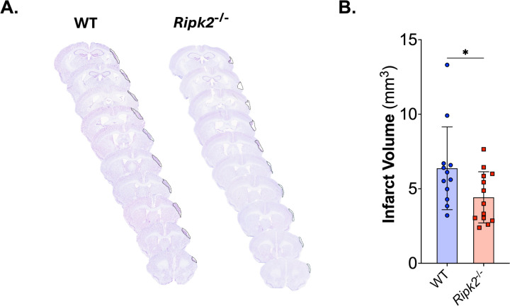
Besides the loss of blood and oxygen reaching the ischemic tissue, many secondary effects of ischemic stroke can cause additional tissue death, including inflammation, oxidative stress, and proteomic disturbances. Receptor-interacting serine/threonine kinase 2 (RIPK2) is an important mediator in the post-stroke inflammatory cascade that responds to signals and molecular patterns released by dead or dying cells in the ischemic area. We hypothesize that RIPK2 signaling worsens injury and neurological recovery post-stroke and that global deletion of Ripk2 will be protective following ischemic stroke in aged mice. Aged (18-24 months) male mice were subjected to permanent middle cerebral artery occlusion (pMCAO). Vertical grid, weight grip, and open field were conducted at baseline and on days 1, 2, 3, 8, 15, and 22 post-stroke. Cognitive tests (novel object recognition and Y-maze) were performed at baseline and day 28 post-stroke. Infarct size was measured using cresyl violet staining, and reactive gliosis was measured using Iba1 and GFAP staining at day 28 post-stroke. Global deletion of Ripk2 (Ripk2 -/- ) in aged mice resulted in smaller infarct volume and improved performance on vertical grid and weight grip tests compared to aged wildtype (WT) mice. Additionally, aged Ripk2 -/- mice had less Iba1 staining in the ipsilateral cortex than the aged WT control mice. This study further elucidates the role of RIPK2 signaling in the ischemic cascade and expands our knowledge of RIPK2 in stroke to aged mice. These results support the hypothesis that RIPK2 signaling worsens injury post-stroke and may be an attractive candidate for therapeutic intervention.

Keywords: RIPK2; aging; ischemic stroke; microgliosis; neuroinflammation.


Conflict of interest statement

Declaration of competing interests The authors declare no competing financial interests.

Figures

Figure 1:
Figure 1:. Ripk2−/− mice have reduced infarct volume at 28 days than aged WT mice.
A) Representative cresyl violet stained sections of brains from either wildtype (WT) or Ripk2 deficient (Ripk2−/−) mice. Sections cut at 30 µm using a cryostat, stained with cresyl violet, and imaged using an Aperio slide scanner. B) Quantification of infarct volume shows significant reduction in infarct volume in the aged Ripk2−/− mice compared to WT mice. n = 12–13/group. Differences determined by two-tailed, unpaired t-test; t(23) = 2.128, p = 0.0443.
Figure 2:
Figure 2:. Ripk2−/− aged mice show improved function on behavior tests compared to aged WT controls.
A) Time to descend the vertical grid at baseline was significantly different between groups, with Ripk2−/− mice taking more time to descend the grid. B) When normalized to baseline data for each genotype, Ripk2−/− mice show less deficit in descending the vertical grid than WT controls at days 1, 3, 8, 15, and 22. C) Ripk2−/− mice had lower weight grip scores than WT controls at baseline. D) Ripk2−/− mice have higher weight grip scores normalized to baseline than WT controls at days 3, 8, 15, and 22 following pMCAO. E) Ripk2−/− mice traveled less distance than WT controls at baseline in the open field test. F) No difference between genotypes in baseline normalized total distance traveled in the open field test following pMCAO. n = 12–13/group. Differences determined by two-tailed, unpaired t-test (A, C, E). Differences determined by two-way, repeated measures ANOVA with Tukey’s post-hoc (B, D, F). * = p < 0.05, ** = p < 0.01, *** = p < 0.001, **** = p < 0.0001.
Figure 3:
Figure 3:. Cognitive testing reveals Ripk2−/− mice display cognitive deficits before stroke and do not show worsened cognitive deficits post-stroke.
A) Schematic depiction of the novel object recognition (NOR) test during the training (left) and testing (right) phases. B-D) Baseline NOR training (B), testing (C), and discrimination index (D) conducted in naïve animals. E-G) NOR testing with the training phase (E) conducted on day 27 and testing (F) on day 28, resulting in a discrimination index (G). H-J) Y maze cognitive test performed 28 d post-stroke. H) Schematic of the Y maze test paradigm. I) The number of alterations per animal. J) The percent alterations per animal. n=12–13/group. (B-C, E-F) Differences were determined by two-way ANOVA with Šídák’s post-hoc. (D, G, I-J) Differences were determined by Student’s t-test. * p < 0.05, ** p < 0.01, *** p < 0.01, **** p < 0.0001.
Figure 4:
Figure 4:. Ripk2−/− mice have less Iba1 staining than WT control mice in the ipsilateral cortex following pMCAO.
A) Representative 30 μm sections show Iba1 staining following pMCAO in WT (left) and Ripk2−/− mice (right). B) Images taken from the contralateral (top) and ipsilateral (bottom) cortex. Scale bar is 200 μm. C) Quantification of Iba1 immunostaining shown in panel B. WT mice show increased Iba1 staining in the ipsilateral cortex compared to the contralateral cortex, and Ripk2−/− mice show decreased Iba1 staining in the ipsilateral cortex compared to WT controls. D) Images taken from the contralateral (top) and ipsilateral (bottom) subcortex. E) Quantification of Iba1 immunostaining shown in panel D shows no differences between genotypes or hemispheres in Iba1 immunostaining in the subcortex. n = 6/group. Differences determined by two-way ANOVA with Tukey’s post-hoc. * p < 0.05, ** p < 0.01.
Figure 5:
Figure 5:. Both WT and Ripk2−/− mice show increased GFAP staining in the ipsilateral cortex with no difference between genotypes.
A) Representative 30 μm sections show GFAP staining following pMCAO in WT (left) and Ripk2−/− mice (right). B) Images taken from the contralateral (top) and ipsilateral (bottom) cortex. Scale bar is 200 μm. C) Quantification of GFAP immunostaining shown in panel B. Both WT and Ripk2−/− mice show increased GFAP staining in the ipsilateral cortex compared to the contralateral cortex. D) Images taken from the contralateral (top) and ipsilateral (bottom) subcortex. E) Quantification of GFAP immunostaining shown in panel D shows no differences between genotypes or hemispheres in GFAP immunostaining in the subcortex. n = 6/group. Differences determined by two-way ANOVA with Tukey’s post-hoc. * p < 0.05, **** p < 0.0001.

References

    1. Martin SS, Aday AW, Almarzooq ZI, et al. 2024. Heart Disease and Stroke Statistics: A Report of US and Global Data From the American Heart Association. Circulation [Internet]. 2024 [cited 2024 Apr 8];149(8). - PubMed
    1. Tissue Plasminogen Activator for Acute Ischemic Stroke. The New England Journal of Medicine. 1995;333(24):7. - PubMed
    1. Jovin TG, Chamorro A, Cobo E, et al. Thrombectomy within 8 Hours after Symptom Onset in Ischemic Stroke. N Engl J Med. 2015;372(24):2296–2306. - PubMed
    1. Candelario-Jalil E, Dijkhuizen RM, Magnus T. Neuroinflammation, Stroke, Blood-Brain Barrier Dysfunction, and Imaging Modalities. Stroke. 2022;53(5):1473–1486. - PMC - PubMed
    1. Stuckey SM, Ong LK, Collins-Praino LE, et al. Neuroinflammation as a Key Driver of Secondary Neurodegeneration Following Stroke? IJMS. 2021;22(23):13101. - PMC - PubMed

Publication types

LinkOut - more resources