[Preprint]. 2025 Feb 1:2025.01.31.635969.
doi: 10.1101/2025.01.31.635969.

In vivo targeted gene delivery using Adenovirus-antibody molecular glue conjugates

Affiliations

In vivo targeted gene delivery using Adenovirus-antibody molecular glue conjugates

Paul J Rice-Boucher et al. bioRxiv. .

Update in

Abstract

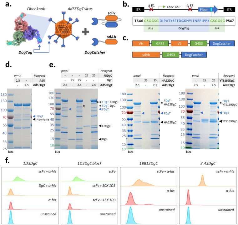
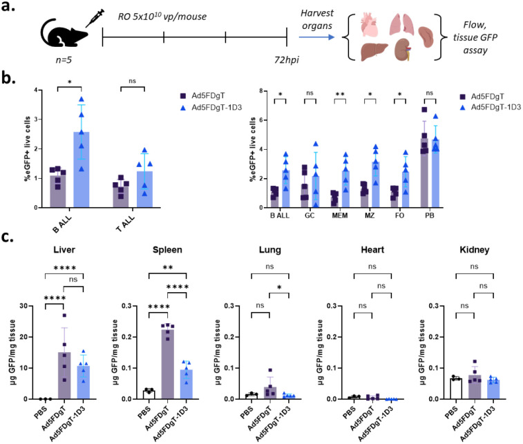
Safe and efficient nucleic acid delivery to targeted cell populations remains a significant unmet need in the fields of cell and gene therapy. Towards this end, we pursued Adenoviral vectors genetically modified with the "DogTag" molecular glue peptide, which forms a spontaneous covalent bond with its partner protein, "DogCatcher". Genetic fusion of DogCatcher to single-domain or single-chain antibodies allowed covalent tethering of the antibody at defined locales on the vector capsid. This modification allowed simple, effective and exclusive targeting of the vector to cells bound by the linked antibody. This dramatically enhanced gene transfer into primary B and T cells in vitro and in vivo in mice. These studies form the basis of a novel method for targeting Adenovirus that is functional in stringent in vivo contexts and can be combined with additional well characterized Adenovirus modifications towards applications in cell engineering, gene therapy, vaccines, oncolytics, and others.

Keywords: DogCatcher; DogTag; Gene delivery; SpyCatcher; SpyTag; adenovirus; molecular glue.


Conflict of interest statement

Declaration of interests: Hongjie Guo is the founder and Chief Scientific Officer of Tiger Biologics, LLC, a protein production company. Paul J. Rice-Boucher. David T. Curiel, and Zhi Hong Lu are co-inventors on a patent application describing the use of the Ad-Ab system for B cell targeting and engineering.

Figures

Figure 1:
Figure 1:
Development and characterization of Ad-Ab targeting. a. Schematic overview of system design. DogTag is genetically inserted into the Ad fiber knob (Ad5FDgT), while DogCatcher is fused to antibody species. Mixing of these reagents results in permanent linkage of the virus and antibody at the fiber knob locale. Fiber model generated with AlphaFold2 and visualized with UCSF ChimeraX,. b. Ad5FDgT genome overview. Ad5FDgT is based on an E1/E3 deleted Ad5 with the CMV promoter driving eGFP expression from the E1 region. DogTag is inserted with minimal flex linkers at the HI loop of the fiber knob domain. c. Antibody-DogCatcher fusion designs. Antibody domains are separated from DogCatcher by a flexible (G4S)3 linker d. SDS-PAGE analysis of control Ad5 and Ad5FDgT. In all cases the molar amount of antibody and virus refers to DogCatcher and DogTag, respectively. e. Gel shift analysis of Ad5FDgT binding to DogCatcher and antibody-DogCatcher fusions. Left, binding to bacterially produced DogCatcher and an sdAb-DogCatcher fusion. Middle and right, binding to representative scFv-DogCatcher fusions. f. Flow cytometry analysis of representative scFvs in murine splenocytes. 1D3DgC and 18B12DgC stains were gated on B220+ populations, while 2.43DgC staining was gated on the CD8+ population. 1D3DgC specificity was assessed by blocking with excess full length 1D3 antibody.
Figure 2:
Figure 2:
In vitro characterization of Ad-Ab targeting. a. Conceptual workflow. Primary lymphocytes are magnetically isolated from mouse splenocytes or human PBMCs then cultured with activating agents. On the day of infection Ad5FDgT is conjugated with the appropriate antibody at the indicated DogCatcher:DogTag molar ratios, then used to infect cells. b. Ad-Ab infectivity enhancement in primary murine B cells (top right), murine T cells (bottom left), and human B cells (bottom right). For each antibody, the group with the maximum mean infectivity was compared to the no antibody group using a standard unpaired two-tailed t-test. Welch’s correction was used in cases where the F-test revealed significant differences in variances. DgC = DogCatcher, n=9 replicates from 3 experiments in mouse B cells, n=6 from 2 experiments in mouse T cells. YTS169DgC = α-mCD8α scFv, n=4–5 from 2 experiments in mouse B cells, n=6 from 2 experiments in mouse T cells, n=5 from 3 experiments in human B cells. 2.43 = α-mCD8α scFv, n=4–5 from 2 experiments in mouse B cells, n=6 from 2 experiments in mouse T cells. F8DgC = α-mCD40 sdAb, n=9 from 3 experiments in mouse B cells, n=6 from 2 experiments in mouse T cells. 1D3DgC = α-mCD19 scFv, n=4 from 2 experiments in mouse B cells, n=6 from 2 experiments in mouse T cells. 18B12DgC = α-mCD20 scFv, n=4–5 from 2 experiments in mouse B cells, n=6 from 2 experiments in mouse T cells. FMC63DgC = α-hCD19 scFv, n=5 from 3 experiments in human B cells. Rtxv1DgC (Rituximab) = α-mCD20 scFv, n=4 from 2 experiments in human B cells. HA22DgC = α-hCD22 scFv, n=5 from 3 experiments in human B cells. Created with BioRender.com.
Figure 3:
Figure 3:
In vivo characterization of Ad-Ab targeting. a. Experiment design. C57BL/6J mice were injected RO with 5×10¹⁰ vp/mouse of either Ad5FDgT or Ad5FDgT conjugated with 1D3DgC. Major organs were harvested 72h later for eGFP expression analyses. b. Flow cytometry results. Left, eGFP expression in B and T cells. Right, eGFP expression in B cell subsets. GC = germinal center, MEM = memory, MZ = marginal zone, FO = follicular, PB = plasmablast. In all cases non-conjugated and conjugated groups were compared in each population using a standard unpaired two-tailed t-test. Welch’s correction was used in cases where the F-test revealed significant differences in variances. eGFP expression in mice injected with PBS was used to normalize the data in all cases. c. Quantitative eGFP analysis in major tissues. Tissues were homogenized in lysis buffer and eGFP expression was analyzed via fluorimetry. eGFP was normalized to total tissue protein content as assessed by BCA. In all cases ordinary one-way ANOVA with Tukey’s correction for multiple comparisons was used to compare groups where n=5 male mice. In the liver, lung and kidney values were log transformed to normalize variances. Created with BioRender.com.
Figure 4:
Figure 4:
Development of purified Ad-Ab complexes. a. Purification workflow. Virus particles are purified from cell lysates via cesium chloride (CsCl) ultracentrifugation, then briefly dialyzed against 1X PBS to remove excess salts. Conjugation is then carried out, followed by a second CsCl purification, dialysis and final storage. b. Viral band images in CsCl gradients after second ultracentrifugation. c. SDS-PAGE analysis of purified Ab-Ab complexes. d. Western blot against the HAdV-C5 fiber tail of purified Ad-Ab complexes. e. In vivo analysis of purified Ad-Ab complexes. C57BL/6J mice aged 6–9 weeks were injected on three separate occasions with 5×10¹⁰ of the indicated vectors. In the first experiment n=2 female mice per group were used. In the second experiment n=2 male mice per group were used, and a single Ad5-FDgT-YTS169 sample was removed from analysis due to very low viability found during flow analysis. In the fourth experiment n=4 mice per group were used split equally between male and female mice. Three days later splenocytes were assessed for eGFP expression using flow cytometry. Livers were assessed for tissue eGFP expression as well. Data from all experiments were combined and analyzed using ordinary one-way ANOVA with Tukey’s correction for multiple comparisons. Created with BioRender.com.

Similar articles

References

    1. Bulcha T. J., Wang Y., Ma H., Tai L. W. P. & Gao G. Viral vector platforms within the gene therapy landscape. Signal Transduction and Targeted Therapy 6 (2021). 10.1038/s41392-021-00487-6 - DOI - PMC - PubMed
    1. Lostalé-Seijo I. & Montenegro J. Synthetic materials at the forefront of gene delivery. Nature Reviews Chemistry 2, 258–277 (2018). 10.1038/s41570-018-0039-1 - DOI
    1. Dunbar E. C. et al. Gene therapy comes of age. Science 359, eaan4672 (2018). 10.1126/science.aan4672 - DOI - PubMed
    1. Wang D., Zhang F. & Gao G. CRISPR-Based Therapeutic Genome Editing: Strategies and In Vivo Delivery by AAV Vectors. Cell 181, 136–150 (2020). 10.1016/j.cell.2020.03.023 - DOI - PMC - PubMed
    1. Wilbie D., Walther J. & Mastrobattista E. Delivery Aspects of CRISPR/Cas for in Vivo Genome Editing. Accounts of Chemical Research 52, 1555–1564 (2019). 10.1021/acs.accounts.9b00106 - DOI - PMC - PubMed

Publication types

LinkOut - more resources