[Preprint]. 2025 Jun 21:2025.01.30.635805.
doi: 10.1101/2025.01.30.635805.

An HIV-1 Reference Epitranscriptome

Affiliations

An HIV-1 Reference Epitranscriptome

Michael S Bosmeny et al. bioRxiv. .

Abstract

Post-transcriptional modifications to RNA, which comprise the epitranscriptome, play important roles in RNA metabolism, gene regulation, and human disease, including viral pathogenesis. Modifications to the RNA viral genome and transcripts of human immunodeficiency virus 1 (HIV-1) have been reported and investigated in the context of virus and host biology. However, the diversity of experimental approaches used has made clear correlations across studies, as well as the significance of the HIV-1 epitranscriptome in biology and disease, difficult to assess. Therefore, we established a reference HIV-1 epitranscriptome. We sequenced the model NL4-3 HIV-1 genome from infected primary CD4+ T cells and the Jurkat cell line using the latest nanopore chemistry, optimized RNA preparation methods, and the most current and readily available base-calling algorithms. A highly reproducible sense and a preliminary antisense HIV-1 epitranscriptome were created, where N6-methyladenosine (m6A), 5-methylcytosine (m5C), pseudouridine (psi), inosine, and 2'-O-methyl (Nm) modifications could be identified by rapid multiplexed base-calling. We observed that sequence and neighboring modification contexts induced modification miscalling, which could be corrected with synthetic HIV-1 RNA fragments. We validated m6A modification sites with STM2457, a small molecule inhibitor of methyltransferase-like 3 (METTL3). We find that modifications are quite stable under combination antiretroviral therapy (cART) treatment, in primary CD4+ T cells, and in HIV-1 virions. Sequencing samples from people living with HIV (PLWH) revealed conservation of m6A modifications. However, analysis of spliced transcript variants suggests transcript-dependent modification levels. Our approach and reference data offer a straightforward benchmark that can be adopted to help advance rigor, reproducibility, and uniformity across HIV-1 epitranscriptomics studies. They also provide a roadmap for the creation of reference epitranscriptomes for many other viruses or pathogens.


Figures

Figure 1.
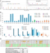
Figure 1.. Probing and correction of HIV-1 base modifications called by nanopore direct RNA sequencing.
(A) Nanopore modification-calling results for HIV-1 viral RNA extracted from Jurkat cell cultures treated with STM2457, a drug that inhibits METTL3 m6A modification activity. The position of each high frequency m6A nucleotide in the HIV-1 genome is indicated on the x-axis. (B) Comparison of modifications at DRACH motif sites where m6A was called at high frequency. The nucleotide position of the m6A modification within the DRACH motif is indicated on the x-axis. Upper plot: comparison of Jurkat cell samples infected with HIV-1 without (back row) or with (front row) 30 μM STM2457 treatment. Lower graph: comparison of Jurkat cell sample infected with HIV-1 before (back row) and after (front row) baseline correction. (C-E) Comparison of modification calling between NL4–3 from Jurkat cells (C) and two synthetic HIV-1 RNA fragments, one unmodified (D) and one bearing m6A (E) at two DRACH motifs. The nucleotide position corresponding to the NL4–3 genome is indicated on the x-axis. All modifications called in panel D are incorrect while modifications called in panel E, besides m6A at position 8975 and 8989, are incorrect.
Figure 2.
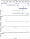
Figure 2.. Baseline-corrected nanopore modification calling results for HIV-1 viral RNA from Jurkat cells.
The HIV-1 genome architecture is illustrated above. Modifications are shown if, after baseline correction, the average modification frequency was at least 10%. m6A (A, blue), m5C (B, green), pseudouridine (psi) (C, yellow), inosine (D, purple), and 2’-O-methylation (E, orange). Inset in panel A is a close-up of the 3’ end of the NL4–3 HIV-1 genome where m6A is most densely called. Results are the average of three separate biological replicates. Error bars are standard deviation.
Figure 3.
Figure 3.. Differential modification frequencies in HIV-1 RNA splice isoforms.
Comparison of modification frequency in each splice isoform as compared to the full-length (unspliced) isoform. X-axis represents individual nucleotides in the HIV-1 genome. Y-axis represents percentage of reads that contained the mutation of interest for that nucleotide, relative to the unspliced form. Bar color indicates modification type: m6A (blue), m5C (green), pseudouridine (yellow), inosine (purple), and 2’-O-methyl (orange). Only modifications with a difference of 10% from the unspliced RNA in at least one isoform are shown. Shaded background shows portion of the full transcript retained in the spliced isoform. Splice isoforms are subsets of direct-RNA sequencing sample 7C. The A6 splice site is exclusive to the HXB2 genome and therefore not indicated here.
Figure 4.
Figure 4.. A preliminary HIV-1 antisense epitranscriptome, the effect of cART and cell type on HIV-1 modification calling, and conservation of m6A in HIV-1 genomes from PLWH.
(A) Nanopore modification-calling for HIV-1 antisense RNA from Jurkat cells. m6A (blue), m5C (green), pseudouridine (yellow), inosine (purple), and 2’-O-methylation (orange). Inset shows a close-up of the asp gene. (B) Nanopore modification-calling results for HIV-1 RNA taken from Jurkat cells treated without (darker color) or with (lighter color) cART treatment. Error bars represent the standard deviation from three separate biological replicates. (C) Nanopore modification-calling results for HIV-1 RNA taken from Jurkat cells, primary CD4+ T cells infected in vitro, supernatant of primary CD4+ T cells infected in vitro, and CD4+ T cells from PLWH samples. In each nucleotide position cluster, the first bar is Jurkat cell samples infected with HIV-1, the second bar is CD4+ T cells from healthy donors and infected with HIV-1, the third bar is the supernatant from the CD4+ T cells from healthy donors, and the fourth, fifth, and sixth bars are samples taken from CD4+ T cells from PLWH donors. (D) Top: comparison of m6A modifications called from HIV-1 RNA from Jurkat cells against preservation of the DRACH motifs for these m6A modifications sequenced from three PLWH samples. Bottom: analysis of conservation of known m6A modification sites in a larger dataset of HIV-1 mutations. Positions containing m6A between nucleotide positions 8000 and 9171 were identified and the average and median events for these positions are reported.

References

    1. Abudayyeh O.O., Gootenberg J.S., Franklin B., Koob J., Kellner M.J., Ladha A., Joung J., Kirchgatterer P., Cox D.B.T., and Zhang F. (2019). A cytosine deaminase for programmable single-base RNA editing. Science 365, 382–386. - PMC - PubMed
    1. Ageely E.A., Chilamkurthy R., Jana S., Abdullahu L., O’Reilly D., Jensik P.J., Damha M.J., and Gagnon K.T. (2021). Gene editing with CRISPR-Cas12a guides possessing ribose-modified pseudoknot handles. Nature communications 12, 6591. - PMC - PubMed
    1. Aksamentov I., Roemer C., Hodcroft E., and Neher R. (2021). Nextclade: clade assignment, mutation calling and quality control for viral genomes. Journal of Open Source Software 6, 3773.
    1. Asang C., Erkelenz S., and Schaal H. (2012). The HIV-1 major splice donor D1 is activated by splicing enhancer elements within the leader region and the p17-inhibitory sequence. Virology 432, 133–145. - PubMed
    1. Baek A., Lee G.E., Golconda S., Rayhan A., Manganaris A.A., Chen S., Tirumuru N., Yu H., Kim S., Kimmel C., et al. (2024). Single-molecule epitranscriptomic analysis of full-length HIV-1 RNAs reveals functional roles of site-specific m(6)As. Nature microbiology 9, 1340–1355. - PMC - PubMed

Publication types