[Preprint]. 2025 Jun 27:2025.02.05.636339.
doi: 10.1101/2025.02.05.636339.

Chemotherapeutic 6-thio-2'-deoxyguanosine selectively targets and inhibits telomerase by inducing a non-productive telomere-bound telomerase complex

Affiliations

Chemotherapeutic 6-thio-2'-deoxyguanosine selectively targets and inhibits telomerase by inducing a non-productive telomere-bound telomerase complex

Samantha L Sanford et al. bioRxiv. .

Abstract

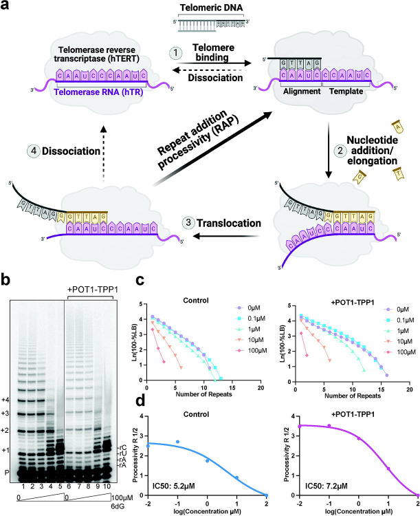
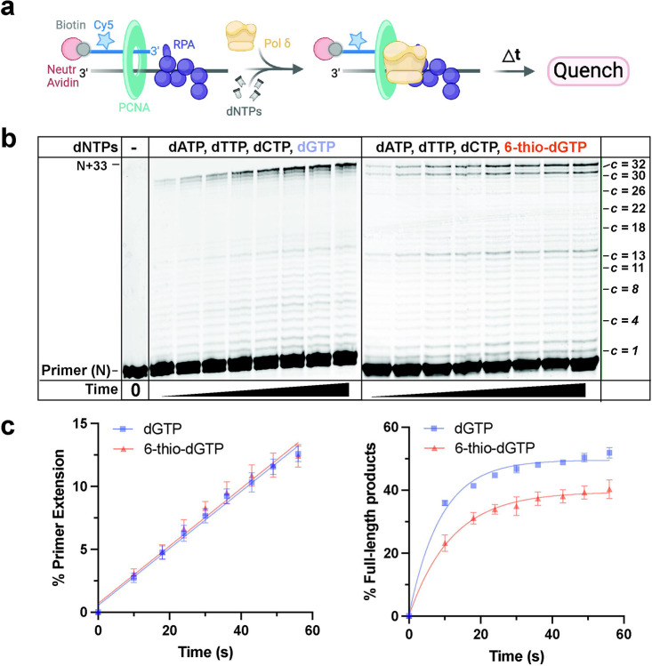
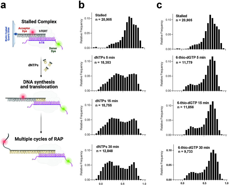
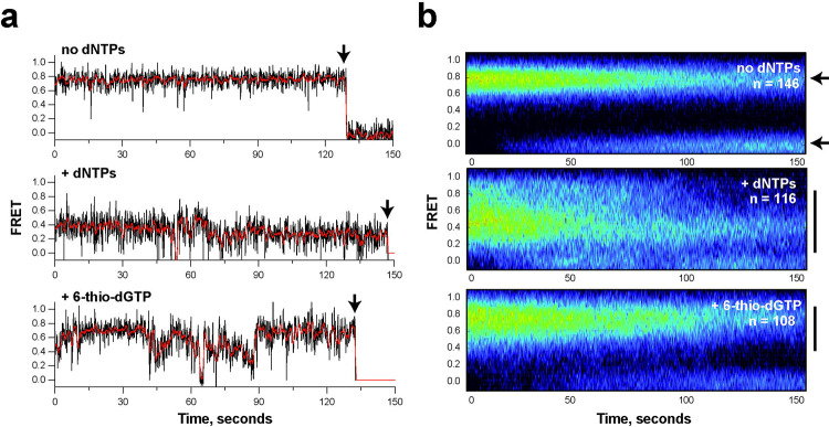
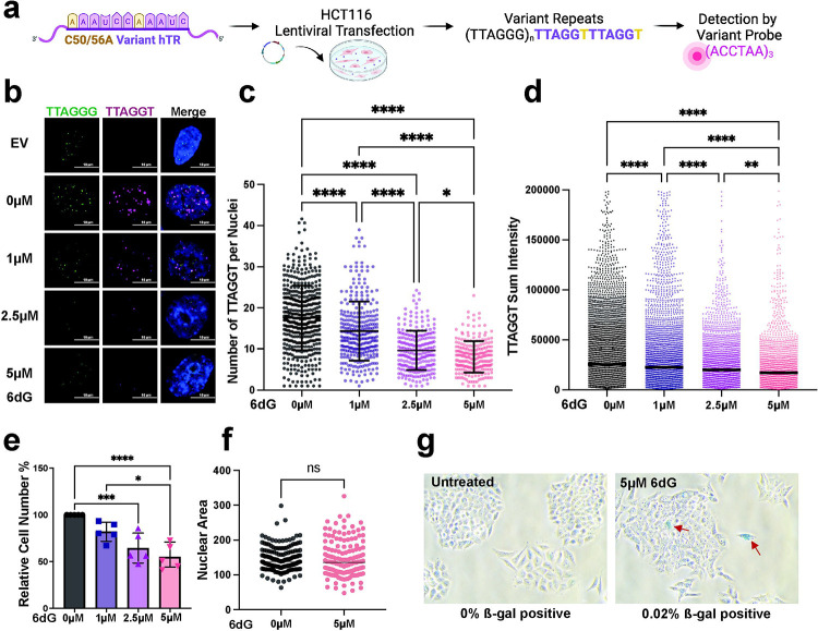
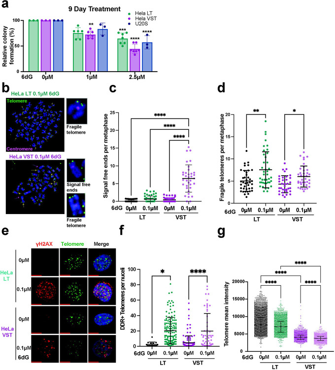
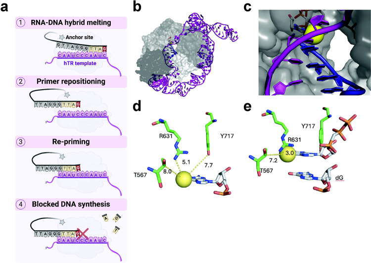
Most cancers upregulate the telomere lengthening enzyme telomerase to achieve unlimited cell division. How chemotherapeutic nucleoside 6-thio-2'-deoxyguanosine (6-thio-dG) targets telomerase to inhibit telomere maintenance in cancer cells and tumors was unclear. Here, we demonstrate that telomerase insertion of 6-thio-dGTP prevents synthesis of additional telomeric repeats but does not disrupt telomerase binding to telomeres. Specifically, 6-thio-dG inhibits telomere extension after telomerase translocates along its product DNA to reposition the template, inducing a non-productive complex rather than enzyme dissociation. Furthermore, we provide direct evidence that 6-thio-dG treatment inhibits telomere synthesis by telomerase in cancer cells. In agreement, telomerase-expressing cancer cells harboring critically short telomeres are more sensitive to 6-thio-dG and show a greater induction of telomere losses compared to cancer cells with long telomere reserves. Our studies reveal that telomere length and telomerase status determine 6-thio-dG sensitivity and uncover the molecular mechanism by which 6-thio-dG selectively inhibits telomerase synthesis of telomeric DNA.


Conflict of interest statement

DECLARATION OF INTERESTS The authors have no competing interests to declare.

Figures

Fig.1
Fig.1. Telomerase processivity factors POT1-TPP1 cannot rescue 6-thio-dGTP inhibition of telomerase RAP.
a, Cartoon of telomerase catalytic cycle. The numbers represent each step in the cycle. Gray indicates the telomeric DNA primer (see Supplementary Table 1). Purple indicates the telomerase RNA template and hTERT. Yellow indicates the newly added nucleotides. b, Direct telomerase assays were conducted in the absence or presence of 500 nM POT1 and 500 nM TPP1, as indicated, and 5 nM 32P-end labeled primer (TTAGGGTTAGCGTTAGGG) designed to position POT1 at the 10 nt primer 5′ end. Reactions contained cellular-concentration dNTPs and 0–100μM 6-thio-dGTP. Numbers on the left indicate number of added repeats, letters on the right indicate template base and P indicates unextended 18-mer primer. c, Processivity (R 1/2) was calculated on the basis of total products normalized to loading control. d, IC50 values calculated from processivity R 1/2.
Fig. 2
Fig. 2. 6-thio-dGTP fails to inhibit replicative DNA polymerase δ holoenzyme.
a, Schematic of assay to monitor primer extension by Pol δ holoenzymes during a single binding encounter with a DNA substrate (BioCy5P/T, Extended Data Fig. 2a). PCNA (green) is assembled onto the substrate (250 nM) orientated toward the primer/template junction. Following the addition of physiological dNTP concentrations, DNA synthesis is initiated by adding 8.8 nM Pol δ. b, Representative 16% denaturing gel of the primer extension products. The sizes of the primer (N) and the full-length product (N+33) are indicated on the left and the dNTP insertion step at each template C (c) is indicated on the right. c, Quantification of DNA synthesis. The left panel shows the percent total primer extension plotted as a function of time. Data points after time = 0 s are fit to a linear regression. The right panel shows the percent of full-length products plotted as a function of reaction time. Data from reactions with natural dNTPs are shown in blue, and data from reactions in which 6-thio-dGTP replaced dGTP are shown in orange. Each data point represents the average ± S.E.M. of three independent reactions.
Fig. 3
Fig. 3. Terminal 6-thio-dG bases impair telomerase RAP but not telomerase binding.
a, Telomerase reactions conducted with 6-thio-dG at different positions on the telomeric substrate primer. P marks the unextended primer and numbers indicate the number of added repeats. b, Cartoons of telomeric primer (gray) with 6-thio-dG (red), telomerase RNA template (purple), and incoming dNTP (green). c, Quantitation of percent primer extension. Statistical significance was determined by one-way ANOVA (**P < 0.005; ****P < 0.0001). Sequence of 3’terminal telomeric repeat in the primer following (GGTTAG)2 is shown on the Y axis (see Supplementary Table 1). Purple G’s indicate 6-thio-dG modification. d, Binding reactions were conducted with 2.5nM fluorescein labeled telomere substrate with a 3’ terminal dG or 6-thio-dG (blue) (see Supplementary Table 1), 1–20 nM Halo-telomerase conjugated with JF-635 dye (red), for 30 min at room temperature, and separated by EMSA to visualize telomerase-bound substrate (pink). e, Quantification of % DNA bound versus Halo-telomerase concentration. Data represent the mean ± s.d. from three independent experiments.
Fig. 4
Fig. 4. Single molecule analysis reveals a non-productive telomerase complex after 6-thio-dG addition.
a, Schematic of experimental setup. LD555-labeled telomerase is pulled down on Cy5-labeled (TTAGGG)3 substrate and immobilized on a quartz slide via a 5’ biotin. The close proximity of the dyes permits FRET. Telomerase-catalyzed addition of dNTPs to the telomere substrate increases the distance between the dyes causing a decrease in FRET dependent on the number of repeats added. b, FRET histograms of complexes prior to telomerase activity (top “stalled”), and collected 5, 15, and 30 minutes after dNTP addition as indicated. Shift from high to low FRET states reports on telomerase movement away from the acceptor dye during telomere elongation. c, FRET histograms of complexes collected prior to (top) and collected 5, 15, and 30 minutes after dATP, dTTP and 6-thio-dGTP addition.
Fig. 5
Fig. 5. Single molecule analysis reveals telomere DNA dynamics within non-productive telomerase complexes after 6-thio-dG addition.
a, Representative traces of stalled single telomerase-DNA complexes (top), in the presence of dNTPs (middle panel), or with dATP, dTTP and 6-thio-dGTP (bottom panel). Data were collected approximately 15 minutes after addition of dNTPs. Raw data collected at 8 Hz framerate are shown in black and a one second moving average is overlaid in red. Black arrows indicate irreversible photobleaching of the FRET dyes. b, Heat map analysis of the time dependent FRET signal of more than one hundred individual telomerase-DNA complexes in each experimental condition. Small black arrows in top panel indicate FRET state of stalled complex (upper arrow) and FRET when dyes have photobleached. In the presence of dNTPs (middle) or dATP, dTTP and 6-thio-dGTP (bottom), broadening of the FRET distribution (solid black lines) indicates DNA dynamics within the telomerase-DNA complexes.
Fig. 6
Fig. 6. 6-thio-dG suppresses telomerase elongation of telomeres in cells.
a, Schematic for expression of C50/56A hTR variant harboring the template sequence 3’-AAAUCCAAAUC-5’ via lentiviral transduction into HCT116 cells, and detection of newly added (TTAGTT)n repeats with a 5’-Cy3-(ACCTAA)3-3’ probe by FISH. b, Representative images of interphase cells stained with PNA probes for wildtype and variant sequence 6 days post-transduction with empty vector (EV) or C50/56A hTR, and after 72 hours treatment with 0, 1, 2.5 or 5 μM 6-thio-dG (6dG). c, Quantification of the number of variant telomeric foci per nuclei from panel b. Error bars represent the mean ± s.d. from n cells analyzed as indicated by dots from 3 independent experiments. Statistical significance was determined by one-way ANOVA (*P < 0.05; ****P < 0.0001). d, Quantification of the variant telomere sum intensity. Error bars represent the mean ± s.d. of n variant telomere foci analyzed as indicated by dots, from 3 independent experiments. Statistical significance was determined by one-way ANOVA (**P < 0.01; ****P < 0.0001). e, HCT116 cell counts obtained 72 hours after treatment with the indicated 6-thio-dG concentrations, relative to untreated cells. Error bars represent the mean ± s.d. from 4 independent experiments. Statistical significance was determined by one-way ANOVA (*P < 0.05; ***P < 0.001; ****P < 0.0001). f, Size of nuclear area (μm2) obtained 72 hours after 0 or 5 μM 6-thio-dG treatment. Error bars represent the mean ± s.d. from the indicated n number of nuclei analyzed as indicated by dots. Statistical analysis by two-tailed t-test (ns = not significant). g, Representative images of β-galactosidase (β-gal) staining 72 hours after 0 or 5 μM 6-thio-dG treatment. Arrows indicate β-gal positive cells of 500 cells analyzed per condition. Scale bar = 100 μm.
Fig. 7
Fig. 7. Telomerase expressing cancer cells with shortened telomeres are hypersensitive to 6-thio-dG.
a, Colony formation efficiency of HeLa LT, HeLa VST and U2OS cells following 9 days of treatment with 1 or 2.5 μM 6-thio-dG (6dG), relative to untreated cells. Error bars represent the mean ± s.d. from the number of independent experiments indicated by the dots. Statistical significance was determined by one-way ANOVA (**P < 0.01; ***P < 0.001; ****P < 0.0001). b, Representative images of FISH staining with WT telomere probes of metaphase chromosomes from HeLa LT and HeLa VST cells after 20 days of treatment with 0.1 μM 6-thio-dG. Images were scored for telomeric signal-free ends (yellow arrowheads) and fragile telomeres (green arrowheads). Green foci = telomeres and pink foci = centromeres. Scale bars, 10 μm. c-d, Quantification of telomeric signal-free ends (c) and fragile telomeres (d) per metaphase from panel b in HeLa LT and HeLa VST cells after 20 days of treatment with 0 or 0.1 μM 6-thio-dG. Error bars represent mean ± s.d. of n metaphases, as indicated by dots, analyzed from three independent experiments, normalized to the chromosome number. Statistical analysis by 2-way Anova (ns, not significant; *P < 0.05; **P < 0.01; ****P < 0.0001). e, Representative images of γH2AX foci (red) and telomeric foci (green) stained by WT telomere FISH of HeLa LT and HeLa VST after 20 days of treatment with 0 or 0.1 μM 6-thio-dG. Colocalization shown as yellow foci. Scale bars, 10 μm. f, Quantification average number of telomeres staining positive for γH2AX per cell for HeLa LT and HeLa VST cells after 20 days of treatment with 0 or 0.1 μM 6-thio-dG. Error bars represent the mean ± s.d. from n number of nuclei analyzed as indicated by dots from three independent experiments. Statistical significance determined by One-way ANOVA (**P < 0.01; ****P < 0.0001). g, Quantification of WT telomere FISH signal intensity from HeLa LT and HeLa VST interphase cells after 20 days of treatment with 0.1 μM 6-thio-dG. Statistical significance determined by Two-way ANOVA (****P < 0.0001).
Figure 8.
Figure 8.. Model for 6-thio-dGTP telomerase inhibition.
a, Working model for how 6-thio-dGTP inhibits telomerase activity, see text for details. Telomerase hTERT in gray, anchor site depicted with the gray star, hTR template in purple, newly added dNTPs in yellow and 6-thio-dG in red. b, Global structure of TERT (protein shown as a gray surface, hTR shown as purple sticks, telomere DNA shown in blue). c, Closeup view of telomerase active site, showing cavity along the major groove side of the telomere. 6-thio-dGTP is modeled in for reference, with the thio group shown as a yellow sphere. Models were generated from PDB code 7BG9 and 6USR. d, Model of 6-thio-dG at the primer terminus of a telomere (thio group shown as a yellow sphere, amino acids shown as green sticks, and DNA shown as white sticks). The closest active site contacts are shown. The thio group is well accommodated by any protein residues. e, Model of incoming 6-thio-dGTP, with closest contacts displayed. The thio group is ~3 angstrom away from R631, but it does not clash with its position and could potentially act as a stabilizing interaction.

References

    1. de Lange T. Shelterin-Mediated Telomere Protection. Annu Rev Genet 52, 223–247 (2018). - PubMed
    1. d’Adda di Fagagna F. et al. A DNA damage checkpoint response in telomere-initiated senescence. Nature 426, 194–8 (2003). - PubMed
    1. Jaskelioff M. et al. Telomerase reactivation reverses tissue degeneration in aged telomerase-deficient mice. Nature 469, 102–6 (2011). - PMC - PubMed
    1. Takai H., Smogorzewska A. & de Lange T. DNA damage foci at dysfunctional telomeres. Curr Biol 13, 1549–56 (2003). - PubMed
    1. Shay J.W. & Wright W.E. Senescence and immortalization: role of telomeres and telomerase. Carcinogenesis 26, 867–74 (2005). - PubMed

Publication types

LinkOut - more resources