[Preprint]. 2025 Feb 9:2025.02.09.637297.
doi: 10.1101/2025.02.09.637297.

Structural basis for HIV-1 capsid adaption to rescue IP6-packaging deficiency

Affiliations

Structural basis for HIV-1 capsid adaption to rescue IP6-packaging deficiency

Yanan Zhu et al. bioRxiv. .

Update in

Abstract

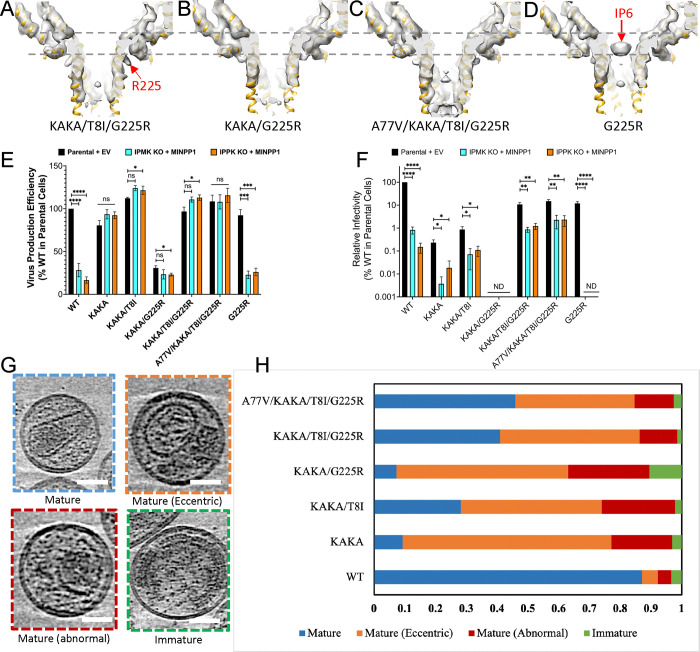
Inositol hexakisphosphate (IP6) promotes HIV-1 assembly via its interaction with the immature Gag lattice, effectively enriching IP6 within virions. During particle maturation, the HIV-1 protease cleaves the Gag polyproteins comprising the immature Gag lattice, releasing IP6 from its original binding site and liberating the capsid (CA) domain of Gag. IP6 then promotes the assembly of mature CA protein into the capsid shell of the viral core, which is required for infection of new target cells. Recently, we reported HIV-1 Gag mutants that assemble virions independently of IP6. However, these mutants are non-infectious and unable to assemble stable capsids. Here, we identified a mutation in the C-terminus of CA - G225R - that restores capsid formation and infectivity to these IP6-packaging-deficient mutants. Furthermore, we show that G225R facilitates the in vitro assembly of purified CA into capsid-like particles (CLPs) at IP6 concentrations well below those required for WT CLP assembly. Using single-particle cryoEM, we solved structures of CA hexamer and hexameric lattice of mature CLPs harbouring the G225R mutation assembled in low-IP6 conditions. The high-resolution (2.7 Å) cryoEM structure combined with molecular dynamics simulations of the G225R capsid revealed that the otherwise flexible and disordered C-terminus of CA becomes structured, extending to the pseudo two-fold hexamer-hexamer interface, thereby stabilizing the mature capsid. This work uncovers a structural mechanism by which HIV-1 adapts to a deficiency in IP6 packaging. Furthermore, the ability of G225R to promote mature capsid assembly in low-IP6 conditions provides a valuable tool for capsid-related studies and may indicate a heretofore unknown role for the unstructured C-terminus in HIV-1 capsid assembly.


Figures

Figure 1 |
Figure 1 |. Characterization of Gag mutants defective in IP6 binding and a compensatory mutation G225R.
(A-D) Tomographic slices of WT, K227A, K158A/K227A (KAKA) and KAKA/T8I VLPs respectively. Scale bar, 50nm. (E-H) CryoET STA structures of immature Gag hexamers from WT, K227A, KAKA and KAKA/T8I VLPs superimposed with the model (PDB: 7ASH). Dashed grey lines mark the height position for IP6. I) Specific infectivity of KAKA and KAKA/T8I in the presence and absence of G225R measured in TZM-bl cells at 36–48 hours post-infection. Data are the mean of at least three independent replicates for each mutant and error bars depict +/− SEM. Statistical analysis was performed using GraphPad Prism. Statistical significance was determined by a one-sample Student’s t-test with a hypothetical value set to 100 when comparing groups to WT. All other comparisons were made using unpaired Student’s t-tests. (p-value summary: >0.05 = not significant; <0.05 = *; <0.01 = **; <0.001 = ***; <0.0001 = ****. J) Representative replication kinetics of KAKA and KAKA/T8I mutants in MT-4 cells showing a delay in viral replication relative to WT. K) Representative re-passage of KAKA and KAKA/T8I in fresh MT-4 cells showing an increase in replication kinetics relative to the initial passage.
Figure 2 |
Figure 2 |. G225R does not restore IP6 recruitment in IP6 deficient mutants.
(A-D) CryoET STA structures of immature Gag from KAKA/T8I/G225R, KAKA/G225R, A77V/KAKA/T8I/G225R and G225R VLPs. E) Cell lysates and concentrated virus lysates were collected after a 24-hour incubation following media change after co-transfection of pNL4–3 with 500 ng of empty vector (EV) or MINPP1 expression vector (IPMK KO and IPPK KO). Cell and virus Gag levels were quantified by western blot. F) Virus from cells transfected as above was collected 24 hours post-transfection and assessed for specific infectivity on TZM-bl cells. Infectivity was normalized to the infectivity of WT produced from parental cells. Data are the mean of at least three independent replicates for each mutant and error bars depict +/− SEM. Statistical analysis was performed using GraphPad Prism. Statistical significance was determined by a one-sample Student’s t-test with a hypothetical value set to 100 when comparing groups to WT. All other comparisons were made using unpaired Student’s t-tests. (p-value summary: >0.05 = not significant; <0.05 = *; <0.01 = **; <0.001 = ***; <0.0001 = ****. (G) A Gallery of distinct morphologies of HIV-1 particles produced from HEK-293 cells, shown in tomographic slices. Particle morphologies are classified as indicated. The slice thickness is 4.36 nm. Scale bar, 50 nm. (H) Distribution of particle morphologies of WT (n=442) and KAKA (n=162), KAKA/T8I (n=181), KAKA/G225R (n=152), KAKA/T8I/G225R (n=66), A77V/KAKA/T8I/G225R (n=687) mutant Gag virions.
Figure 3 |
Figure 3 |. G225R increases the efficiency of capsid formation in vitro.
(A-D) Sup-pellet arrays for the in vitro assembly of CA WT, KAKA, G225R and KAKA/G225R at 0, 5, 15, 50, 150 μM and 5 mM IP6 concentrations. (E) Negative stain images of CA WT, KAKA, G225R and KAKA/G225R with IP6 concentrations from left to right are 0, 5, 50 and 150μM respectively. (F) The numbers of assembled cones and tubes in each micrographs of WT and mutant CA at different IP6 concentrations (n=5). Scale bars, 100nm
Figure 4 |
Figure 4 |. CryoEM structure of CA KAKA/G225R hexamer.
(A) A representative cryoEM micrograph of in vitro assembled KAKA/G225R capsid with 100 μM IP6. (B) The overview KAKA/CA G225R density map at 2.7 Å resolution superimposed with the refined molecular model (PDB 9I8I), with CA NTD colored blue and CA CTD colored gold. (C) The central slice of KAKA/G225R CA density map. (D) The central slice of KAKA/G225R CA hexamer density map superimposed with the refined model with IP6 density colored pink. R18 sidechain is indicated. (E) The top view of KAKA/G225R CA hexamer density map superimposed with the refined model, showing β-hairpins in an open conformation. (F) Comparison of β-hairpin among HIV-1 CA hexamers from crystal of WT (M) (PDB 5HGN, gold) and HIV-1 (O) (PDB 7T12, pink), cryoEM of WT CLP (PDB 7URN, grey) and KAKA/G225R (PDB 9I8I, blue). (G) Comparison of CA NTD-CTD interfaces among HIV-1 KAKA/G225R, HIV-1 WT (M) and HIV-1 (O). (H) An enlarged view of NTD-CTD interfaces. Helices H4 from CA1 NTD and H8 from CA2 CTD are labelled. (I) Comparison of CA monomers among HIV-1 KAKA/G225R (PDB 9I8I, blue), HIV-1 (M) WT (PDB 5HGN, gold) and HIV-1 (O) (PDB 7T12, pink). Inset: an enlarged view of the C-terminus, additionally overlapped with an NMR HIV-1 WT CTD144–231 model (PDB 2KOD, grey). The last resolved residue G223 in the cryoEM structure (blue) is marked. Scale bar, 50 nm
Figure 5 |
Figure 5 |. CryoEM structures of CA KAKA/G225R tri-hexamer.
(A) The overview KAKA/CA G225R tri-hexamer density map at 3.63 Å resolution superimposed with the refined molecular model (PDB 9I8I). Three hexamer models are colored blue, green and purple. The dimer and trimer interfaces are marked with red and black dashed circles. (B) Comparison of dimer interfaces between WT (PDB 6SKN, green) and KAKA/G225R (PDB 9I8I, blue). (C) ) Comparison of trimer interfaces between WT (PDB 6SKN, green) and KAKA/G225R (PDB 9I8I, blue). (D-E) Two 3D classes of CA KAKA/G225R tri-hexamer maps, superimposed with model (PDB 9I8I), viewed from CTD side (inside of capsid). Class 2 (E) distinguishes from Class 1 (D) by additional densities (segmented in red) occupying the space from H11 to the dimer interface. (F) Interactions observed between C-terminal region (220–231, red) and residues at the dimer interface in unbiased molecular dynamics simulation of the KAKA/G225R CA trimer of dimers superimposed with the density map. Interacting residues are labelled.
Figure 6 |
Figure 6 |. KAKA/G225R CA dimer interfaces compared to assembly interfaces from full size capsid cone.
(A) All cases of capsomer interfaces in the full WT capsid (left) overlayed on the KAKA/G225R CA trimer of dimers (right). NTD is colored tan on hexamers and green on pentamers; CTD is colored dark blue.(B) Distance and orientation measurements of the CTD structural elements at the dimer interface for the KAKA/G225R CA trimer of dimers (ToD) or the WT capsomer interfaces involving hexamers (H) and pentamers (P). Helices for a WT hexamer-hexamer-hexamer are colored in teal and helices for the KAKA/G225R CA ToD are colored in purple. Dimer interface residues M185 and W184 are visualized in licorice representation.

References

    1. Bell N. M. & Lever A. M. HIV Gag polyprotein: processing and early viral particle assembly. Trends Microbiol 21, 136–144 (2013). 10.1016/j.tim.2012.11.006 - DOI - PubMed
    1. Zhao G. et al. Mature HIV-1 capsid structure by cryo-electron microscopy and all-atom molecular dynamics. Nature 497, 643–646 (2013). 10.1038/nature12162 - DOI - PMC - PubMed
    1. Jang S. & Engelman A. N. Capsid-host interactions for HIV-1 ingress. Microbiol Mol Biol Rev 87, e0004822 (2023). 10.1128/mmbr.00048-22 - DOI - PMC - PubMed
    1. Dick R. A. et al. Inositol phosphates are assembly co-factors for HIV-1. Nature 560, 509–512 (2018). 10.1038/s41586-018-0396-4 - DOI - PMC - PubMed
    1. Mallery D. L. et al. IP6 is an HIV pocket factor that prevents capsid collapse and promotes DNA synthesis. Elife 7 (2018). 10.7554/eLife.35335 - DOI - PMC - PubMed

Publication types

LinkOut - more resources