[Preprint]. 2025 Jan 30:2025.01.30.635751.
doi: 10.1101/2025.01.30.635751.

Heterochronous laminar maturation in the human prefrontal cortex

Affiliations

Heterochronous laminar maturation in the human prefrontal cortex

Valerie J Sydnor et al. bioRxiv. .

Abstract

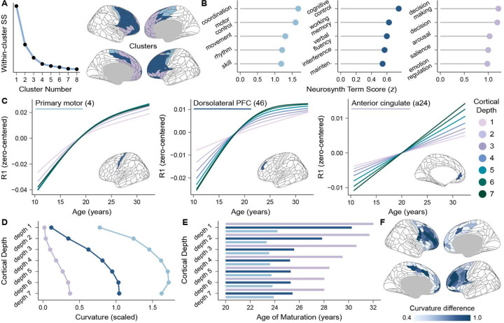
The human prefrontal cortex (PFC) exhibits markedly protracted developmental plasticity, yet whether reductions in plasticity occur synchronously across prefrontal cortical layers is unclear. Animal studies have shown that intracortical myelin consolidates neural circuits to close periods of plasticity. Here, we use quantitative myelin imaging collected from youth (ages 10-32 years) at ultra-high field (7T) to investigate whether deep and superficial PFC layers exhibit different timeframes of plasticity. We find that myelin matures along a deep-to-superficial axis in the PFC; this axis of maturational timing is expressed to a different extent in cytoarchitecturally distinct regions along the frontal cortical hierarchy. By integrating myelin mapping with electroencephalogram and cognitive phenotyping, we provide evidence that deep and superficial prefrontal myelin dissociably impact timescales of neural activity, task learning rates, and cognitive processing speed. Heterochronous maturation across deep and superficial layers is an underrecognized mechanism through which association cortex balances cognitively-relevant increases in circuit stability and efficiency with extended neuroplasticity.


Conflict of interest statement

Competing Interests The authors declare no competing interests.

Figures

Figure 1.
Figure 1.. R1 captures myeloarchitectural variation throughout the youth cortex.
A) A volumetric R1 map from a representative individual is shown along with projections of R1 to that individual’s cortical surface at superficial (left) and deep (right) depths. To provide intuition into the sampling of R1 at 7 intracortical depths, a histochemical myelin stain of prefrontal area 44 is included, along with the labeling of cortical layers 1–6 and white matter (WM). The estimated locations of the 7 cortical depths under study, which range from 20% to 80% of total cortical thickness, are indicated. The myelin stain is reproduced from Palomero-Gallagher and Zilles, 2019. B) A map of regional average R1 in the 7T youth sample. R1 is a quantitative MRI measure with units of sec−1. C) R1 progressively decreased along the sensorimotor-association axis (cortical map from Sydnor et al.) demonstrating hierarchical variation in this myelin-sensitive measure. D) R1 positively correlated with myelin basic protein (MBP) gene expression (cortical map from Wagstly et al.), demonstrating alignment between R1 and a transcriptomic readout of myelin level. E) Regional differences in R1 were coupled to differences in the myelin water fraction (MWF; cortical map from Liu et al.), confirming spatial correspondence between R1 and an independent myelin mapping technique. F) R1 increases in the frontal lobe with increasing intracortical depth. R1 is shown in frontal regions at each intracortical depth (right) along with corresponding depth-specific violin plots (left). The violin plots summarize the distribution of regional R1 at each depth; white diamonds represent average frontal R1 by depth. G) A Pearson’s correlation matrix comparing frontal lobe R1 between pairs of cortical depths reveals that there is high congruence but not equivalence in the regional distribution of R1 at each depth. H) Depth-dependent profiles of R1 differ between three laminarly distinct regions, including the primary motor cortex (area 4), the dorsolateral prefrontal cortex (area 46), and the anterior cingulate (area a24). Regional differences in depth-wise R1 profiles were observable both in group-averaged data (top) and data from individual participants (bottom; three participants shown). Left and right hemisphere depth profiles are plotted separately for each region.
Figure 2.
Figure 2.. A deep-to-superficial axis of frontal cortex R1 maturation.
A) Developmental trajectories of frontal lobe R1, modeled using GAMM smooth functions, are shown for 6 intracortical depths. Developmental trajectories are overlaid on participant-level data; longitudinal imaging sessions are connected. Growth bars demarcating age windows of significant R1 increase are also shown, shaded by the first derivative (Δ R1/Δ age) of the age spline. The F-statistic and the p-value of the age smooth term from each GAMM are indicated; F-statistics progressively increase in magnitude from superficial cortex (depth 1) to deep cortex (depth 7). B) The average rate at which R1 increased with age gets faster when moving from superficial to deep depths, indicative of larger magnitude developmental change in deeper cortex. Cortical plots (right) display the average rate of R1 increase in each frontal region at each depth. The corresponding depth plot (left) quantifies the average rate of R1 increase across all frontal regions at each cortical depth. The rate of increase was calculated as the average first derivative of the age spline across the age range studied. C) The age at which R1 matured in each region gets younger when moving from superficial to deep depths, suggestive of earlier maturational timing in deeper portions of cortex. Cortical plots (right) show the age of R1 maturation in each region at each depth. The depth plot (left) quantifies the average age of maturation across frontal regions at each depth. The age of R1 maturation was operationalized as the youngest age at which the first derivative of the age spline was no longer significantly different from zero, based on a 95% simultaneous interval.
Figure 3.
Figure 3.. Hierarchical development of frontal R1 throughout the cortical ribbon.
A) Each frontal lobe region is ranked along the sensorimotor-association (S-A) axis. The S-A axis is a hierarchical axis of cortical organization that captures the stereotyped patterning of feature heterogeneity from primary and unimodal cortices involved in perception and action (sensorimotor pole) to heteromodal and paralimbic cortices involved in executive and socioemotional processing (association pole). The S-A axis was derived in Sydnor et al. by averaging rank orderings of the 10 listed cortical feature maps. B) Developmental correlations quantifying the association between regional rates of R1 increase (average first derivative) and regional S-A axis ranks within the frontal lobe are presented for each cortical depth. Correlations were negative, indicating that the average rate of R1 increase was largest at the motor pole of the frontal S-A axis and progressively declined towards its association pole. The strength of the negative correlation increased from deeper cortex (depth 7) to superficial cortex (depth 1). C) A scatterplot of the developmental correlation with the S-A axis for intracortical depth 1 illustrates hierarchical myelination of superficial cortices during youth. D) Each frontal lobe region is ranked along an axis of cytoarchitectural variation obtained by Paquola et al. using the BigBrain 3D histological atlas, a post-mortem atlas of cell body staining. Cytoarchitectural variation was characterized by cross correlating depth-wise straining intensity profiles between different cortical locations, thereby indexing regional differences in neuron density and soma size throughout the cortical ribbon. Cytoarchitectural variation captures a continuum of cortical types along a eulaminate to dys/agranular axis characterized by decreasing laminar differentiation. E) Developmental correlations quantifying the association between regional rates of R1 increase and position on the frontal axis of cytoarchitecture variation are shown for all depths. Correlations were positive, linking accelerated myelination to eulaminate cortices and slower myelination to dys/agranular cortices. F) A scatterplot of the developmental correlation with the cytoarchitectural axis is displayed for intracortical depth 1, highlighting differences in R1 development depending on a region’s cytoarchitectural profile.
Figure 4.
Figure 4.. Depth-wise R1 maturational profiles diverge between functionally distinct cortical areas.
A) A clustering analysis identified zones of the frontal lobe that exhibit similar R1 maturational profiles across all 7 intracortical depths. Clusters were identified by calculating the curvature of each region’s 7 depth-dependent splines and applying k-means clustering to the curvature values. Three clusters were extracted based on the convergence of 30 indices for determining the optimal number of clusters; the suitability of a three-clustering solution was confirmed with a scree plot of the total within-cluster sum of squares (SS). B) Functional decoding of identified clusters classified motor, cognitive control, and salience/emotion clusters. The primary functions supported by cortical regions included in each of the clusters were identified using meta-analytic maps of over 100 psychological terms derived from Neurosynth. C) Developmental trajectories of R1 are shown for all 7 cortical depths in the primary motor cortex (area 4; left), the dorsolateral prefrontal cortex (area 46; middle) and the anterior cingulate cortex (area a24; right). Developmental trajectories are GAMM smooth estimates that are independently zero-centered along the y-axis. Regional plots illustrate three different modes of depth-dependent R1 change characteristic of motor, cognitive control, and salience/emotion clusters. D) Developmental spline curvature values are displayed for all 7 intracortical depths for each cluster. High curvature values, seen in all depths of the motor cluster and deep depths of the cognitive control cluster, reflect non-linear, curved splines that plateau. Low curvature values, characteristic of all depths in the salience/emotion cluster and superficial depths of the cognitive cluster, signify relatively more linear spline fits. E) The average age at which R1 matured is charted for all intracortical depths in each cluster. Ages of maturation got progressively older between motor, cognitive control, and salience/emotion clusters and showed the greatest depth-wise variability within the cognitive cluster. F) Cortical regions of the cognitive cluster generally exhibited the greatest difference in developmental spline curvature values between deep depths (4–7) and superficial depths (1–3), indicative of laminar variability in the shape of R1 trajectories. The difference in curvature values between deep and superficial depths is plotted on the cortex as the curvature difference.
Figure 5.
Figure 5.. Developmental findings are robust in sensitivity analyses.
Regional and depth-wise patterns of frontal cortex R1 development are robust to controls for individual differences in i) biological sex, ii) cortical thickness, iii) cortical curvature, iv) structural data quality, and v) cortex partial volume effects. Key results are shown for each of these five sensitivity analyses; all results strongly converge with findings from the main analysis. A) Depth plots display the average rate of R1 increase in the frontal lobe at each of the 7 intracortical depths (colored circles; average first derivative). The corresponding cortical maps present the average rate of R1 increase in each frontal region in the most superficial depth (1) and deepest depth (7). Percentages indicate the percent of frontal lobe regions that exhibited a significant increase in R1 at the corresponding depth. B) Depth plots chart the average age at which R1 matured in the frontal lobe at each of the 7 intracortical depths (colored circles; first age at which the derivative was no longer significant). The corresponding cortical maps show the age of R1 maturation in each frontal region in depths 1 and 7. C) Developmental trajectories of R1 at all 7 intracortical depths are shown for the primary motor cortex (area 4), the dorsolateral prefrontal cortex (dlPFC; area 46) and the anterior cingulate cortex (ACC; area a24).
Figure 6.
Figure 6.. Frontal cortex R1 is associated with EEG-derived neural dynamics.
A) Frontal EEG electrodes were mapped from the scalp to the cortex to derive a surface-based atlas of EEG electrode positions. Depth-dependent R1 and the aperiodic exponent were averaged across spatially proximal electrodes within the ventrolateral prefrontal cortex (PFC), dorsolateral PFC, superior PFC, and primary motor cortex. B) The aperiodic exponent was calculated from the slope of the 1/f power spectral density (approximated by the dotted line for illustration). The exponent quantifies how quickly power decays with increasing frequency. The average power spectrum across frontal electrodes is shown for individuals 10–16 and 24–32 years old. C) The aperiodic exponent decreased from age 10 to the early 20s in the ventrolateral PFC, dorsolateral PFC, superior PFC, and primary motor cortex. Developmental trajectories (GAMM-predicted exponent values) for all four territories are shown overlaid on participant-level data. Trajectories are colored according to the EEG electrode atlas in panel A. Trajectories were derived from GAMMs fit independently for each cortical territory prior to visualization in the same graph. D) Depth-specific GAMMs provide evidence for stronger associations of the aperiodic exponent with deep than superficial R1 across the lateral PFC. Independent GAMMs were fit to model relationships between the exponent and deep R1 or superficial R1 in each frontal territory, controlling for age. The F-statistic derived from the GAMM term relating the exponent to R1 is plotted. F-statistics are larger in deep than in superficial cortex. E) GAMMs with a depth interaction term reveal a significantly stronger negative association between R1 and the aperiodic exponent in deep as compared to superficial cortex. R1-exponent associations are shown for deep and superficial cortex in the ventrolateral PFC (left) and the superior PFC (right); data points represent partial residuals from a depth interaction model that controlled for age.
Figure 7.
Figure 7.. Prefrontal cortex R1 is linked to learning rate and processing speed.
A) Participants completed a two-stage sequential decision-making task to earn rewards. During the first stage of each trial, participants selected one of two spaceships that transitioned with fixed probabilities to either a common planet (70% of transitions) or rare planet (30% of transitions). During the second stage of each trial, participants selected one of two aliens on the planet for the chance to receive a reward; the probability of receiving a reward (P(reward)) from each alien fluctuated randomly to incentivize continuous learning of changing contingencies. B) Learning rates and response times significantly increased within-person (grey lines) between stages 1 and 2 of the task, as assessed with paired-samples t-tests. C) Learning rates increased linearly with age on stage 1 and 2 of the task, although substantial inter-individual variability is present at all ages. D) Response times decreased non-linearly with age on stage 1 and 2 of the task. E) Cortical plots are shown that highlight frontal regions where across-depth R1 was significantly associated with i) stage 1 learning rates, ii) stage 2 learning rates, iii) stage 1 response times, and iv) stage 2 responses times. Significant regions are colored. Below each cortical plot, the relationship between each cognitive measure and R1 in the dorsolateral prefrontal cortex (dlPFC) is shown in a partial residual plot. F) Neurosynth-based functional decoding of brain regions that exhibited a significant association between R1 and stage 2 learning rates (purple regions in Eii) emphasizes their role in cognitive control, decision making, and reinforcement learning processes. G) Associations between frontal R1 and response times (RT) were larger when engaging higher-order cognitive control (stage 1, stage 2, anti-saccade) than when performing a simple visual-motor response (visually-guided saccade, vgs). The cortical brain plots highlight frontal regions with a significant relationship between R1 and task-specific response times; significant regions are in black. Boxplots summarize the distribution of regional F-statistics derived from GAMMs relating frontal R1 to RT on each task. Regional F-statistics are smaller for the vgs. Boxplot limits correspond to the first and third quantiles with whiskers extending up to 1.5x interquartile range.

Similar articles

References

    1. Donahue C. J., Glasser M. F., Preuss T. M., Rilling J. K. & Essen D. C. V. Quantitative assessment of prefrontal cortex in humans relative to nonhuman primates. PNAS 115, E5183–E5192 (2018). - PMC - PubMed
    1. Buckner R. L. & Krienen F. M. The evolution of distributed association networks in the human brain. Trends Cogn Sci 17, 648–665 (2013). - PubMed
    1. Sydnor V. J. et al. Neurodevelopment of the association cortices: Patterns, mechanisms, and implications for psychopathology. Neuron 109, 2820–2846 (2021). - PMC - PubMed
    1. Larsen B. & Luna B. Adolescence as a neurobiological critical period for the development of higher-order cognition. Neurosci Biobehav Rev 94, 179–195 (2018). - PMC - PubMed
    1. Sydnor V. J. et al. Intrinsic activity development unfolds along a sensorimotor–association cortical axis in youth. Nat Neurosci 26, 638–649 (2023). - PMC - PubMed

Publication types

LinkOut - more resources