

This is a preprint.
Tissue-specific and spatially dependent metabolic signatures perturbed by injury in skeletally mature male and female mice
- PMID: 39975211
- PMCID: PMC11838485
- DOI: 10.1101/2024.09.30.615873
Tissue-specific and spatially dependent metabolic signatures perturbed by injury in skeletally mature male and female mice
Abstract
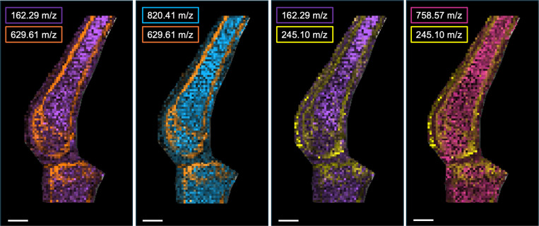
Joint injury is a risk factor for post-traumatic osteoarthritis. However, metabolic and microarchitectural changes within the joint post-injury in both sexes remain unexplored. This study identified tissue-specific and spatially-dependent metabolic signatures in male and female mice using matrix-assisted laser desorption ionization-mass spectrometry imaging (MALDI-MSI) and LC-MS metabolomics. Male and female C57Bl/6J mice were subjected to non-invasive joint injury. Eight days post-injury, serum, synovial fluid, and whole joints were collected for metabolomics. Analyses compared between injured, contralateral, and naïve mice, revealing local and systemic responses. Data indicate sex influences metabolic profiles across all tissues, particularly amino acid, purine, and pyrimidine metabolism. MALDI-MSI generated 2D ion images of bone, the joint interface, and bone marrow, highlighting increased lipid species in injured limbs, suggesting physiological changes across injured joints at metabolic and spatial levels. Together, these findings reveal significant metabolic changes after injury, with notable sex differences.
Keywords: MALDI imaging; metabolomics; osteoarthritis; sex differences.
Conflict of interest statement
Competing interests: Dr. June owns stock in Beartooth Biotech. Drs. June and Brahmachary own stock in OpenBioWorks. Neither company was involved in this study. Remaining authors have no conflicts of interest to disclose.
Figures


Similar articles
-
Subchondral bone structure and synovial fluid metabolism are altered in injured and contralateral limbs 7 days after non-invasive joint injury in skeletally-mature C57BL/6 mice.Osteoarthritis Cartilage. 2022 Dec;30(12):1593-1605. doi: 10.1016/j.joca.2022.09.002. Epub 2022 Sep 20. Osteoarthritis Cartilage. 2022. PMID: 36184957 Free PMC article.
-
Spatially resolved metabolic distribution for unraveling the physiological change and responses in tomato fruit using matrix-assisted laser desorption/ionization-mass spectrometry imaging (MALDI-MSI).Anal Bioanal Chem. 2017 Feb;409(6):1697-1706. doi: 10.1007/s00216-016-0118-4. Epub 2016 Dec 8. Anal Bioanal Chem. 2017. PMID: 27933363 Free PMC article.
-
Comprehensive MALDI mass spectrometry imaging of tumor regions post-neoadjuvant therapy.Anal Bioanal Chem. 2025 Apr;417(10):2039-2046. doi: 10.1007/s00216-025-05785-4. Epub 2025 Feb 20. Anal Bioanal Chem. 2025. PMID: 39976685
-
Matrix assisted laser desorption/ionization-mass spectrometry imaging (MALDI-MSI) for direct visualization of plant metabolites in situ.Curr Opin Biotechnol. 2016 Feb;37:53-60. doi: 10.1016/j.copbio.2015.10.004. Epub 2015 Nov 22. Curr Opin Biotechnol. 2016. PMID: 26613199 Review.
-
Recent advances in matrix-assisted laser desorption/ionisation mass spectrometry imaging (MALDI-MSI) for in situ analysis of endogenous molecules in plants.Phytochem Anal. 2018 Jul;29(4):351-364. doi: 10.1002/pca.2759. Epub 2018 Apr 17. Phytochem Anal. 2018. PMID: 29667236 Review.
References
-
- Brown T.D., Johnston R.C., Saltzman C.L., Marsh J.L., Buckwalter J.A., Posttraumatic osteoarthritis: a first estimate of incidence, prevalence, and burden of disease, J Orthop Trauma 20(10) (2006) 739–44. - PubMed
-
- Gottlob C.A., Baker C.L. Jr., Pellissier J.M., Colvin L., Cost effectiveness of anterior cruciate ligament reconstruction in young adults, Clin Orthop Relat Res (367) (1999) 272–82. - PubMed
-
- Griffin L.Y., Albohm M.J., Arendt E.A., Bahr R., Beynnon B.D., Demaio M., Dick R.W., Engebretsen L., Garrett W.E. Jr., Hannafin J.A., Hewett T.E., Huston L.J., Ireland M.L., Johnson R.J., Lephart S., Mandelbaum B.R., Mann B.J., Marks P.H., Marshall S.W., Myklebust G., Noyes F.R., Powers C., Shields C. Jr., Shultz S.J., Silvers H., Slauterbeck J., Taylor D.C., Teitz C.C., Wojtys E.M., Yu B., Understanding and preventing noncontact anterior cruciate ligament injuries: a review of the Hunt Valley II meeting, January 2005, Am J Sports Med 34(9) (2006) 1512–32. - PubMed
-
- Lohmander L.S., Englund P.M., Dahl L.L., Roos E.M., The long-term consequence of anterior cruciate ligament and meniscus injuries: osteoarthritis, Am J Sports Med 35(10) (2007) 1756–69. - PubMed
Publication types
Grants and funding
LinkOut - more resources
Full Text Sources