[Preprint]. 2025 Feb 3:2025.02.03.636156.
doi: 10.1101/2025.02.03.636156.

Endoderm differentiates into a transient epidermis in the mouse perineum

Affiliations

Endoderm differentiates into a transient epidermis in the mouse perineum

Christine E Larkins et al. bioRxiv. .

Update in

Abstract

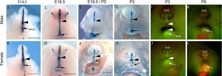
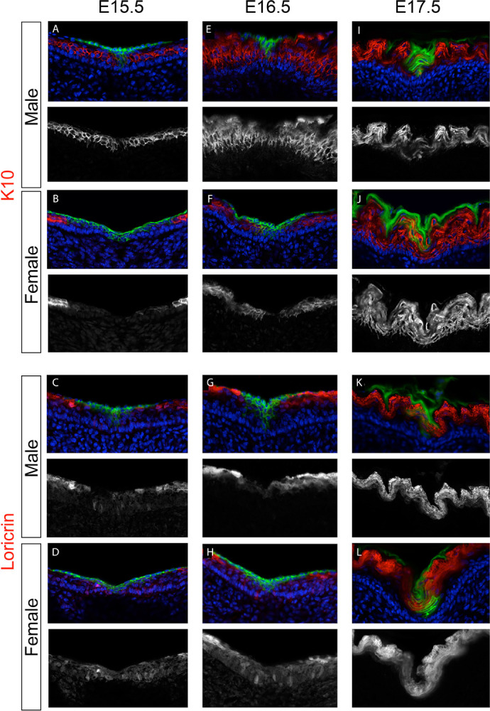
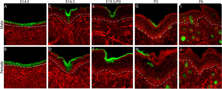
In eutherian mammals, the embryonic cloaca is partitioned into genitourinary and anorectal canals by the urorectal septum. At the caudal end of the mouse embryo, the urorectal septum contributes to the perineum, which separates the anus from the external genitalia. Growth of the urorectal septum displaces cloacal endoderm to the surface of the perineum, where it is incorporated into epidermis, an enigmatic fate for endodermal cells. Here we show that endodermal cells differentiate into true epidermis in the perineum, expressing basal, spinous, and granular cell markers. Endodermal epidermis is lost through terminal differentiation and desquamation postnatally, when it is replaced by ectoderm. Live imaging and single-cell tracking reveal that ectodermal cells move at a faster velocity in a lateral-to-medial direction, converging towards the narrow band of endoderm between the anus and external genitalia. Although the perineum is sexually dimorphic, similar spatiotemporal patterns of cell movement were observed in males and females. These results demonstrate that cloacal endoderm differentiates into a non-renewing, transient epidermis at the midline of the perineum. Differential movement of endodermal and ectodermal cells suggests that perineum epidermis develops by convergent extension. These findings provide a foundation for further studies of perineum development and of sex-specific epidermal phenotypes.


Conflict of interest statement

Competing Interests None

Figures

Figure 1.
Figure 1.
Endoderm cell lineage tracing in the perineum of male and female mice. (A-H) ShhGFPcre/+; R26RlacZ/+ embryos and pups stained with X-gal to show Shh-expressing cells and their descendants in the endodermal lineage of the perineum from E14.5 to P3. Black arrowheads mark the base of the genital tubercle/phallus and black arrows indicate the position of the anus. Scale bar is 1mm. (I-L) ShhGFPcre/+; R26RmTmG/+ males and females at P3 and P6 showing GFP expression in the Shh-expressing cells and their descendants. White arrowheads mark GFP-positive cells at the midline of the perineum; white arrows mark the position of the anus; ExG, external genitalia.
Figure 2.
Figure 2.
The endodermal lineage of the perineum expresses epidermal differentiation markers. (A-L) ShhGFPcre/+; R26RmTmG/+ males and females at E15.5, E16.5, and E17.5 showing immunofluorescence of K10 (A,B,E,F,I,J) and loricrin (C,D,G,H,K,L). Green signal indicates GFP expression in endoderm. Red signal is pseudocolored immunofluorescence of K10 and loricrin. Blue indicates Hoechst staining of cell nuclei. Greyscale panels show single-channel immunofluorescence.
Figure 3.
Figure 3.
Terminal differentiation leads to loss of endoderm from the epidermis. (A-J) Transverse sections through the perineal skin of male and female ShhGFPcre/+; R26RmTmG/+ mice from E14.5-P6. (A,B) At E14.5, GFP-expressing endodermal cells are found in the basal and suprabasal layers of epidermis in the medial region of the perineum. (C,D) At E16.5, GFP-positive endoderm is throughout all epidermal layers at the midline, but laterally, the majority of endodermal cells are located suprabasally. (E,F) At E19.5-P0, the endoderm lineage is almost entirely in the stratum corneum and no GFP-positive cells are detectable in the basal layers. (G-J) At P6, a small domain of endodermal cells is visible in the stratum corneum at the midline of the perineum epidermis (G,H), but these are no longer detectable at P6 (I,J). GFP expression in the in the hair follicles is seen in the dermis.
Figure 4.
Figure 4.
Live imaging of cell movements in the endoderm and surrounding ectoderm of the perineum. (A,B) Time lapse of a male and a female ShhGFPcre/+; R26RmTmG/+ perineum. Cell tracks are marked with colored dots and lines. Cells were followed on both sides of the midline of the perineum, although only one side is shown here. (C) Graph showing the average mediolateral velocity of ectoderm and endoderm cells. Note that ectoderm cells have a significantly higher mediolateral velocity compared to endoderm cells *P < 0.05 (P = 0.015); **P < 0.01 (P = 0.003), using a two-tailed t test.

References

    1. Arai K.Y., Hara T., Nagatsuka T., Kudo C., Tsuchiya S., Nomura Y., Nishiyama T., 2017. Postnatal changes and sexual dimorphism in collagen expression in mouse skin. PLoS One 12, e0177534. - PMC - PubMed
    1. Azzi L., El-Alfy M., Martel C., Labrie F., 2005. Gender differences in mouse skin morphology and specific effects of sex steroids and dehydroepiandrosterone. J Invest Dermatol 124, 22–27. - PubMed
    1. Blanpain C., Fuchs E., 2009. Epidermal homeostasis: a balancing act of stem cells in the skin. Nat Rev Mol Cell Biol 10, 207–217. - PMC - PubMed
    1. Chi L., Liu C., Gribonika I., Gschwend J., Corral D., Han S.-J., Lim A.I., Rivera C.A., Link V.M., Wells A.C., Bouladoux N., Collins N., Lima-Junior D.S., Enamorado M., Rehermann B., Laffont S., Guéry J.-C., Tussiwand R., Schneider C., Belkaid Y., 2024. Sexual dimorphism in skin immunity is mediated by an androgen-ILC2-dendritic cell axis. Science 384, eadk6200. - PMC - PubMed
    1. Clayton E., Doupe D.P., Klein A.M., Winton D.J., Simons B.D., Jones P.H., 2007. A single type of progenitor cell maintains normal epidermis. Nature 446, 185–189. - PubMed

Publication types

LinkOut - more resources