

Abdominal CT scan findings of a child with hepatic fascioliasis: A case report on rarely reported emerging disease
- PMID: 39975650
- PMCID: PMC11835549
- DOI: 10.1016/j.radcr.2025.01.030
Abdominal CT scan findings of a child with hepatic fascioliasis: A case report on rarely reported emerging disease
Abstract
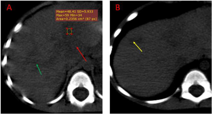
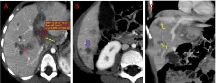
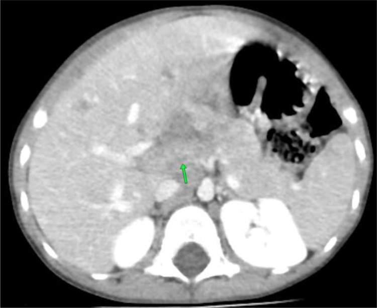
Fasciolosis is a zoonotic infection caused by trematodes fasciola hepatica and fasciola gigantic, and humans are incidental hosts. Although infrequently reported in developed nations, it is common in developing countries. Few cases have been reported in Africa, specifically in Ethiopia. This article reports a case of a 4-year-old Ethiopian child who presented with right upper quadrant abdominal pain. His complete blood count showed eosinophilia, and imaging demonstrated lesions at peripheral subcapsular parenchyma and central along the biliary tree. Serologic tests confirmed liver fluke infection with fasciola hepatica indirect hemagglutination test titer of 1/4000. Computed tomography imaging appearances of hepatic fasciolosis depend on the phase and course of the disease and should be considered in differential diagnosis of lesions along the biliary tree.
Keywords: Biliary phases; CT findings; Case report; Ethiopia; Fasciolosis.
© 2025 Published by Elsevier Inc. on behalf of University of Washington.
Figures
Similar articles
-
Fascioliasis complicated by acute necrotizing pancreatitis in an Ethiopian child - a case report on a rare complication of a rarely reported emerging disease.IJID Reg. 2022 Mar 24;3:135-137. doi: 10.1016/j.ijregi.2022.03.016. eCollection 2022 Jun. IJID Reg. 2022. PMID: 35755466 Free PMC article.
-
Imaging findings of human hepatic fascioliasis: a case report and review of the literature.J Med Case Rep. 2021 Jun 24;15(1):324. doi: 10.1186/s13256-021-02945-9. J Med Case Rep. 2021. PMID: 34162437 Free PMC article. Review.
-
Clinical presentation and management of Fasciola hepatica infection: single-center experience.World J Gastroenterol. 2011 Nov 28;17(44):4899-904. doi: 10.3748/wjg.v17.i44.4899. World J Gastroenterol. 2011. PMID: 22171131 Free PMC article.
-
Fascioliasis: A challenging differential diagnosis for radiologists.J Radiol Case Rep. 2019 Jan 31;13(1):11-16. doi: 10.3941/jrcr.v13i1.3451. eCollection 2019 Jan. J Radiol Case Rep. 2019. PMID: 31565163 Free PMC article.
-
Neurological and ocular fascioliasis in humans.Adv Parasitol. 2014;84:27-149. doi: 10.1016/B978-0-12-800099-1.00002-8. Adv Parasitol. 2014. PMID: 24480313 Review.
References
-
- Bayu B., Sebhat A., Alemseged W., Jemal a., Molla G., Tsegaw F., et al. Cases of human fascioliasis in North-West Ethiopia. Ethiop J Health Develop. 2005;19(3):237–240.
Publication types
LinkOut - more resources
Full Text Sources
