[Preprint]. 2025 Jan 29:rs.3.rs-5627712.
doi: 10.21203/rs.3.rs-5627712/v1.

Human Organ Chips Reveal New Inflammatory Bowel Disease Drivers

Affiliations

Human Organ Chips Reveal New Inflammatory Bowel Disease Drivers

Alican Özkan et al. Res Sq. .

Abstract

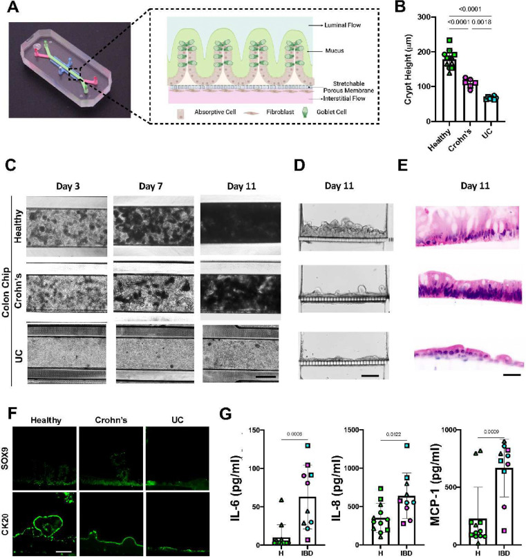
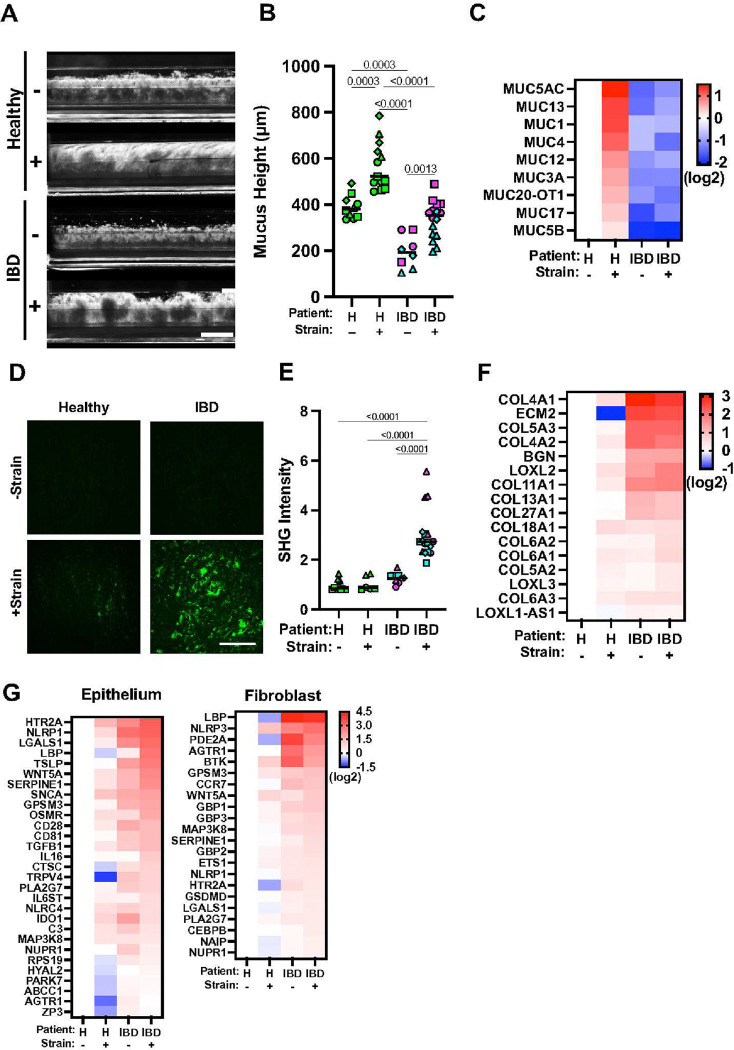
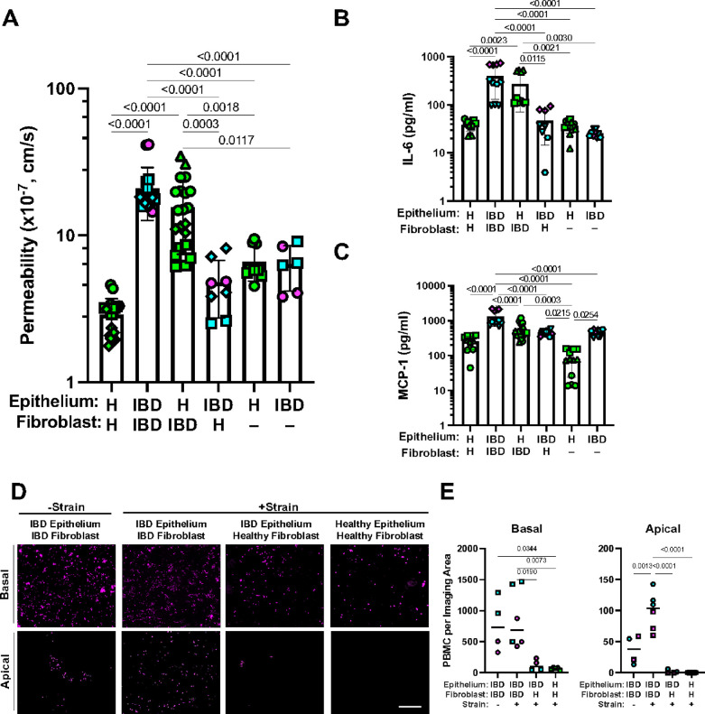
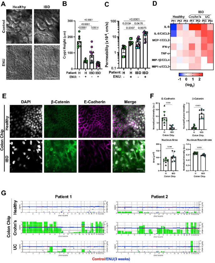
Inflammatory bowel disease (IBD) patients exhibit compromised intestinal barrier function and decreased mucus accumulation, as well as increased inflammation, fibrosis, and cancer risk, with symptoms often being exacerbated in women during pregnancy. Here, we show that these IBD hallmarks can be replicated using human Organ Chips lined by IBD patient-derived colon epithelial cells interfaced with matched fibroblasts cultured under flow. Use of heterotypic tissue recombinants revealed that IBD fibroblasts are the primary drivers of multiple IBD symptoms. Inflammation and fibrosis are accentuated by peristalsis-like motions in IBD Chips and when exposed to pregnancy-associated hormones in female IBD Chips. Carcinogen exposure also increases inflammation, gene mutations, and chromosome duplication in IBD Chips, but not in Healthy Chips. These data enabled by human Organ Chip technology suggest that the intestinal stroma, sex hormones, and peristalsis-associated mechanical deformations play a key role in driving inflammation, fibrosis, and disease progression in male and female IBD patients.

Keywords: Carcinogenesis; Crohn’s Disease; Epithelial-Stromal Interactions; Organ-on-a-chip; Ulcerative Colitis; Women’s Health.


Conflict of interest statement

Competing interests D.E.I. holds equity in Emulate, chairs its scientific advisory board and is a member of its board of directors. The other authors declare no competing interests.

Figures

Figure 1:
Figure 1:. Primary human Colon Chips generated with epithelium and fibroblasts isolated from Healthy and IBD patient samples.
A) Photograph of the commercially available Organ Chip device (left) and a cross-sectional illustration of the epithelial-stromal tissue interface (right) formed with patient-derived cells, which is composed of a fibroblast rich stroma that is perfused with medium to mimic interstitial fluid flow separated by a flexible, porous, ECM-coated membrane from a colon epithelium above. The epithelium, which is composed of absorptive cells and Goblet cells, secretes a thick mucus layer into the lumen of the apical channel that also experiences fluid flow. The entire tissue-tissue interface can be stretched and relaxed rhythmically to mimic peristalsis-like motions by applying cyclic suction to hollow side chambers in the flexible device (not shown). B) Height of crypts formed in Colon Chips measured at 11 days. Numbers indicate P values between compared groups, as determined by one-way ANOVA test (n=3 Healthy, green; 2 Crohn’s, magenta; and 2 UC, cyan). C) Representative bright field microscopic images of Healthy and IBD Chips created with cells from Crohn’s or UC patients viewed from above on day 3, 7, and 11. A functional epithelial monolayer progressively accumulated mucus, which appears as opaque blackened fuzzy material, in the apical channel of all chips over the 11 day time course; however, mucus accumulation was suppressed in the IBD Chips with the UC Chips displaying a greater reduction (bar, 500 μm). D) Brightfield image of vertical cross-sections through healthy and IBD Colon chips showing that the crypt-like epithelial structures are shorter in the Crohn’s and UC Colon Chips (bar, 100 μm). E) Histological H&E stained vertical cross-sections through healthy and IBD Colon chips confirming the presence of columnar colon epithelium containing goblet cells in healthy epithelium, and a significant reduction in the height of the epithelial monolayer with reduced numbers of goblet cells in the IBD Chips (bar, 50 μm). F) Immunofluorescence microscopic views showing distribution of SOX9 and CK20 in crypts in the colon epithelium of Healthy and IBD Chips (bar, 50 μm). G) Inflammatory cytokine protein levels measured in the basal outflows of Healthy (H) and IBD Chips on culture day 11 (n = 3–5 chips/ condition; chips were created with cells from 3 healthy (green), 2 Crohn’s (magenta), and 2 UC (cyan) patient donors, with each symbol represents a chip created with cells from a different patient. Numbers indicate P values between compared groups, as determined by two-tailed student’s t-test. All data represent mean α SD; p values are shown in all figures.
Figure 2:
Figure 2:. Peristalsis-like cyclic strain influences mucus production and fibrosis in Colon Chips.
A) Representative side-view images microscopy of Healthy or IBD Chips with (+) and without (−) exposure to mechanical deformations visualizing mucus layer accumulation (white diffuse material) in Healthy Chips using dark-field microscopy. Note that limited mucus was produced by IBD epithelium (bar, 1 mm). B) Quantification of the height of the mucus layer overlying Healthy and IBD epithelium on-chip when cultured under the conditions in A. Numbers indicate P values between compared groups, as determined by one-way ANOVA test. (n = 3 healthy, green; 2 Crohn’s, magenta; and 2 UC, cyan). C) Heat map of genes showing that peristalsis-like mechanical stimulation (+) increases expression of genes related to mucus production in Healthy Chips, but not IBD Chips (n = 3 healthy, 1 Crohn’s and 2 UC). D) Second harmonic microscopic images of the fibroblast stroma in Healthy versus IBD Chips showing greater collagen fibril accumulation (green) with mechanical strain (+ Strain) in both chips, and that this response is greatly accentuated in IBD Chips (bar, 200 μm). E) Collagen fibril signal intensity in the stroma of Healthy versus IBD Chips quantified in second harmonic microscopic images. Numbers indicate P values between compared groups, as determined by one-way ANOVA test (n = 3 healthy, green; 2 Crohn’s, magenta; and 2 UC, cyan). F) Heat map of genes showing that peristalsis-like cyclic strain enhances expression of genes associated with fibrillar collagen production in IBD fibroblasts, but not in healthy fibroblasts (n = 3 healthy, 1 Crohn’s and 2 UC). G) Heat maps of showing that cyclic strain enhances greater expression of IBD-associated inflammatory genes in IBD Chips compared to Healthy Chips both in epithelium and fibroblasts (n=3 Healthy, 1 Crohn’s, and 2 UC).
Figure 3:
Figure 3:. Effects of ovarian hormones in Female Colon Chips.
A) Production of pro-inflammatory cytokine and chemokine proteins in Apical and Basal channels of Healthy versus IBD Chips 7 days post treatment with different female hormone exposures (E2, MPA, and PAHs, as indicated). Note that exposure to female hormones significantly increased expression of inflammatory factors in both epithelium and fibroblast of IBD Colon Chips whereas they suppressed their production in Healthy Chips. B) Collagen fibril signal intensity in the stroma of Healthy versus IBD Chips quantified in second harmonic microscopic images showing that only exposure to pregnancy-like hormonal conditions produced a major increase in stromal fibrillogenesis, and this only was observed in IBD fibroblasts and not healthy fibroblasts from IBD patients. Numbers indicate P values between compared groups, as determined by one-way ANOVA test (n=4–6 chips of 1 Crohn’s and 1 Healthy each from 2 independent experiments). C) Second harmonic microscopic images of fibrillar collagen (green) produced by fibroblasts showing that the E2 + MPA produce a small increase in fibrillogenesis in IBD Chips, while simulating pregnancy by exposure to E2 + MPA + PAH results in dramatic collagen fibril accumulation, and that neither of these responess is observed in Healthy Chips (bar, 200 μm; n=4–6 chips of 1 Crohn’s and 1 Healthy each from 2 independent experiments).
Figure 4:
Figure 4:. Tissue recombinant chips reveal that the IBD fibroblasts drive compromise of the permeability barrier and inflammation in the epithelium.
A) Graphs showing that intestinal barrier permeability to cascade blue is much higher in homotypic IBDChips compared to Healthy Chips and that tissue recombinants of IBD fibroblasts with healthy (H) epithelium results in a similar high level of barrier compromise on-chip. This is not observed in the IBD or healthy epithelium in the absence of fibroblasts or in IBD epithelium with healthy fibroblasts. Numbers indicate P values between compared groups, as determined by one-way ANOVA test (n = 3 healthy, green; 2 Crohn’s, magenta; and 2 UC, cyan). B) Quantification of cytokine protein levels revealed that combination of IBD fibroblasts with healthy epithelium results in increased production of IL-6 and MCP-1 in Colon Chips, whereas this was not seen in healthy epithelium alone or homotypic Colon Chips. Numbers indicate P values between compared groups, as determined by one-way ANOVA test (n = 3 healthy, green; 1 Crohn’s, magenta; and 2 UC, cyan). C) IL-6 and MCP-1 production by IBD epithelium also required the presence of IBD fibroblasts. Numbers indicate P values between compared groups, as determined by one-way ANOVA test (n = 3 healthy, green; 1 Crohn’s, magenta; and 2 UC, cyan). D) Peristalsis-like cyclic strain preferentially stimulates migration of PBMCs (magenta) from the fibroblast stroma in the basal channel (Basal) to the epithelium in the apical channel (Apical) in IBD Chips but not in heterotypic chips with IBD epithelium and healthy fibroblasts or in Healthy Chips. E) Quantification of the results from D confirmed that IBD Chips exhibited higher baseline recruitment of PBMCs to the surface of the fibroblast stroma in the basal channel and that this was not altered by mechanical strain, whereas IBD epithelium combined with healthy stroma or Healthy Chips only displayed significantly less PBMC recruitment. In contrast, the number of PBMCs that migrated to the epithelium in the Apical channel more than doubled in IBD Chips when they were exposed to peristalsis-like mechanical deformations, and overall migration of PBMCs was minimal when IBD fibroblasts were absent. Numbers indicate P values between compared groups, as determined by one-way ANOVA test (n = 2 healthy, green; 1 Crohn’s, magenta; and 1 UC, cyan).
Figure 5:
Figure 5:. Exposure to carcinogen preferentially promotes mutagenesis and early steps in carcinogenesis in IBD Chips.
A) DIC images of the epithelium when viewed from above in Healthy (H) and IBD Chips after 21 days of culture in the presence or absence of the carcinogen (ENU) showing that undulating crypt-like structures are lost in both Healthy and IBD Chips after exposure to ENU (bar, 500 μm). B) Quantification of results from A showing that ENU decreases height of the epithelium in both healthy and IBD Colon Chips. Numbers indicate P values between compared groups, as determined by one-way ANOVA test (n = 3 healthy, green; 2 Crohn’s, magenta; and 2 UC, cyan). C) Epithelial barrier permeability increased as a result of exposure to ENU in Healthy and IBD Chips. Numbers indicate P values between compared groups, as determined by one-way ANOVA test. (n = 3 healthy, green; 2 Crohn’s, magenta; and 2 UC, cyan). D) Heatmaps showing inflammatory cytokine protein levels measured in outflows from the apical channel of Healthy and IBD Chips 3 weeks post-ENU exposure. Importantly, ENU exposure significantly increased inflammatory cytokine production in IBD Chips made with cells from both Crohn’s and UC patients compared to healthy patients. E) Confocal microscopic top-view images showing β-catenin and E-cadherin localization and nuclear staining (DAPI) in the epithelium within Healthy and IBD Chips 3 weeks post-ENU exposure (bar, 20 μm). F) Quantification of protein staining intensity and morphology reveal that ENU exposure decreases E-cadherin levels and increases nuclear localization of β-catenin in IBD patient-derived Colon Chips compared to Healthy Chips. ENU exposure also resulted in greater nucleus size and less roundness. Numbers indicate P values between compared groups, as determined by Student’s t-test (n = 3 healthy, green; 2 Crohn’s, magenta; and 2 UC, cyan). G) Plots showing copy number changes across all human chromosomes in Healthy and IBD Chips pre- (red) and 3 weeks post-ENU exposure (blue). Green bars indicate amplification of gene sequences for the whole chromosome. Each symbol represents an individual Healthy or IBD patient-derived chip.

References

    1. Friedrich M. et al. IL-1-driven stromal–neutrophil interactions define a subset of patients with inflammatory bowel disease that does not respond to therapies. Nat Med 27, 1970–1981 (2021). - PMC - PubMed
    1. Goodman W. A., Erkkila I. P. & Pizarro T. T. Sex matters: impact on pathogenesis, presentation and treatment of inflammatory bowel disease. Nature Reviews Gastroenterology & Hepatology 2020 17:12 17, 740–754 (2020). - PMC - PubMed
    1. Watermeyer G. Pregnancy and inflammatory bowel disease. South African Gastroenterology Review 5, 4–6 (2007).
    1. Sarvestani S. K. et al. Cancer-predicting transcriptomic and epigenetic signatures revealed for ulcerative colitis in patient-derived epithelial organoids. Oncotarget 9, 28717–28730 (2018). - PMC - PubMed
    1. Ingber D. E. Cancer as a disease of epithelial–mesenchymal interactions and extracellular matrix regulation. Differentiation 70, 547–560 (2002). - PubMed

Publication types

LinkOut - more resources