. 2025 Apr;12(15):e2412559.
doi: 10.1002/advs.202412559. Epub 2025 Feb 20.

Lactylation-Driven HECTD2 Limits the Response of Hepatocellular Carcinoma to Lenvatinib

Affiliations

Lactylation-Driven HECTD2 Limits the Response of Hepatocellular Carcinoma to Lenvatinib

Runyu Dong et al. Adv Sci (Weinh). 2025 Apr.

Abstract

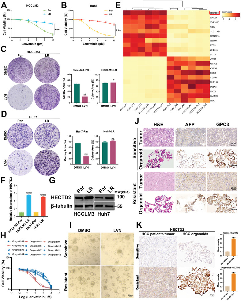
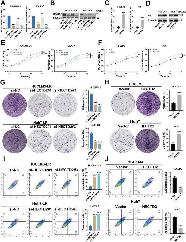
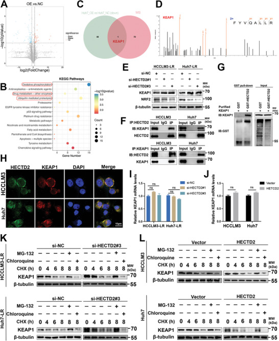
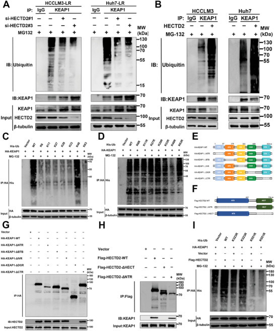
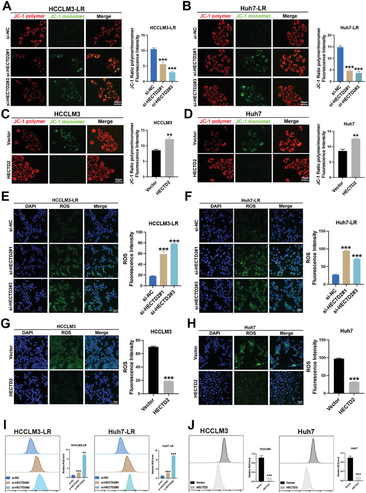
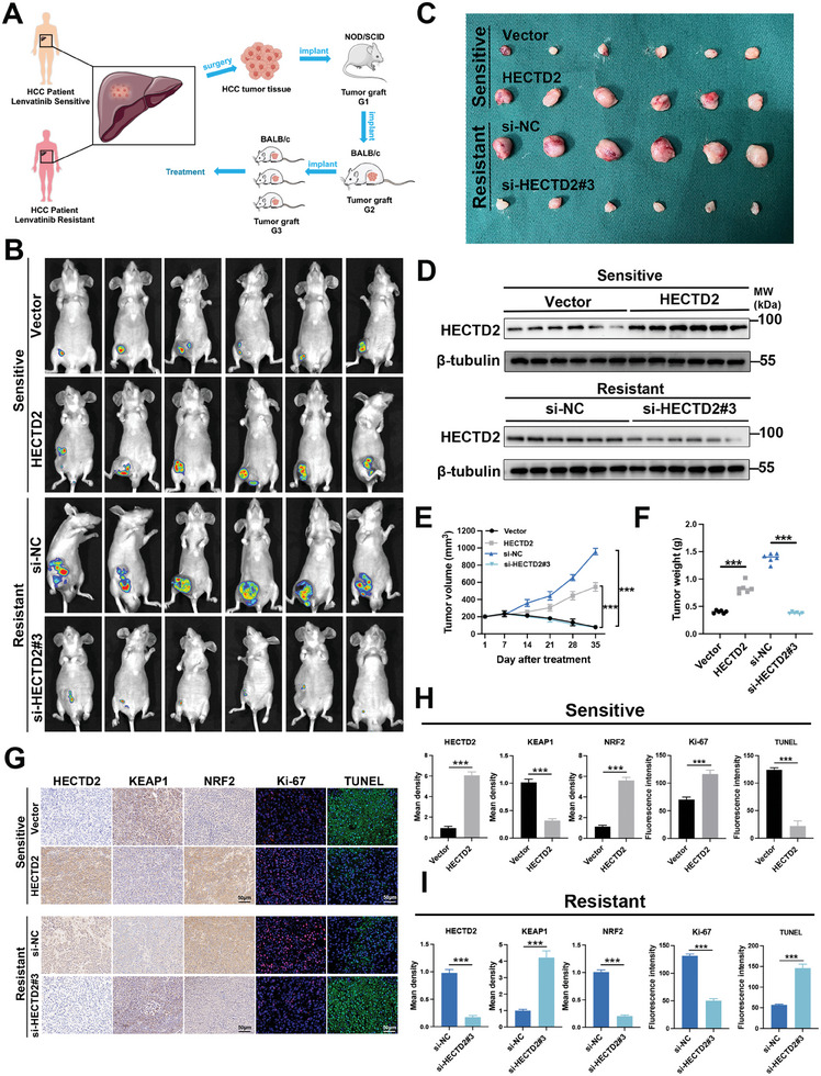
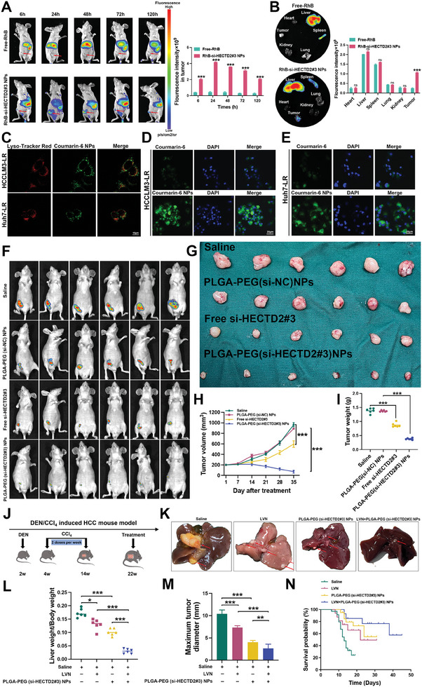
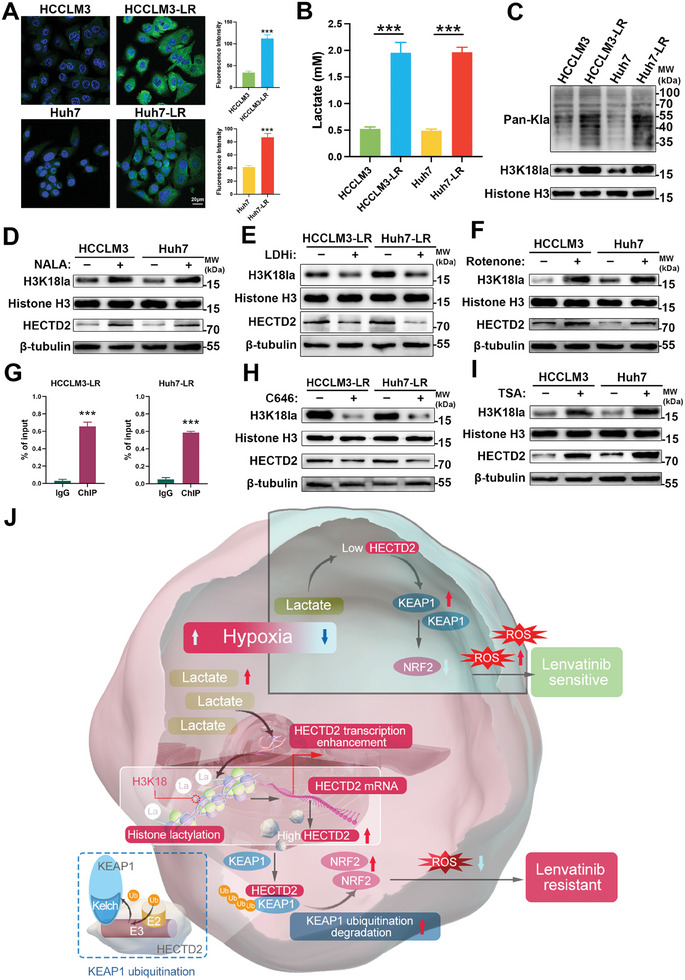
Drug resistance remains a major hurdle for the therapeutic efficacy of lenvatinib in hepatocellular carcinoma (HCC). However, the underlying mechanisms remain largely undetermined. Unbiased proteomic screening is performed to identify the potential regulators of lenvatinib resistance in HCC. Patient-derived organoids, patient-derived xenograft mouse models, and DEN/CCl4 induced HCC models are constructed to evaluate the effects of HECTD2 both in vitro and in vivo. HECTD2 is found to be highly expressed in lenvatinib-resistant HCC cell lines, patient tissues, and patient-derived organoids and xenografts. In vitro and in vivo experiments demonstrated that overexpression of HECTD2 limits the response of HCC to lenvatinib treatment. Mechanistically, HECTD2 functions as an E3 ubiquitin ligase of KEAP1, which contributes to the degradation of KEAP1 protein. Subsequently, the KEAP1/NRF2 signaling pathway initiates the antioxidative response of HCC cells. Lactylation of histone 3 on lysine residue 18 facilitates the transcription of HECTD2. Notably, a PLGA-PEG nanoparticle-based drug delivery system is synthesized, effectively targeting HECTD2 in vivo. The NPs achieved tumor-targeting, controlled-release, and biocompatibility, making them a promising therapeutic strategy for mitigating lenvatinib resistance. This study identifies HECTD2 as a nanotherapeutic target for overcoming lenvatinib resistance, providing a theoretical basis and translational application for HCC treatment.

Keywords: HECTD2; histone lactylation; nanoparticles; oxidative stress; ubiquitination.


Conflict of interest statement

The authors declare no conflict of interest.

Figures

Figure 1
Figure 1
HECTD2 expression level is associated with lenvatinib resistance in HCC. A,B) Lenvatinib‐resistant HCCLM3 and Huh7 cells (HCCLM3‐LR and Huh7‐LR) and the corresponding parental cells (HCCLM3‐Par and Huh7‐Par) were treated with different doses of lenvatinib for 3 days and cell viability was examined to compare the half‐maximal inhibitory concentration (IC50) values between lenvatinib‐resistant cells and parental cells. The IC50 value of HCCLM3‐Par is 6.93 µm, IC50 of HCCLM3‐LR is 20.15 µm. The IC50 value of Huh7‐Par is 4.51 µm, IC50 of Huh7‐LR is 15.30 µm. C,D) HCCLM3‐LR, Huh7‐LR, and the corresponding parental cells were treated with lenvatinib (LVN) or DMSO for two weeks and colony formation assays were performed to compare the cell growth between lenvatinib‐resistant cells and parental cells. E) Unbiased proteomic screening of lenvatinib‐resistant and parental HCC cells showed that HECTD2 was among the top ten upregulated proteins in lenvatinib‐resistant HCC cells. F) qRT‐PCR was performed to show the mRNA expression levels of HECTD2 in lenvatinib‐resistant and parental HCC cells. G) Western blotting was conducted to assess the protein expression levels of HECTD2 in lenvatinib‐resistant and parental HCC cells. H) Therapeutic effects of lenvatinib on HCC organoids. Red lines indicated lenvatinib‐sensitive organoids (n = 3); Blue lines indicated lenvatinib‐resistant organoids (n = 9). I) Representative images of lenvatinib‐sensitive and lenvatinib‐resistant organoids. J) H&E evaluation and IHC staining with AFP and GPC3 of lenvatinib‐sensitive and lenvatinib‐resistant HCC patients and the corresponding organoids. K) IHC staining of HECTD2 in lenvatinib‐sensitive and lenvatinib‐resistant HCC patients and the corresponding organoids. **p < 0.01; ***p < 0.001; ns, no statistical significance.
Figure 2
Figure 2
HECTD2 restricts the response of HCC to lenvatinib. A,B) Knockdown efficiency of HECTD2 in HCCLM3‐LR and Huh7‐LR cells was assessed using qRT‐PCR and western blotting. C,D) Overexpression efficiency of HECTD2 in HCCLM3 and Huh7 cells was assessed using qRT‐PCR and western blotting. E) The proliferative ability of HECTD2‐silenced HCCLM3‐LR and Huh7‐LR cells was evaluated using CCK‐8 assays. F) The proliferative ability of HECTD2‐overexpressing HCCLM3 and Huh7 cells was evaluated using CCK‐8 assays. G) Colony formation assays were performed to examine the cell proliferation of HECTD2‐silenced HCCLM3‐LR and Huh7‐LR cells. H) Colony formation assays were performed to examine the cell proliferation of HECTD2‐overexpressing HCCLM3 and Huh7 cells. I) Apoptosis rate of HCCLM3‐LR and Huh7‐LR cells with HECTD2 knockdown was evaluated by flow cytometry. J) Apoptosis rate of HCCLM3 and Huh7 cells with HECTD2 overexpression was evaluated by flow cytometry. All experiments were performed under lenvatinib treatment. **p < 0.01; ***p < 0.001; ns, no statistical significance.
Figure 3
Figure 3
HECTD2 interacts with KEAP1. A) The differentially expressed protein profile in lenvatinib‐sensitive Huh7 cells with or without HECTD2 overexpression. B) HECTD2‐associated molecular events were demonstrated using KEGG analysis on the differentially expressed protein profile. C) Venn graph showing the differentially expressed protein profile upon HECTD2 overexpression and HECTD2‐interacting protein profile. D) Mass spectrometry result of KEAP1 from the immunoprecipitation products of HECTD2. E) Western blotting analysis was performed to detect the protein expression levels of KEAP1 and NRF2 in HECTD2‐silenced HCCLM3‐LR and Huh7‐LR cells. F) Co‐IP results showing the interaction between KEAP1 and NRF2 in HCC cells. G) GST‐pulldown assays showing that HECTD2 interacted with KEAP1. H) Immunofluorescence showing the colocalization of HECTD2 and KEAP1 in the cytoplasm. I,J) The mRNA expression levels of KEAP1 upon HECTD2 knockdown or overexpression. K,L) KEAP1 protein levels were evaluated in lenvatinib‐resistant and lenvatinib‐sensitive HCC cells after CHX, MG‐132, or chloroquine treatment. ns, no statistical significance.
Figure 4
Figure 4
HECTD2 serves as an E3 ubiquitin ligase of KEAP1. A) KEAP1 ubiquitination and KEAP1 protein levels were examined in HCCLM3‐LR and Huh7‐LR cells with HECTD2 knockdown. B) KEAP1 ubiquitination and KEAP1 protein levels were assessed in HCCLM3 and Huh7 cells with HECTD2 overexpression. C,D) The specific type of polyubiquitination linkage of KEAP1 was determined by mutating each lysine residue or six out of the seven lysine residues of ubiquitin (K6, K11, K27, K29, K33, K48, and K63) to arginine. E) Different truncations of KEAP1. F) Different truncations of HECTD2. G) Interaction between HECTD2 and different truncations of KEAP1 was examined. H) Interaction between KEAP1 and different truncations of HECTD2 was evaluated. I) The specific KEAP1 lysine residue that mediated KEAP1 ubiquitination was investigated using K323 or K551 mutated KEAP1 protein.
Figure 5
Figure 5
HECTD2 sustains lenvatinib resistance through attenuating oxidative stress. A,B) JC‐1 staining assays of HECTD2‐silenced HCCLM3‐LR and Huh7‐LR cells. C,D) JC‐1 staining assays of HECTD2‐overexpressed HCCLM3 and Huh7 cells. E,F) ROS levels of HECTD2‐silenced HCCLM3‐LR and Huh7‐LR cells. G,H) ROS levels of HECTD2‐overexpressed HCCLM3 and Huh7 cells. I) Flow cytometry assays were performed to compare the ROS levels of HECTD2‐silenced HCCLM3‐LR and Huh7‐LR cells. J) Flow cytometry assays were performed to compare the ROS levels of HECTD2‐overexpressed HCCLM3 and Huh7 cells. **p < 0.01; ***p < 0.001.
Figure 6
Figure 6
HECTD2 modulates KEAP1/NRF2 signaling and promotes lenvatinib resistance in vivo. A) Construction of lenvatinib‐sensitive and lenvatinib‐resistant PDX models. B) In vivo imaging of the xenografts from lenvatinib‐sensitive PDX models with HECTD2 overexpression and lenvatinib‐resistant PDX models with HECTD2 knockdown. C) Photographs of the tumors from lenvatinib‐sensitive PDX models with HECTD2 overexpression and lenvatinib‐resistant PDX models with HECTD2 knockdown. D) HECTD2 protein level of the PDX models. E) Tumor volume curves of the PDX models. F) Tumor weight of the PDX models. G) IHC staining results of HECTD2, KEAP1, NRF2 in the xenografts, and immunofluorescence results showing Ki‐67 and TUNEL of the xenografts. H,I) Quantitative results of HECTD2, KEAP1, NRF2, Ki‐67 and TUNEL in the xenografts. ***p < 0.001.
Figure 7
Figure 7
Targeting HECTD2 using PLGA‐PEG(si‐HECTD2#3) NPs effectively retards lenvatinib resistance in vivo. A) The accumulation of free‐RhB and RhB‐NPs at the tumor sites were visualized and quantified. B) The accumulation of free‐RhB and RhB‐NPs in the heart, liver, spleen, lung, kidney, and tumor. C) Lysosomal escape experiments were performed to evaluate the effects of NPs on the escape of loaded drug from lysosomes in HCCLM3 and Huh7 cells. D,E) The effects of NPs on cellular uptake of siRNAs in HCCLM3 and Huh7 cells. F) In vivo imaging of the xenografts treated with saline, PLGA‐PEG(si‐NC) NPs, free si‐HECTD2#3, and PLGA‐PEG(si‐HECTD2#3) NPs. G) Photographs of the tumors treated with saline, PLGA‐PEG(si‐NC) NPs, free si‐HECTD2#3, and PLGA‐PEG(si‐HECTD2#3) NPs. H) Volume curves of the tumors from different groups. I) Weight of the tumors from different groups. J) Construction of DEN/CCl4 induced HCC mouse model. K) Representative photographs of tumors from different groups. L) Ratios of liver to body weight (%). M) Maximum diameter of tumors from different groups. N) Survival analysis of the mice from different groups. *p < 0.05; **p < 0.01; ***p < 0.001.
Figure 8
Figure 8
Histone lactylation drives HECTD2 transcription. A) Pimonidazole was used for hypoxia detection in lenvatinib‐sensitive HCCLM3 and Huh7 cells and HCCLM3‐LR and Huh7‐LR cells. B) Lactate levels in lenvatinib‐sensitive HCCLM3 and Huh7 cells and HCCLM3‐LR and Huh7‐LR cells. C) Pan‐Kla and H3K18la levels in lenvatinib‐sensitive HCCLM3 and Huh7 cells and HCCLM3‐LR and Huh7‐LR cells. D) After administration of NALA, H3K18la and HECTD2 levels in HCCLM3 and Huh7 cells were examined. E) H3K18la and HECTD2 levels in HCCLM3‐LR and Huh7‐LR cells were investigated after treatment of LDHi. F) Rotenone was added to HCCLM3 and Huh7 cells, and the levels of H3K18la and HECTD2 were detected. G) ChIP‐qPCR assays were performed to show the enrichment of H3K18la in the HECTD2 promoter region in HCCLM3‐LR and Huh7‐LR cells. H) After administration of C646, H3K18la and HECTD2 levels in HCCLM3‐LR and Huh7‐LR cells were compared. I) H3K18la and HECTD2 levels in HCCLM3 and Huh7 cells were evaluated after TSA treatment. J) The schematic graph of this study. ***p < 0.001.

Similar articles

References

    1. Bray F., Laversanne M., Sung H., Ferlay J., Siegel R. L., Soerjomataram I., Jemal A., CA A Cancer J. Clin. 2024, 74, 229. - PubMed
    1. Singal A. G., Kanwal F., Llovet J. M., Nat. Rev. Clin. Oncol. 2023, 20, 864. - PubMed
    1. Kudo M., Finn R. S., Qin S., Han K.‐H., Ikeda K., Piscaglia F., Baron A., Park J.‐W., Han G., Jassem J., Blanc J. F., Vogel A., Komov D., Evans T. R. J., Lopez C., Dutcus C., Guo M., Saito K., Kraljevic S., Tamai T., Ren M., Cheng A.‐L., Lancet 2018, 391, 1163. - PubMed
    1. Ladd A. D., Duarte S., Sahin I., Zarrinpar A., Hepatology 2023, 1, 926. - PubMed
    1. Lloyd S. E., Maytham E. G., Pota H., Grizenkova J., Molou E., Uphill J., Hummerich H., Whitfield J., Alpers M. P., Mead S., Collinge J., PLoS Genet. 2009, 5, e1000383. - PMC - PubMed

MeSH terms

LinkOut - more resources