. 2025 Dec;40(1):2466841.
doi: 10.1080/14756366.2025.2466841. Epub 2025 Feb 20.

Development of ketobenzothiazole-based peptidomimetic TMPRSS13 inhibitors with low nanomolar potency

Affiliations

Development of ketobenzothiazole-based peptidomimetic TMPRSS13 inhibitors with low nanomolar potency

Alexandre Joushomme et al. J Enzyme Inhib Med Chem. 2025 Dec.

Abstract

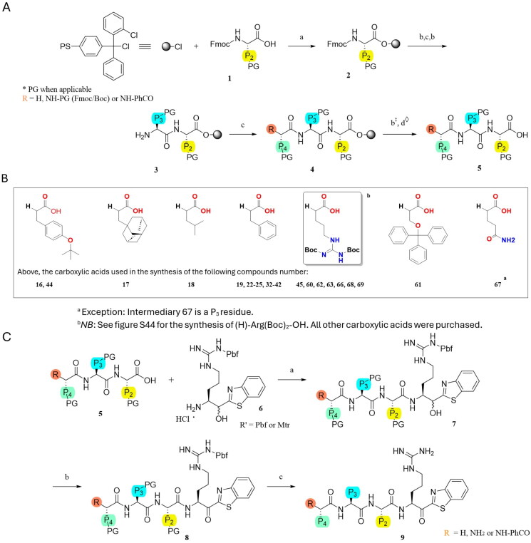
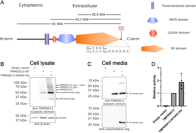
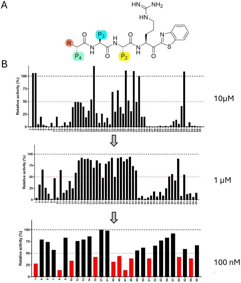
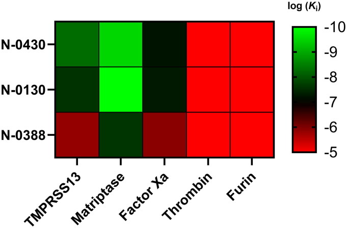
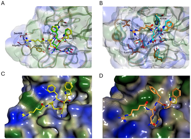
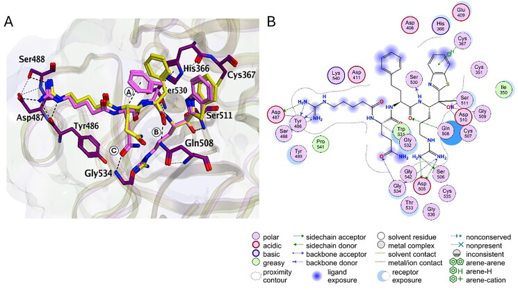
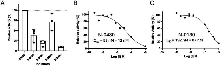
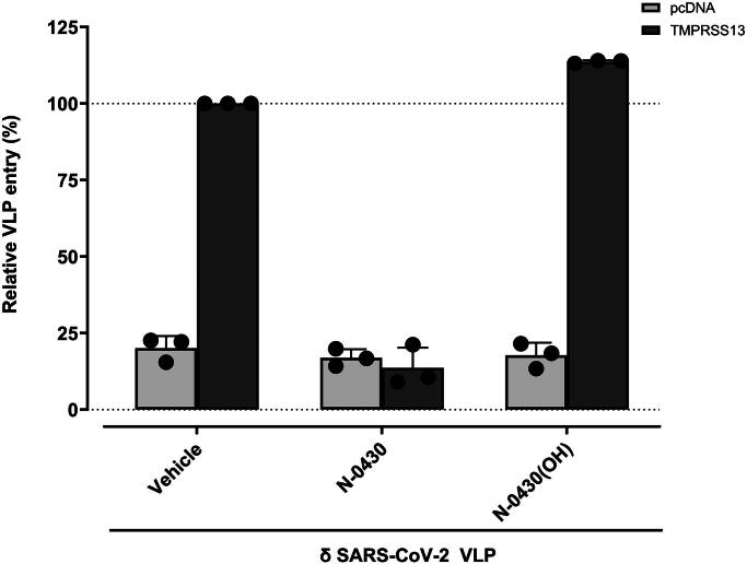
TMPRSS13, a member of the Type II Transmembrane Serine Proteases (TTSP) family, is involved in cancer progression and in respiratory virus cell entry. To date, no inhibitors have been specifically developed for this protease. In this study, a chemical library of 65 ketobenzothiazole-based peptidomimetic molecules was screened against a proteolytically active form of recombinant TMPRSS13 to identify novel inhibitors. Following an initial round of screening, subsequent synthesis of additional derivatives supported by molecular modelling revealed important molecular determinants involved in TMPRSS13 inhibition. One inhibitor, N-0430, achieved low nanomolar affinity towards TMPRSS13 activity in a cellular context. Using a SARS-CoV-2 pseudovirus cell entry model, we further demonstrated the ability of N-0430 to block TMPRSS13-dependent entry of the pseudovirus. The identified peptidomimetic inhibitors and the molecular insights into their potency gained from this study will aid in the development of specific TMPRSS13 inhibitors.

Keywords: Peptidomimetic; SARS-CoV-2; TMPRSS13; compound screening; protease inhibitor.


Figures

Scheme 1.
Scheme 1.
Synthesis. (A) General synthesis of peptide moieties: (a) Fmoc-AA (2 eq.), DIPEA (3 eq.), DCM (b) Piperidine 20%/DMF (c) Fmoc-AA (3 eq.) or R-COOH (3 eq.) (sB), HATU (3 eq.), DIPEA (5 eq.), DMF (d) 20% HFIP/DCM, r.t. (‡: When applicable; ◊: Capping group PhCO was added on the N-terminus part using standard peptide coupling conditions c). (B) Carboxylic acids structures coupled at P4 for the respective compounds. (C) Warhead coupling to the protected peptides: (a) 7 (1,1), HATU (1,1 eq.), DIPEA (5 eq.), DMF (b) DMP (1,5 eq.), DCM, 0 °C (c) TFA/TIPS/H2O (95:2.5:2.5).
Figure 1.
Figure 1.
Schematic representation of TMPRSS13 and expression of active recombinant TMPRSS13 forms in HEK293SL cells. (A) Representation of TMPRSS13. Depicted are the different domains of TMPRSS13, the theoretical molecular weight of specific segments, and a close-up view of the his-tagged mutated sequence. In red are noted the amino acids which are subject to spontaneous cleavage following expression and enrichment in the extracellular media (spectra in supplementary data 1). The theoretical weights of 25.5 kDa for the catalytic domain, 43.1 kDa for the extracellular part and 61 kDa for the total protein are given as an indication of what the weights would be without glycosylation. (B) HEK293SL cells were transfected with pcDNA3.1 (empty vector), the full length TMPRSS13 (TMPRSS13-WT) and the his-tagged TMPRSS13 (TMPRSS13-AFAAS-His). Proteins extracted from cell lysates were separated using 15% SDS-PAGE gels under reducing conditions. The separated proteins were then analysed by Western blotting. Proteins were detected with the rabbit C-terminal TMPRSS13 antibody (PA5-30935), targeting the catalytic region of TMPRSS13, and the mouse anti-β-actin antibody (12262S). (C) HEK293SL cells were transfected with an empty vector, the full length TMPRSS13 (TMPRSS13 WT) and the his-tagged TMPRSS13 (TMPRSS13-AFAAS-His). Media concentrates were separated by SDS-PAGE under reducing conditions using 15% gels and then analysed through Western blotting. Proteins were detected with the rabbit C-terminal TMPRSS13 antibody (PA5-30935), targeting the catalytic region of TMPRSS13, or the rabbit anti-His antibody (2365S). In these western blots, FL means Full length, Glyc means Glycosylated, Phos means phosphorylated, and SP is the serine protease domain. (D) Media samples from transfected HEK193SL cells were assessed for TMPRSS13 enzymatic activity using the Boc-RVRR-AMC substrate (100 µM). TMPRSS13 cleaves after an R-R site, releasing the AMC fluorophore. Fluorescence was measured every minute for 60 min. Results were normalised to WT TMPRSS13 specific activity.
Figure 2.
Figure 2.
Screening of a 65-compound ketobenzothiazole-based library against TMPRSS13. (A) Scaffold of ketobenzothiazole-based inhibitors. P1 is an Arginine, P2, P3, and P4 were modified with different amino acids. In C-terminal position, a ketobenzothiazole, allows trapping of serine of the catalytic pocket. In N-terminal position, R in orange can be NH2, H or NH-PhCO. (B) Compounds that were active at 10 µM were screened at 1 µM then at 100 nM against recombinant TMPRSS13. Compounds affecting > 50% activity were selected for further characterisation.
Figure 3.
Figure 3.
Selectivity heatmap of peptide inhibitors against fives targets. The heatmap displays the selectivity profiles of three compounds (N-0430, N-0130, and N-0388) tested against TMPRSS13, matriptase, Factor Xa, thrombin, and furin. The colour scale on the right ranges from red (log(Ki) = -5) to green (log(Ki) = -10), indicating the logarithmic inhibitory constant (Ki) values for each compound-enzyme pair. Higher log(Ki) values (red) represent weaker inhibition, while lower log(Ki) values (green) indicate stronger inhibition. Data are available in Table S2.
Figure 4.
Figure 4.
Molecular modelling. (A) Docking of N-0130 ((H)RQFR-Kbt, yellow for compound and dark yellow for ligands) and N-0388 ((H)QFR-Kbt, green for compound and dark green for ligands) to TMPRSS13. (B) Docking of N-0130 ((H)RQFR-Kbt, orange) and N-0388 ((H)QFR-Kbt, cyan) to matriptase. (C) Docking of N-0130 ((H)RQFR-Kbt, yellow) in TMPRSS13. (D) Docking of N-0130 ((H)RQFR-Kbt, orange) in Matriptase. For all panels, hydrophilic part of inhibitor is depicted in blue, hydrophobic in green, and residues involved in interactions are illustrated with dashed lines with each inhibitor are represented in stick form and labelled.
Figure 5.
Figure 5.
Representation of the interactions between N-0430 and TMPRSS13. Representations are in 3D (A) and 2D (B). N-0430 is shown in light pink stick mode and TMPRSS13 shown in purple stick mode. For comparison, N-0130 is superimposed and represented in yellow stick.
Figure 6.
Figure 6.
Evaluation of the potency in a cellular context. Vero cells were transfected with WT TMPRSS13, and activity was monitored using Boc-RVRR-AMC as a fluorogenic. (A) Relative activity of TMPRSS13 in the presence of 10 µM of compounds (N-0159, N-0430, N-0130, N-0388 and N-0430-OH) or vehicles (DMSO), displaying relative activity as a percentage of DMSO. (B) Relative activity of TMPRSS13 as a function of N-0430 concentration. (C) Relative activity of TMPRSS13 as a function of N-0130 concentration.
Figure 7.
Figure 7.
δ SARS-CoV-2 VLPs entry in VERO E6 cells transfected with TMPRSS13 in presence of N-0430, N-0430(OH) and vehicles. VLPs infection in the presence of 10 µM of N-0430 or N-0430(OH) relative to the vehicle-treated condition (DMSO condition). Cells were transfected with TMPRSS13 WT 24 h before incubation with VLP. Compounds were added 3 h before and during the 72 h VLP incubation on cells. The background was subtracted from all particles using a VLP without surface glycoprotein for both experiments. Each experiment was repeated at least three times with vehicle condition, N-0430 and N-0430(OH), with five technical replicates per biological replicate.

Update of

Similar articles

Cited by

References

    1. Patel S. A critical review on serine protease: Key immune manipulator and pathology mediator. Allergol Immunopathol (Madr). 2017;45(6):579–591. - PMC - PubMed
    1. Antalis TM, Bugge TH, Wu Q.. Membrane-anchored serine proteases in health and disease. Prog Mol Biol Transl Sci. 2011;99:1–50. - PMC - PubMed
    1. Szabo R, Bugge TH.. Membrane-anchored serine proteases as regulators of epithelial function. Biochem Soc Trans. 2020;48(2):517–528. - PMC - PubMed
    1. Webb SL, Sanders AJ, Mason MD, Jiang WG.. Type II transmembrane serine protease (TTSP) deregulation in cancer. Front Biosci. 2011;16(2):539–552. - PubMed
    1. Bugge TH, Antalis TM, Wu Q.. Type II transmembrane serine proteases. J Biol Chem. 2009;284(35):23177–23181. - PMC - PubMed

MeSH terms

LinkOut - more resources