. 2025 Sep;254(9):1055-1067.
doi: 10.1002/dvdy.70004. Epub 2025 Feb 20.

β-Catenin localization in the ctenophore Mnemiopsis leidyi suggests an ancestral role in cell adhesion and nuclear function

Affiliations

β-Catenin localization in the ctenophore Mnemiopsis leidyi suggests an ancestral role in cell adhesion and nuclear function

Brian M Walters et al. Dev Dyn. 2025 Sep.

Abstract

Background: The emergence of multicellularity in animals marks a pivotal evolutionary event, which was likely enabled by molecular innovations in the way cells adhere and communicate with one another. β-Catenin is significant to this transition due to its dual role as both a structural component in the cadherin-catenin complex and as a transcriptional coactivator involved in the Wnt/β-catenin signaling pathway. However, our knowledge of how this protein functions in ctenophores, one of the earliest diverging metazoans, is limited.

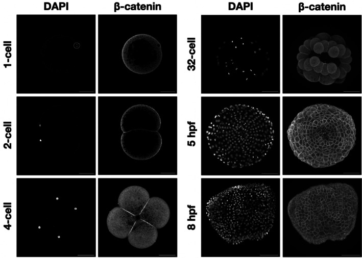
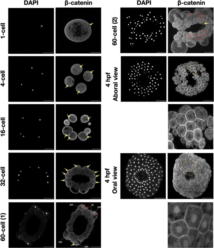
Results: To study β-catenin function in the ctenophore Mnemiopsis leidyi, we generated affinity-purified polyclonal antibodies targeting Mlβ-catenin. We then used this tool to observe β-catenin protein localization in developing Mnemiopsis embryos. In this article, we provide evidence of consistent β-catenin protein enrichment at cell-cell interfaces in Mnemiopsis embryos. Additionally, we found β-catenin enrichment in some nuclei, particularly restricted to the oral pole around the time of gastrulation. The Mlβ-catenin affinity-purified antibodies now provide us with a powerful reagent to study the ancestral functions of β-catenin in cell adhesion and transcriptional regulation.

Conclusions: The localization pattern of embryonic Mlβ-catenin suggests that this protein had an ancestral role in cell adhesion and may have a nuclear function as well.

Keywords: Wnt signaling; cellfate specification; cell‐adhesion; ctenophore; fate specification; mnemiopsis; β‐catenin.


Figures

FIGURE 1
FIGURE 1
The dual role of β‐catenin in cell adhesion and transcriptional activation. APC, Adenomatous Polyposis Coli; CK1α, Casein Kinase 1α; Dsh, Disheveled; GSK‐3β, Glycogen Synthase Kinase 3β; LEF, Lymphoid Enhancer Factor; LRP, Low‐density lipoprotein Receptor‐related Protein; TCF, T‐Cell Factor; β‐cat, β‐catenin; α‐cat, α‐catenin.
FIGURE 2
FIGURE 2
The predicted β‐catenin architecture in M. leidyi. (A) Mlβ‐cat protein contains 11 of the 12 Arm domains identified with high confidence flanked by an N‐Terminal and a C‐terminal region. The asterisk above domain 7 indicates predicted partial (2 out of 3 alpha helices) domain conservation. The amino acids necessary for the interaction with GSK3β and CK1α are conserved, as well as the α‐cat binding site. The two crucial lysines (K), essential for the interaction with a classical cadherin, are also conserved. (B) Predicted tertiary structure of Mlβ‐cat protein with Arm domains denoted by color. Red through pink domains were identified through domain prediction software; silver, gray, and white domains fall within the Arm repeat superfamily but were identified manually. The blue Arm domain (domain 7) was identified with low confidence likely due to the missing alpha helix. (C) pLDDT plot for predicted structure. A dip in confidence values for residues between 472 and 478 corresponds with a disordered region that truncates an alpha helix in Arm domain 7.
FIGURE 3
FIGURE 3
Mlβ‐cat antibody specifically recognizes endogenous protein. (A) Western blot analysis of M. leidyi cell lysates using affinity‐purified anti‐Mlβ‐cat antibodies. The specificity of the antibody was tested against whole‐cell lysates from both M. leidyi embryos and adults. A single band slightly larger than the expected size (~100 kDa) was detected for both samples. (B) Immunostaining of M. leidyi embryos using affinity‐purified anti‐Mlβ‐cat polyclonal antibodies. The top panels show nuclei stained by DAPI; the bottom panels show results of Mlβ‐cat staining with and without primary antibody (negative control). Images are maximal projection. Scale bar, 50 μM.
FIGURE 4
FIGURE 4
Mlβ‐catenin localizes at cell–cell contacts during M. leidyi embryonic development. Immunostaining of M. leidyi embryos at different stages using affinity‐purified β‐cat antibodies. The left column panels exhibit nuclei stained by DAPI; the right column panels exhibit results of Mlβ‐cat antibody staining. Images for 1 and 2‐cell stages are z‐stack projected images. All other images are maximal projection. Note that enriched localization of β‐catenin only occurs at cell–cell contacts. Scale bar, 50 μM.
FIGURE 5
FIGURE 5
The localization of β‐catenin in M. leidyi embryos. Immunostaining of M. leidyi embryos at different stages using affinity‐purified β‐cat antibodies. The left columns show nuclei stained by DAPI, the right columns show the results of Mlβ‐cat antibody staining. Zoomed‐in insets (dotted boxes) for both the oral and aboral 4 hpf are included below the respective samples. The 1‐cell, 4‐cell, 16‐cell, 32‐cell, 60‐cell (1), and 4 hpf aboral view are z‐stack projected images. The 60‐cell (2) and 4 hpf oral views are maximal projection images. The yellow arrows show the absence of β‐catenin nuclear translocation; while the red arrows indicate β‐catenin nuclear translocation. Scale bar, 50 μM.

References

    1. Richter DJ, King N. The genomic and cellular foundations of animal origins. Annu Rev Genet. 2013;47:509‐537. doi: 10.1146/annurev-genet-111212-133456 - DOI - PubMed
    1. Ros‐Rocher N, Pérez‐Posada A, Leger MM, Ruiz‐Trillo I. The origin of animals: an ancestral reconstruction of the unicellular‐to‐multicellular transition. Open Biol. 2021;11(2):200359. doi: 10.1098/rsob.200359 - DOI - PMC - PubMed
    1. Shapiro L. The multi‐talented β‐catenin makes its first appearance. Structure. 1997;5(10):1265‐1268. doi: 10.1016/S0969-2126(97)00278-5 - DOI - PubMed
    1. Valenta T, Hausmann G, Basler K. The many faces and functions of Î 2‐catenin. EMBO J. 2012;31(12):2714‐2736. doi: 10.1038/emboj.2012.150 - DOI - PMC - PubMed
    1. Nollet F, Kools P, Van Roy F. Phylogenetic analysis of the cadherin superfamily allows identification of six major subfamilies besides several solitary members. J Mol Biol. 2000;299(3):551‐572. doi: 10.1006/jmbi.2000.3777 - DOI - PubMed

LinkOut - more resources