. 2025 Feb 20;167(1):50.
doi: 10.1007/s00701-025-06432-8.

How I do it: surgical techniques for vagus nerve stimulation in pediatric drug-resistant epilepsy

Affiliations

How I do it: surgical techniques for vagus nerve stimulation in pediatric drug-resistant epilepsy

Santiago Ezequiel Cicutti et al. Acta Neurochir (Wien). .

Abstract

Background: Vagus nerve stimulation (VNS) is a neuromodulatory treatment for refractory epilepsy, involving electrical stimulation of the vagus nerve via a subcutaneously implanted pulse generator.

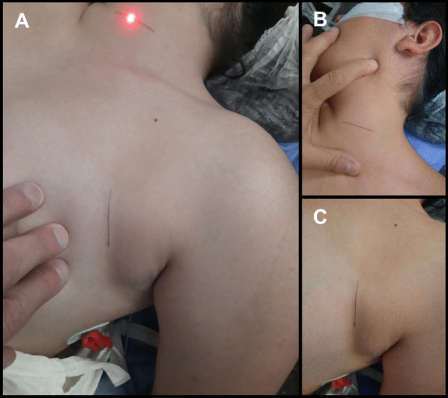
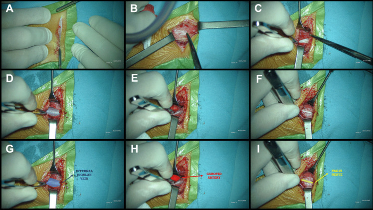
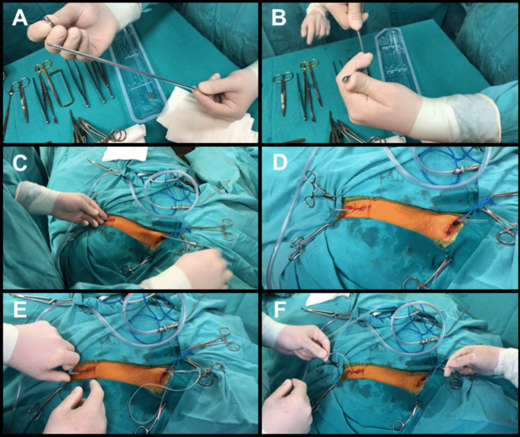
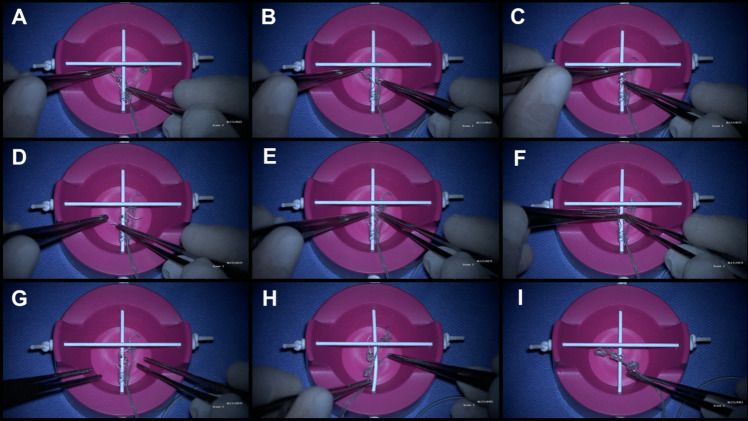
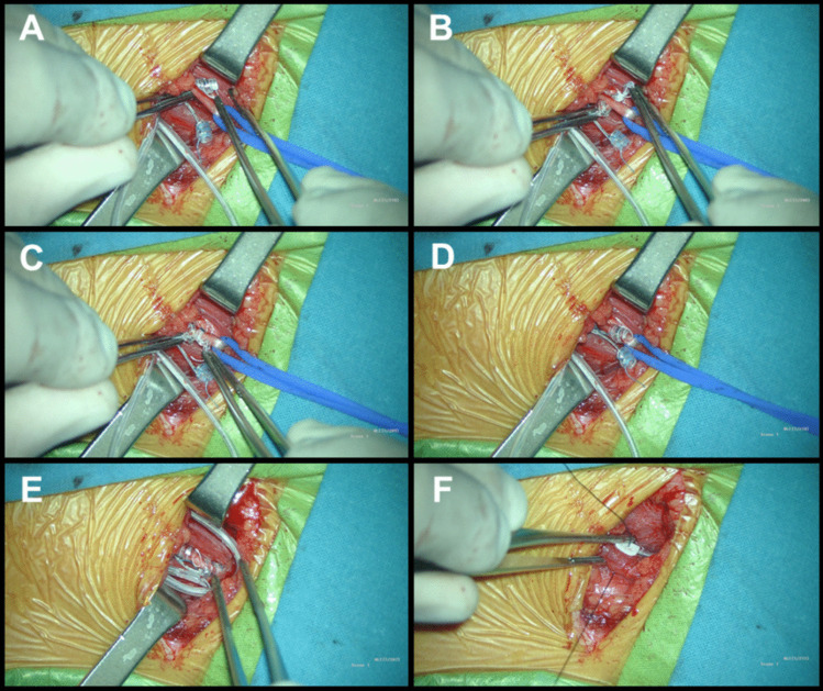
Method: We present a step-by-step description of the VNS implantation technique, incorporating tips and tricks from an epilepsy surgeon with over 20 years of experience (MB).

Conclusion: Proper patient selection, meticulous surgical technique, and comprehensive postoperative care, supported by educational resources such as surgical technique articles, videos, and simulators, are crucial for minimizing complications and optimizing patient outcomes.

Keywords: Drug-resistant epilepsy; Neurosurgery; Surgical technique; Vagus nerve stimulation.


Conflict of interest statement

Declarations. Ethical approval: All procedures performed in studies involving human participants were in accordance with the Declaration of Helsinki and adhered to the Good Clinical Practice Guidelines (Resolution 1480/11) of the Argentine Ministry of Health. Furthermore, approval for the protocol was obtained by the Ethics and Research Committee of "Juan P. Garrahan" Pediatric Hospital. Informed consent: Informed consent was obtained from all individual participants included in the study. Competing interests: The authors declare no competing interests.

Figures

Fig. 1
Fig. 1
Patient Positioning and incisions: A The patient is positioned supine on the operating room table with the head on a donut pillow or a headrest, extended and turned, approximately, 30 degrees to the right. The neck is extended with a shoulder roll in order to ease the passage of the tunneling rod. B 2–3 cm transverse incision in a skin fold on the left side of the neck, located between the mastoid and the clavicle, extending to the medial border of the sternocleidomastoid muscle. C 2.5 to 3 cm-long incision following a skin crease line is performed, approximately 5 cm below the clavicle and 5 cm medially to the shoulder
Fig. 2
Fig. 2
Neck incision and Vagus Nerve Dissection: A 2–3 cm transverse incision in a skin fold on the left side of the neck, located between the mastoid and the clavicle, extending to the medial border of the sternocleidomastoid muscle. B The subcutaneous dissection exposes the platysma, which can be divided following the directions of its fibers. C Muscle plane showing the omohyoid and other strap muscles inferiorly and medially, whereas the sternocleidomastoid is located laterally. D and G The carotid sheath is opened, showing the internal jugular vein laterally. E and H The carotid sheath is opened, showing the carotid artery medially. F and I The vagus nerve is located between the carotid artery medially and the internal jugular vein laterally
Fig. 3
Fig. 3
Subcutaneous tunneling: A Manufacturer-supplied tunneling device, which includes an inner and an outer tunneling tube, and a bullet tip threaded onto the end of the inner tube. B Curvature of the tunneler to optimize the trajectory. C Tunneling from the neck incision towards the axillary incision. D The bullet tip is removed and the inner shaft is withdrawn, leaving the clear hollow sheath. E The free end of the electrode is inserted in the sheath and drawn, with the sheath, from the neck to the chest incision. F Electrode tunneled from the neck incision to the chest incision
Fig. 4
Fig. 4
Simulator: A The electrode is positioned on the right side, passing underneath the nerve. B Once positioned beneath the nerve, forceps are used to reach the suture at the end of the helix, while the other forceps holds the opposite end of the suture on the right side of the nerve. Subsequently, controlled tension is applied to open the helices in the direction of the stimulator’s rotation, ensuring a precise fit on the nerve. C The central turn of the spiral is placed first. D By pulling the lower suture, the lower helix is placed around the nerve. E and F The upper helix is passed under the nerve, following the curvature of the electrode. G Electrode correctly positioned. H A common mistake is attempting to place the helices from the left side of the vagus nerve. Since the system is designed for right-handed surgeons, starting from the left side makes the placement very challenging, if not impossible. I Another common mistake: helices tangled with each other, making placement impossible. They should be stretched before placing them on the nerve
Fig. 5
Fig. 5
Electrode placement: A The electrode is positioned on the right side, passing underneath the nerve. B Once positioned beneath the nerve, forceps are used to reach the suture at the end of the helix, while the other forceps holds the opposite end of the suture on the right side of the nerve. Subsequently, controlled tension is applied to open the helices in the direction of the stimulator’s rotation, ensuring a precise fit on the nerve. C and D In applying each electrode, the central part of the spiral must be positioned first, and then the upper and lower turns are wound around the nerve. The tethering (inferior) anchor is secured around the vagus nerve, and then the positive (middle helical contact) and the negative (upper helical contact) electrodes are positioned. E After the electrodes have been positioned on the nerve, it is necessary to make redundant loops so that movements of the neck do not dislodge the electrodes or put strain on the vagus nerve itself. F The electrode is secured by tacking nonabsorbable sutures to silicon holders at the deep cervical fascia

Similar articles

References

    1. Aalbers MW, Rijkers K, Klinkenberg S, Majoie M, Cornips EM (2015) Vagus nerve stimulation lead removal or replacement: surgical technique, institutional experience, and literature overview. Acta Neurochir (Wien) 157(11):1917–1924. 10.1007/s00701-015-2547-9 - PMC - PubMed
    1. Ansari S, Chaudhri K, Al Moutaery KA (2007) Vagus nerve stimulation: indications and limitations. Acta Neurochir Suppl 97(Pt 2):281–286. 10.1007/978-3-211-33081-4_31 - PubMed
    1. Boon P, De Herdt V, Vonck K, Van Roost D (2007) Clinical experience with vagus nerve stimulation and deep brain stimulation in epilepsy. Acta Neurochir Suppl 97(Pt 2):273–280. 10.1007/978-3-211-33081-4_30 - PubMed
    1. Boon P, Vonck K, Vandekerckhove T et al (1999) Vagus nerve stimulation for medically refractory epilepsy; efficacy and cost-benefit analysis. Acta Neurochir (Wien) 141(5):447–453. 10.1007/s007010050324 - PubMed
    1. Cicutti SE, Gromadzyn GP, Cuello JF, Bartuluchi M (2024) Vagus nerve stimulation in pediatric patients with drug-resistant epilepsy: a step-by-step video. Neurosurg Focus Video 11(1):V2. 10.3171/2024.4.FOCVID244. - PMC - PubMed

LinkOut - more resources