. 2025 Feb 3;66(2):53.
doi: 10.1167/iovs.66.2.53.

Depth and Strain-Dependent Structural Responses of Mouse Lens Fiber Cells During Whole Lens Shape Changes

Affiliations

Depth and Strain-Dependent Structural Responses of Mouse Lens Fiber Cells During Whole Lens Shape Changes

Sepideh Cheheltani et al. Invest Ophthalmol Vis Sci. .

Abstract

Purpose: To study the relationship between whole lens shape changes and fiber cell responses to externally applied loads.

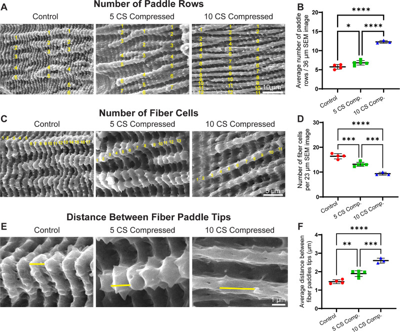
Methods: Freshly dissected mouse lenses were compressed by applying glass coverslips to the lens anterior, followed by fixation to preserve lens shape, and preparation for scanning electron microscopy (SEM). SEM images were collected from the outer cortex to the nucleus, and fiber cell end-to-end curvature and membrane paddle dimensions were measured using ImageJ.

Results: At 23% and 29% axial strain, cortical fiber bundle curvature increased significantly compared to control uncompressed lenses, whereas nuclear fiber bundle curvature was unaffected. Outer cortical fiber cell membrane paddles and protrusions were dramatically distorted in a radial direction, with loss of paddle-associated small protrusions in compressed lenses compared to controls, but nuclear fiber cell morphologies were unchanged. The compression-induced increases in cortical fiber cell curvature and distortion of membrane paddles were reversible, with fiber cell morphologies returning to those of control lenses after the release of load.

Conclusions: Whole lens shape changes due to an increase in axial strain result in increased fiber cell curvature and distorted membrane morphologies in cortical but not nuclear fiber cells, indicating that mechanical strain dissipates with depth. The recovery of normal cortical fiber cell curvature and membrane morphologies after the removal of load and lens rounding back to its original shape suggests elastic properties of the young and mature fiber cells and their membrane paddles.


Conflict of interest statement

Disclosure: S. Cheheltani, None; M.H. Shihan, None; J. Parreno, None; S.K. Biswas, None; W-K. Lo, None; V.M. Fowler, None

Figures

Figure 1.
Figure 1.
Cortical fiber cells of compressed lenses show an increase in curvature compared to control and recovered lenses. (A) SEM images of lens halves for control, compressed lenses (5 CS and 10 CS), and recovered lenses (5 CS recovered, 10 CS recovered). The blue curved lines indicate the contours of cortical and nuclear fibers. Average curvature of (B, C) cortical fiber cells and (D, E) nuclear fiber cells measured using ImageJ for control lenses, 5 CS and 10 CS compressed (Comp.), and recovered lenses. Scale bar:1 mm. Graphs represent mean ± SEM of two measurements per lens from three biological replicates per condition. **P ≤ 0.01; ***P ≤ 0.001; ****P ≤ 0.0001.
Figure 2.
Figure 2.
Representative SEM images of lens fiber cell morphologies in control lenses, lenses under compression and recovered lenses. (A) Low magnification images of lens halves. Yellow box indicates regions of high magnification images for panel B. Scale bar:1 mm. (B) High magnification images of regions at 100 µm from the surface in the lens cortex up to 600 µm from the surface in the lens nucleus. Scale bar: 10 µm.
Figure 3.
Figure 3.
Cortical and mature fiber cells exhibit structural changes under high axial strain (5 and 10 coverslips) compared to control lenses. (A) Representative SEM images of control, 5 CS compressed, and 10 CS compressed lenses from cortical fiber cells (left) to lens nucleus (right). (B) The magnified region cropped out of images in panel A for a better representation of the fiber cell microstructures. The outer cortical fibers in both 5 CS and 10 CS compressed lenses are significantly distorted (green boxes), with mature fiber cells also affected in 10 CS compressed lenses (pink boxes). However, the membrane ridge and groove structures (orange arrowheads) in fiber cells near the nucleus (yellow boxes) remain unchanged across all conditions. Scale bars: 1 mm, 5 µm, and 1 µm.
Figure 4.
Figure 4.
Compressed lenses exhibit altered fiber cell morphology compared to control lenses. (A, B) Average number of paddle rows per 36 µm radial distance of SEM images for control, 5 CS, and 10 CS compressed lenses. (C, D) Number of fiber cells in a 23 µm radial distance of SEM image of control, 5 CS, and 10 CS compressed lenses. (E, F) Distance between paddle tips of adjacent fiber cells in control, 5 CS, and 10 CS compressed lenses. Graphs represent mean ± SEM of measurements from at least three biological replicates per condition. Scale bars: (A) 10 µm, (C) 5 µm, and (E) 1 µm. *P < 0.05; **P < 0.01; ***P <0.001; ****P < 0.0001.
Figure 5.
Figure 5.
The fiber cell morphology is normal in recovered lenses. (A, B) Average number of paddle rows per 36 µm radial distance of SEM images for control, 5 CS, and 10 CS recovered lenses. (C, D) Number of fiber cells in a 23 µm radial distance of SEM images for control, 5 CS and 10 CS recovered lenses. (E, F) Distance between paddle tips of adjacent fiber cells in control, 5 CS, and 10 CS recovered lenses. Graphs represent mean ± SEM of measurements from at least three biological replicates per condition. Scale bars: (A) 10 µm, (C) 5 µm, and (E) 1 µm.
Figure 6.
Figure 6.
Diagram of fiber cell morphology in compressed lenses. (A) The schematic (created with biorender.com) illustrates that lenses become increasingly flattened with higher axial strain compared to control lenses, whereas the nuclear shape remains consistent across all conditions. Arrows on images indicate the direction of lens shape changes due to axial compression (vertical arrows) and equatorial expansion (horizontal arrows). (B) The cortical fibers in control lenses display undulating paddle domains with finger-like protrusions. As axial strain increases (5 CS), the fiber cells stretch radially, causing the paddles to appear smoother with more rounded protrusions. Under even greater strain (10 CS), the paddles adopt a narrower shape with reduced protrusions.

References

    1. Hejtmancik JF, Shiels A.. Overview of the lens. Prog Mol Biol Transl Sci. 2015; 134: 119–127. - PMC - PubMed
    1. Heys KR, Cram SL, Truscott RJ.. Massive increase in the stiffness of the human lens nucleus with age: the basis for presbyopia? Mol Vis. 2004; 10: 956–963. - PubMed
    1. Fisher RF. Elastic properties of the human lens. Exp Eye Res. 1971; 11: 143. - PubMed
    1. Glasser A, Campbell MC.. Presbyopia and the optical changes in the human crystalline lens with age. Vision Res. 1998; 38: 209–229. - PubMed
    1. Glasser A, Campbell MC.. Biometric, optical and physical changes in the isolated human crystalline lens with age in relation to presbyopia. Vision Res. 1999; 39: 1991–2015. - PubMed

LinkOut - more resources