. 2025 Feb 20;21(2):e1012904.
doi: 10.1371/journal.ppat.1012904. eCollection 2025 Feb.

Isolation of a novel human prion strain from a PRNP codon 129 heterozygous vCJD patient

Affiliations

Isolation of a novel human prion strain from a PRNP codon 129 heterozygous vCJD patient

Fuquan Zhang et al. PLoS Pathog. .

Abstract

The epizootic prion disease of cattle, bovine spongiform encephalopathy (BSE), caused variant Creutzfeldt-Jakob disease (vCJD) in humans following dietary exposure. Codon 129 polymorphism of the human prion protein gene (PRNP), encoding either methionine (M) or valine (V), dictates the propagation of distinct human prion strains and up to now all but one neuropathologically confirmed vCJD patients have had a 129MM genotype. Concordant with this genetic association, transgenic modelling has established that human PrP 129V is incompatible with the vCJD prion strain and that depending on codon 129 genotype, primary human infection with BSE prions may, in addition to vCJD, result in sporadic CJD-like or novel phenotypes. In 2016 we saw the first neuropathologically confirmed case of vCJD in a patient with a codon 129MV genotype. This patient's neuropathology and molecular strain type were pathognomonic of vCJD but their clinical presentation and neuroradiological features were more typical of sporadic CJD, suggestive of possible co-propagation of another prion strain. Here we report the transmission properties of prions from the brain and lymphoreticular tissues of the 129MV vCJD patient. Primary transmissions into transgenic mice expressing human PrP with different codon 129 genotypes mainly produced neuropathological and molecular phenotypes congruent to those observed in the same lines of mice challenged with prions from 129MM vCJD patient brain, indicative that the vCJD prion strain was the dominant propagating prion strain in the patient's brain. Remarkably however, some transgenic mice challenged with 129MV vCJD patient brain propagated a novel prion strain type which at secondary passage was uniformly lethal in mice of all three PRNP codon 129 genotypes after similar short mean incubation periods. These findings establish that cattle BSE prions can trigger the co-propagation of distinct prion strains in humans.


Conflict of interest statement

I have read the journal's policy and the authors of this manuscript have the following competing interests: JC is a Director and shareholder and JW is a shareholder of D-Gen Limited, an academic spin-out company (co-owners include MRC, UCL, Imperial College and the Wellcome Trust) working in the field of prion disease diagnosis, decontamination, and therapeutics. The other authors have declared no competing interests.

Figures

Fig 1
Fig 1. Prion transmission rates and findings in transgenic and wild-type mice challenged with 129MV vCJD patient frontal cortex.
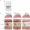
(A) Overview of transmission rates and molecular and neuropathological findings in recipient mice. Mice were intracerebrally inoculated with 1% (w/v) homogenate prepared from 129MV vCJD patient frontal cortex. Total attack rate is defined as the total number of clinically affected and subclinically infected mice as a proportion of the number of inoculated mice after post-inoculation periods extending to 600 days. Subclinical prion infection was assessed by immunohistochemical examination of brain for abnormal PrP deposition and western blot analyses of mouse brain homogenates for detectable PrPSc (by direct western blotting or after NaPTA precipitation if required). The type of PrPSc (PrPSc types 4, 5 and 3*; T4, T5, T3*) seen in infected mice is reported together with mean incubation periods (Mean IP) for clinically affected mice in days (where n ≥ 3 the mean ± SEM is reported otherwise individual incubation times are given). (B) PrP deposition patterns in infected transgenic mouse brains by IHC using anti-PrP monoclonal antibody 3F4. Upper panels show schematic drawings in which the overall spatial distribution and intensity of abnormal PrP deposition is indicated by red shading with PrP plaques shown as red dots. Lower panels show representative PrP IHC in hippocampus (including corpus callosum) and cortex. Scale bar, 300 µm for upper row (hippocampus) and 30 µm for lower row (cortex).
Fig 2
Fig 2. PrPSc types seen in the brain of transgenic mice after primary transmission of 129MV vCJD patient frontal or parietal cortex.
(A) Western blot of proteinase K-digested 10% (w/v) brain homogenates from transgenic mice after challenge with frontal cortex from the 129MV vCJD patient using anti-PrP monoclonal antibody 3F4 and high sensitivity enhanced chemiluminescence. The propagated PrPSc types are shown below each lane. Lane 1, 129MM Tg35c brain, lane 2, 129VV Tg152c brain, lane 3, 129VV Tg152c brain, lane 4, 129MV Tg35c/Tg152c brain. (B) Ratios of the di- and mono-glycosylated protease-resistant PrP glycoforms in PrPSc from transgenic mouse brain after challenge with frontal or parietal cortex from the 129MV vCJD patient in comparison to PrPSc from the 129MV vCJD patient’s frontal cortex and reference cases of frontal cortex from patients with sporadic CJD (PrPSc types 1, 2 or 3; London classification, [26]) and 129MM vCJD patients with type 4 PrPSc. Where sample size is ≥ 3 symbols show mean percentage ± SEM otherwise individual samples are shown. In some cases the error bars are smaller than the symbols used. Squares, human samples, circles, mouse primary transmission samples.
Fig 3
Fig 3. Molecular and neuropathological findings in wild-type FVB/N mice challenged with frontal cortex from vCJD patients with 129MM and 129MV genotypes.
(A) Western blot of proteinase K-digested 10% (w/v) brain homogenates from FVB/N mice using anti-PrP monoclonal antibody 6D11 and high sensitivity enhanced chemiluminescence. Lanes 1 and 2, FVB/N mouse brains after challenge with 129MM vCJD patient frontal cortex from reference cases 1 and 2 respectively, lane 3, FVB/N mouse brain after challenge with 129MV vCJD patient frontal cortex. (B) PrP deposition patterns by IHC in the same transgenic mouse brains shown in panel A using anti-PrP monoclonal antibody 6D11. Upper panels show schematic drawings in which the overall spatial distribution and intensity of abnormal PrP deposition is indicated by red shading with PrP plaques shown as red dots. Lower panels show representative PrP immunohistochemistry in cortex, hippocampus and midbrain. Scale bar 300 µm for upper two rows (cortex and hippocampus) and 60 µm for lower row (midbrain).
Fig 4
Fig 4. Prion transmission rates and findings in transgenic and wild-type mice challenged with 129MV vCJD patient parietal cortex, thalamus or cerebellum.
(A-C) Mice were intracerebrally inoculated with 1% (w/v) homogenate prepared from 129MV vCJD patient parietal cortex (A), thalamus (B) or cerebellum (C). Total attack rate is defined as the total number of clinically affected and subclinically infected mice as a proportion of the number of inoculated mice after post-inoculation periods extending to 600 days. Subclinical prion infection was assessed by immunohistochemical examination of brain for abnormal PrP deposition and western blot analyses of mouse brain homogenates for detectable PrPSc (by direct western blotting or after NaPTA precipitation if required). The type of PrPSc (PrPSc types 4, 5 and 3*; T4, T5, T3*) seen in infected mice is reported together with mean incubation periods (Mean IP) for clinically affected mice in days (where n ≥ 3 the mean ± SEM is reported otherwise individual incubation times are given). NA, not applicable.
Fig 5
Fig 5. Prion transmission rates and findings in transgenic mice challenged with 129MM Tg35c-passaged 129MV vCJD patient frontal cortex.
(A) Overview of transmission rates and molecular and neuropathological findings in recipient mice. Mice were intracerebrally inoculated with 1% (w/v) homogenate prepared from a 129MM Tg35c mouse brain propagating type 4 PrPSc derived from primary transmission of 129MV vCJD patient frontal cortex (shown in Figs 1 and 2). Total attack rate is defined as the total number of clinically affected and subclinically infected mice as a proportion of the number of inoculated mice after post-inoculation periods extending to 600 days. Subclinical prion infection was assessed by immunohistochemical examination of brain for abnormal PrP deposition. The phenotype in infected mice (T4, T5) is reported together with mean incubation period (Mean IP) for clinically affected mice in days (where n ≥ 3 the mean ± SEM is reported). NA, not applicable. (B) PrP deposition patterns in infected transgenic mouse brains by IHC using anti-PrP monoclonal antibody 3F4. Panels show schematic drawings in which the overall spatial distribution and intensity of abnormal PrP deposition is indicated by red shading with PrP plaques shown as red dots.
Fig 6
Fig 6. Primary and secondary transmission of 129MV vCJD patient frontal cortex in 129MM Tg35c mice.
(A) Western blot of proteinase K-digested 10% (w/v) brain homogenates using anti-PrP monoclonal antibody 3F4 and high sensitivity enhanced chemiluminescence. The type of PrPSc seen in infected brain is reported below each lane. Lane 1, reference case of 129MM vCJD patient frontal cortex, lane 2, 129MM Tg35c mouse brain from primary transmission of 129MV vCJD patient frontal cortex, lane 3, 129MM Tg35c mouse brain challenged with 129MM Tg35c-passaged 129MV vCJD patient frontal cortex. (B) PrP deposition patterns in infected 129MM Tg35c mouse brains by IHC using anti-PrP monoclonal antibody 3F4. Upper panels show schematic drawings in which the overall spatial distribution and intensity of abnormal PrP deposition is indicated by red shading with PrP plaques shown as red dots. Lower panels show representative haematoxylin- and eosin-stained sections (H&E) showing spongiform neurodegeneration including florid plaques in the cortex and representative PrP IHC (PrP) with anti-PrP monoclonal antibody 3F4 in cortex, thalamus and midbrain. Scale bar, all lower panels, 100 µm.
Fig 7
Fig 7. Summary of secondary prion transmissions in transgenic and wild-type mice.
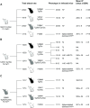
(A-C) Mice were intracerebrally inoculated with 1% (w/v) brain homogenate prepared from prion-infected transgenic mice challenged with 129MV vCJD patient frontal cortex. Patterns of abnormal PrP deposition by IHC and PrPSc types in the infected mice from which the inocula were derived are shown in Figs 1B and 2A, respectively. Total attack rate is defined as the total number of clinically affected and subclinically infected mice as a proportion of the number of inoculated mice after post-inoculation periods extending to 600 days. Subclinical prion infection was assessed by immunohistochemical examination of brain for abnormal PrP deposition and western blot analyses of mouse brain homogenates for detectable PrPSc (by direct western blotting or after NaPTA precipitation if required). The type of PrPSc (PrPSc types 2, 4, 5 and 3*, T2, T4, T5, T3*) seen in infected mice is reported together with mean incubation periods (Mean IP) for clinically affected mice in days (where n ≥ 3 the mean ± SEM is reported otherwise individual incubation times are given). NA, not applicable.
Fig 8
Fig 8. PrPSc types and glycoform ratios in transgenic mice from secondary transmissions.
(A) Western blot of proteinase K-digested 10% (w/v) brain homogenates from transgenic mice or human reference cases of sporadic CJD (sCJD) (PrPSc type 2 129MM and type 3 129MV) using anti-PrP monoclonal antibody 3F4 and high sensitivity enhanced chemiluminescence. The propagated PrPSc type (PrPSc types 2, 3, 3*) is shown below each lane. Lanes 1, 3, 6 and 8, sCJD brain. Lane 2, PrPSc type 3*-positive 129VV Tg152c brain from primary transmission of 129MV vCJD patient frontal cortex. Lanes 4, 5 and 7, secondary transmissions of PrPSc type 3*-positive 129VV Tg152c brain to further transgenic mice; lane 4, 129MM Tg35c brain, lane 5, 129VV Tg152c brain, lane 7, 129MV Tg35c/Tg152c brain. (B) Ratios of the di- and mono-glycosylated protease-resistant PrP glycoforms seen in PrPSc from the brains of transgenic mice, 129MV vCJD patient frontal cortex, 129MM vCJD patient frontal cortex or reference cases of sCJD patient frontal cortex with PrPSc types 1, 2 or 3 (London classification, [26]). Where sample size is ≥ 3 symbols show mean percentage ± SEM otherwise individual samples are shown. In some cases the error bars are smaller than the symbols used. Squares, human samples, circles, mouse primary transmission samples, triangles, mouse secondary transmission samples.
Fig 9
Fig 9. Abnormal PrP deposition in the brain of transgenic mice propagating type 3* PrPSc.
The provenance of the brain sample is designated above each column and the type of PrPSc (PrPSc type 3*; T3*) seen in infected brain is designated below. Upper panels show schematic drawings in which the overall spatial distribution and intensity of abnormal PrP deposition is indicated by red shading with PrP plaques shown as red dots. Lower panels show representative PrP IHC in cortex, hippocampus (including corpus callosum), thalamus and brain stem using anti-PrP monoclonal antibody 3F4. Scale bar, 150 µm for row 1 (cortex) and 300 µm for rows 2 (hippocampus), 3 (thalamus), and 4 (brain stem).
Fig 10
Fig 10. Prion transmission rates and findings in transgenic and wild-type mice challenged with 129MV vCJD patient appendix and mesenteric lymph node.
Mice were intracerebrally inoculated with 1% (w/v) homogenate prepared from 129MV vCJD patient appendix (A and B), and mesenteric lymph nodes (C). The status of whether PrPSc was detectable in the homogenate is shown. Total attack rate is defined as the total number of clinically affected and subclinically infected mice as a proportion of the number of inoculated mice after post-inoculation periods extending to 600 days. Subclinical prion infection was assessed by immunohistochemical examination of brain for abnormal PrP deposition and western blot analyses of mouse brain homogenates for detectable PrPSc after NaPTA precipitation. Incubation periods (IP) for individual clinically affected mice are reported. Survival periods of subclinically infected mice reports the number of days between inoculation and culling due to inter-current illness or termination of the experiment. The survival periods of individual mice culled before termination of the experiment are shown together with the survival period at termination of the experiment, with number of mice culled at this point shown in parentheses. NA, not applicable.
Fig 11
Fig 11. Abnormal PrP deposition in the brain of 129MM Tg35c mice after primary transmission of 129MV vCJD patient appendix.
Mice were intracerebrally inoculated with 1% (w/v) homogenate prepared from 129MV vCJD patient appendix homogenate (negative for detectable PrPSc). The schematic drawing shows the spatial distribution of PrP plaques detected by IHC with anti-PrP monoclonal antibody 3F4 as red dots. Brain sections from areas defined by rectangles in the schematic drawing are shown on the right; representative haematoxylin- and eosin-stained sections (H&E) showing spongiform neurodegeneration and florid plaques in the cortex and representative PrP IHC (PrP) in cortex and mid brain. Scale bar, H&E 50 µm, PrP 200 µm.

References

    1. Collinge J. Variant Creutzfeldt-Jakob disease. Lancet. 1999;354(9175):317–23. doi: 10.1016/S0140-6736(99)05128-4 - DOI - PubMed
    1. Collinge J. Prion diseases of humans and animals: their causes and molecular basis. Annu Rev Neurosci. 2001;24:519–50. doi: 10.1146/annurev.neuro.24.1.519 - DOI - PubMed
    1. Watson N, Brandel JP, Green A, Hermann P, Ladogana A, Lindsay T, et al.. The importance of ongoing international surveillance for Creutzfeldt-Jakob disease. Nat Rev Neurol. 2021;17(6):362–79. doi: 10.1038/s41582-021-00488-7 - DOI - PMC - PubMed
    1. Collinge J, Whitfield J, McKintosh E, Beck J, Mead S, Thomas DJ, et al.. Kuru in the 21st century--an acquired human prion disease with very long incubation periods. Lancet. 2006;367(9528):2068–74. doi: 10.1016/S0140-6736(06)68930-7 - DOI - PubMed
    1. Rudge P, Jaumuktane Z, Adlard P, Bjurstrom N, Caine D, Lowe J, et al.. Iatrogenic CJD due to pituitary-derived growth hormone with genetically determined incubation times of up to 40 years. Brain. 2015;138:3386–99. - PMC - PubMed

MeSH terms