. 2025 Feb 20;16(1):118.
doi: 10.1038/s41419-025-07436-z.

Inactivation of necroptosis-promoting protein MLKL creates a therapeutic vulnerability in colorectal cancer cells

Affiliations

Inactivation of necroptosis-promoting protein MLKL creates a therapeutic vulnerability in colorectal cancer cells

Peijia Jiang et al. Cell Death Dis. .

Abstract

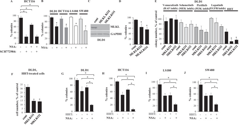
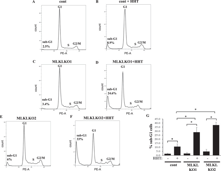
Mortality from colorectal cancer (CRC) is significant, and novel CRC therapies are needed. A pseudokinase MLKL typically executes necroptotic cell death, and MLKL inactivation protects cells from such death. However, we found unexpectedly that MLKL gene knockout enhanced CRC cell death caused by a protein synthesis inhibitor homoharringtonine used for chronic myeloid leukemia treatment. In an effort to explain this finding, we observed that MLKL gene knockout reduces the basal CRC cell autophagy and renders such autophagy critically dependent on the presence of VPS37A, a component of the ESCRT-I complex. We further found that the reason why homoharringtonine enhances CRC cell death caused by MLKL gene knockout is that homoharringtonine activates p38 MAP kinase and thereby prevents VPS37A from supporting autophagy in MLKL-deficient cells. We observed that the resulting inhibition of the basal autophagy in CRC cells triggers their parthanatos, a cell death type driven by poly(ADP-ribose) polymerase hyperactivation. Finally, we discovered that a pharmacological MLKL inhibitor necrosulfonamide strongly cooperates with homoharringtonine in suppressing CRC cell tumorigenicity in mice. Thus, while MLKL promotes cell death during necroptosis, MLKL supports the basal autophagy in CRC cells and thereby protects them from death. MLKL inactivation reduces such autophagy and renders the cells sensitive to autophagy inhibitors, such as homoharringtonine. Hence, MLKL inhibition creates a therapeutic vulnerability that could be utilized for CRC treatment.


Conflict of interest statement

Competing interests: The authors declare no competing interests. Ethics approval and consent to participate: Animal studies were approved by Dalhousie University Committee on Laboratory Animals, protocol 20-081.

Figures

Fig. 1
Fig. 1. MLKL inactivation cooperates with HHT in reducing CRC cell clonogenic survival.
A HCT116 cells were treated with DMSO (−) or 1 μM SCH 772984 (SCH) and/or 3 μM NSA (+), and colonies formed by the cells were counted. B Indicated cell lines were treated with DMSO (−) or 1 μM NSA (+), and colonies formed by the cells were counted. C Control cells (cont) or MLKL-deficient cells MLKL KO1 and MLKL KO2 were tested for MLKL levels by western blotting. GAPDH served as a loading control. D Cells generated in C were assayed as in A. E Indicated cell lines were treated or not with 5 μM vemurafrnib, 1 μM selumetinib, 1 μM pictilisib, 1 μM lapatinib or 10 ng/ml HHT and colonies formed by the cells were counted. F Indicated cell lines were treated or not with 10 ng/ml HHT for 96 h and counted. GJ Indicated cell lines were treated (+) or not (−) with 1 μM NSA and 10 ng/ml HHT, and colonies formed by the cells were counted. The data represent % of colony number formed by the untreated cells (A, B, D, E, GJ) or % of untreatded cells (F). The data in A, GJ are the average of the triplicates plus SD, and each experiment was repeated twice with similar results. The data in E are the average of two independent experiments plus SD. The data in B and D and F are the average of three independent experiments plus SD. *p value < 0.05.
Fig. 2
Fig. 2. MLKL inhibitor NSA cooperates with HHT in blocking CRC cell tumorigenicity in vivo.
A HCT116 cells were injected in the flanks of 28 Nu/Nu Nude mice. Once tumor volumes reached the average volume of 80 mm3, the mice were injected intraperitoneally with DMSO (control), NSA, HHT or with NSA and HHT together. 7 mice were used per group. One mouse had to be sacrificed in the control (drug-untreated) group on day 25, one more mouse in the same group, on 30 and two more mice in this group, on day 37 because their tumor sizes exceeded 1000 mm3 on the respective days. Changes in relative tumor volumes (A) and mouse weight (B) plus SE are shown. Tumor volume observed on the day of the first injection was designated as 1.0 for each mouse, and subsequent changes in each tumor volume were calculated relative to that number. *p < 0.05.
Fig. 3
Fig. 3. MLKL inactivation and HHT cooperate in killing CRC cells by inhibiting their basal autophagy.
A, B Indicated cell lines were treated with DMSO (−) or 10 ng/ml HHT (+) for 24 h in the absence (−) or in the presence (+) of 50 nM bafilomycin A1, and the cells were assayed for LC3B levels by western blotting. C Indicated cell lines were treated with DMSO (−) or 10 ng/ml HHT and/or 3 μM NSA (+) for 24 h in the presence of 50 nM bafilomycin A1 and the cells were assayed for LC3B levels by western blotting. α-tubulin was used as a loading control in AC. D Indicated cells were transfected with the GFP-LC3 expression vector and treated with DMSO (DMSO) or 10 ng/ml HHT (HHT) for 24 in the presence of 50 nM bafilomycin A1. Green puncta per cell were counted. Representative fluorescence microscopy images are shown. Bar - 10 μm. E Quantification of the number of green puncta per cell for the cells treated as in A. The numbers represent the average of the number of puncta per cell plus the SE. 24 untreated and 13 HHT-treated cells were counted in the case of the control clone, 42 untreated and 28 HHT-treated cells in the case of MLKL KO1 cells, and 29 untreated and 34 HHT-treated cells, in the case of MLKL KO2 cells This experiment was repeated twice with similar results. F Indicated cell lines were treated with DMSO (−) or 10 ng/ml HHT (+) for 96 h in the absence (−) or in the presence (+) of 12 μM of control (cont) or cell-permeable Beclin-1 (Beclin-1) peptide and counted. The data in F are the average of three independent experiments plus SD. *p < 0.05.
Fig. 4
Fig. 4. Basal autophagy inhibition is suffcieent for reducing CRC cell clonogenic survival.
A DLD1 cells were transfected with a 100 nM control RNA (cont RNA) or ATG12-specidic siRNA (ATG12 siRNA) 1 or 2 and assayed for the levels of the covalent ATG5-ATG12 complex (ATG5-12) by western blotting. B Cells treated as in A and cultured in the absence (−) or in the presence (+) of 50 nM bafilomycin A1 were assayed for LC3BII levels by western blotting. C Cells treated as in A were assayed for clonogenic survival. D DLD1 cells were treated with DMSO (−) or 20 μM ULK-1 inhibitor (ULK-1 inhib) SBI-0206965 (+) for 24 h in the absence (-) or in the presence (+) of 50 nM bafilomycin A1 were assayed for LC3BII levels by western blotting. E DLD1 cells were treated as in D for 96 h and counted. α-tubulin was used as a loading control in A, B, D. The data in C, E are the average of three independent experiments plus SD. *- p < 0.05.
Fig. 5
Fig. 5
MLKL inactivation renders CRC cell autophagy VPS37A-dependent. Indicated cell lines were transfected with a 100 nM control RNA (cont RNA) or VPS37A-specidic siRNA (VPS37A siRNA) 1 (A, B, E) or 2 (C, D, F), treated with DMSO (−) or 10 ng/ml HHT (+) for 24 h in the presence of 50 nM bafilomycin A1 and assayed for VPS37A (AD) or LC3B (E, F) levels by western blotting. α-tubulin was used as a loading control.
Fig. 6
Fig. 6
HHT contributes to autophagy inhibition in CRC cells by activating p38MAPK. Indicated cell lines were treated with DMSO (−) or 10 ng/ml HHT for 24 h and/or 20 μM p38MAPK inhibitor SB203580 (SB203580) (+) in the presence of 50 nM bafilomycin A1 and assayed for p38MAPK and phospho-38MAPK (A) or LC3B (B, C) levels by western blotting. α-tubulin was used as a loading control in B and C.
Fig. 7
Fig. 7. MLKL inactivation and HHT cooperate in triggering chromosomal DNA fragmentation in CRC cells.
Indicated cell lines were treated with with 10 ng/ml HHT (HHT) or DMSO for 96 h and analyzed for the cell cycle profile by flow cytometry. AF Representative cell cycle profiles are shown. G The average % of the hypodiploid sub-G1 cells for the indicated treatments derived from three independent experiments plus SD is shown. *p < 0.05.
Fig. 8
Fig. 8. MLKL inactivation and HHT cooperate in triggering parthanatos of CRC cells.
MLKL KO1 cells lines were treated with DMSO (cont), 10 ng/ml HHT (HHT), 50 nM olaparib (Olaparib), or both drugs (HHT+ Olaparib) at the indicated concentrations for 96 h and analyzed for the cell cycle profile by flow cytometry. AD Representative cell cycle profiles are shown. E The average % of the hypodiploid sub-G1 cells for the indicated treatments derived from three independent experiments plus SD is shown. *p < 0.05. F MLKL KO1 cells lines were treated with DMSO (−) or 10 ng/ml HHT (+) in the absence (−) or in in the presence (+) of 12 μM of control (cont) or cell-permeable Beclin-1 (Beclin-1) peptide for 96 h and analyzed for poly(ADP-ribose) (PAR) levels by western blotting. α-tubulin was used as a loading control.
Fig. 9
Fig. 9. Low MLKL mRNA expression signifies increased CRC patient survival.
Kaplan–Meier analysis-based estimation of probabilities of patients’ survival depending on the level of MLKL mRNA was performed.

References

    1. Biller LH, Schrag D. Diagnosis and Treatment of Metastatic Colorectal Cancer: A Review. JAMA. 2021;325:669–85. - PubMed
    1. Galluzzi L, Vitale I, Aaronson SA, Abrams JM, Adam D, Agostinis P, et al. Molecular mechanisms of cell death: recommendations of the Nomenclature Committee on Cell Death 2018. Cell Death Differ. 2018;25:486–541. - PMC - PubMed
    1. Sun L, Wang H, Wang Z, He S, Chen S, Liao D, et al. Mixed lineage kinase domain-like protein mediates necrosis signaling downstream of RIP3 kinase. Cell. 2012;148:213–27. - PubMed
    1. Winer ES, DeAngelo DJ. A Review of Omacetaxine: A Chronic Myeloid Leukemia Treatment Resurrected. Oncol Ther. 2018;6:9–20. - PMC - PubMed
    1. Gurel G, Blaha G, Moore PB, Steitz TA. U2504 determines the species specificity of the A-site cleft antibiotics: the structures of tiamulin, homoharringtonine, and bruceantin bound to the ribosome. J Mol Biol. 2009;389:146–56. - PMC - PubMed

Substances