. 2025 Feb 20;16(1):1825.
doi: 10.1038/s41467-025-57006-2.

Molecular basis of foreign DNA recognition by BREX anti-phage immunity system

Affiliations

Molecular basis of foreign DNA recognition by BREX anti-phage immunity system

Alena Drobiazko et al. Nat Commun. .

Abstract

Anti-phage systems of the BREX (BacteRiophage EXclusion) superfamily rely on site-specific epigenetic DNA methylation to discriminate between the host and invading DNA. We demonstrate that in Type I BREX systems, defense and methylation require BREX site DNA binding by the BrxX (PglX) methyltransferase employing S-adenosyl methionine as a cofactor. We determined 2.2-Å cryoEM structure of Escherichia coli BrxX bound to target dsDNA revealing molecular details of BREX DNA recognition. Structure-guided engineering of BrxX expands its DNA specificity and dramatically enhances phage defense. We show that BrxX alone does not methylate DNA, and BREX activity requires an assembly of a supramolecular BrxBCXZ immune complex. Finally, we present a cryoEM structure of BrxX bound to a phage-encoded inhibitor Ocr that sequesters BrxX in an inactive dimeric form. We propose that BrxX-mediated foreign DNA sensing is a necessary first step in activation of BREX defense.


Conflict of interest statement

Competing interests: The authors declare no competing interests.

Figures

Fig. 1
Fig. 1. The BrxX (PglX) methyltransferase specifically recognizes BREX sites in DNA.
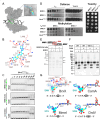
A The Type I BREX locus of E. coli HS, its genomic position, and predicted functions of encoded proteins. B Potassium efflux assay with E. coli BW25113 BREX, BREX+, EcoRV, or ΔlamB cultures infected with phage λ at MOI = 5. A representative result from three independent experiments is shown. C EMSA with 20 nM Cy5-labeled 43 bp dsDNA substrates with no BREX sites (scrambled), or with one or two BREX sites, incubated with an indicated amount of BrxX in the presence or in the absence of 0.5 mM SAM. Green arrows indicate the orientation of BREX sites. Representative gels from experiments performed in triplicate are shown. D Strep-seq analysis of DNA cross-linked to BrxX-Strep 15 min after infection of a BREX+ culture with T7fusion phage at MOI = 1. Reads from the enriched sample were mapped to T7 genome and the signal was normalized to the bulk DNA level and genome size to obtain CPM values. Positions of BREX sites (GGTAAG) on the top and bottom DNA strands are indicated with red and blue dots, correspondingly. E Cross-correlation analysis (normalized coverage of the forward and reverse reads) for a representative peak in the T7 genome. F EMSA with 20 nM of 40 bp Cy5-labeled dsDNA substrate with methylated or non-methylated BREX site incubated with indicated amounts of BrxX. Representative gels from triplicate experiments are shown.
Fig. 2
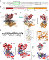
Fig. 2. Architecture of the BrxX-DNA complex.
A The 43 bp head-to-tail (H2T) DNA fragment used for cryoEM. The six-base pair BREX recognition sequence is shown in green, dark gray shows the resolved (modeled) bases whereas light gray shows unresolved bases. Black rectangle highlights the second (unbound) BREX site. B Domain arrangement of the BrxX monomer: N-terminal domain (NTD) is shown in dark red, methyltransferase domain (MTD) in pink, target recognition domain (TRD) in yellow, and C-terminal domain in orange (CTD). This color scheme is used throughout the manuscript. C CryoEM map, side view, of the BrxX-DNA complex. The sharpened map, contoured at 5σ, is colored according to the diagram above and shown within the contour of unsharpened map (also at 5σ, transparent contour). Only the density within the sharpened map was modeled, and resulting 25 bp of DNA was built. D A cartoon representation of an atomic model of the BrxX-DNA complex. SAM (cyan) and magnesium ion (purple) are shown as van der Waals sphere representations. The two inset regions highlight the flexible loops between the MTD and the TRD (i) and the metal-binding site in the CTD (ii). E A structural comparison of the 25 bp DNA bound to BrxX as seen in the cryoEM structure (gray tubes) to idealized B-form DNA (black cartoon). BrxX-bound DNA deviates from a linear trajectory by ~33° with widened major and minor grooves. F Surface coloring based on Coulombic electrostatic potential calculated by ChimeraX where red and blue show, respectively, negative and positive potentials.
Fig. 3
Fig. 3. Molecular basis of BREX site recognition by BrxX.
A A scheme of interactions between BrxX and the six-base pair BREX site. Distances in Angstrom are indicated. An asterisk indicates that the main chain and not the side chain is involved. Colors correspond to amino acids from MTD (pink) or TRD (yellow). B Molecular basis for site-specific recognition of the BREX site. Each base pair is presented separately and oriented with major groove on top. Distances in Angstrom are indicated. Non-target strand indicated by a prime symbol. C A species-specific MTD loop recognizes the sixth base pair. A structural superposition is shown of loop 587–601 of E. coli HS BrxX MTD (rose) and equivalent loops in Salmonella typhimurium (purple) and Escherichia fergusonii (magenta) based on AlphaFold DB models AF-A0A5A8QD96-F1 and AF-B7L3T0-F1 respectively. D Side-by-side comparison of above loops with amino acids predicted to be important for the recognition of the sixth base pair shown as sticks. Recognition sites for all three BREX systems are shown, and the last base pair of the recognition site is boxed (G6 for E. coli). As the cognate DNA structure is not available for S. typhimurium and E. fergusonii, sixth base pair of E.coli HS BrxX is shown for all three models. In the case of E. fergusonii, a larger purine base would be replaced by a smaller pyrimidine, in line with an observed change in the MTD loop structure.
Fig. 4
Fig. 4. Substitutions in BrxX TRD mediate BREX specificity change and enhance anti-phage defense.
A EOP assay with BREX-sensitive T7Δ0.3 and λvir phages and cells carrying BREX systems encoding indicated BrxX mutants. B EMSA with 20 nM of 40 bp Cy5-labeled dsDNA substrate with intact or scrambled BREX site and WT or mutant BrxX proteins. C Substitutions in BrxX TRD result in relaxed BREX site specificity. Methylation of BREX sites in genomic DNA of BREX+ cells with WT BrxX or indicated BrxX mutants assessed through ONT sequencing. Diagrams demonstrate the density distribution of modified sites for each possible nucleotide variation in the second (above) or fourth (below) position; a 66% threshold (counted as significant) is indicated with dotted lines (see “Methods”). The fraction of sites exceeding the modification threshold and the total number of available sites in the genome are provided in the insets. D EMSA with 20 nM of 40 bp Cy5-labeled dsDNA substrates with varying nucleotides at the fourth position of the BREX site or scrambled BREX site and WT or TRD mutated BrxX proteins.
Fig. 5
Fig. 5. The DNA mimic Ocr displaces DNA from BrxX and locks it in an inactive conformation.
A SEC traces of BrxX, Ocr, or an equimolar BrxX + Ocr mixture injected onto Superdex 200 Increase 10/300 column. SEC profiles of standard calibrants and their corresponding molecular masses are indicated in the background in gray. B EMSA with 20 nM Cy5-labeled 40 bp dsDNA substrate with a single BREX site incubated with a 20-fold (0.4 μM) molar excess of BrxX in the presence of indicated concentrations of Ocr dimer. BrxX was either incubated with DNA for 45 min, followed by an additional 45 min incubation with Ocr (top), or was first incubated with Ocr for 45 min, followed by an additional 45 min incubation with DNA (bottom). C Competition between DNA and Ocr for binding to BrxX. 20 nM Cy5-labeled 40 bp dsDNA substrate bearing one BREX site was incubated with a 20-fold (0.4 μM) molar excess of BrxX, followed by an indicated amount of Ocr dimer (top) or of a non-labeled dsDNA substrate (bottom). D CryoEM maps of the BrxX-Ocr complex. The autosharpened map, contoured at 7σ, is colored according to BrxX domains coloring scheme (Fig. 2) and shown within the transparent contour of the unsharpened map (at 5σ). Ocr monomers are shaded in blue; the lower-resolution BrxX monomer is shown in white. E Surface coloring based on Coulombic electrostatic potential (as implemented in ChimeraX), where red is negative potential and blue is positive potential. Ocr dimer is shown as representation. F Direct interactions between BrxX and Ocr residues. Interacting residues are shown as balls and sticks; Ocr residues are white while TRD and CTD residues are colored yellow and orange, respectively. G A structural superposition of atomic models of DNA-bound (white) and Ocr-bound (colored by domain) BrxX monomers. Movements are indicated. H Structural superposition of surface models. DNA-bound model is depicted in transparent white and an Ocr-bound depicted colored by domain. Alignment generated via ChimeraX matchmaker tool using the TRD domain boundaries. I EOP assay with WT T7, Ocr-deficient T7Δ0.3, and λvir phages and cells carrying BREX systems encoding indicated BrxX mutants.
Fig. 6
Fig. 6. BrxX alone lacks methyltransferase activity.
A Surface representation of the BrxX monomer shown in gray bound to DNA (black) containing the BREX binding site (green). Inset highlights the only external access point to the SAM molecule enclosed within the catalytic pocket (cyan backbone, colored by heteroatom). B Direct interactions between the SAM molecule and MTD residues. Dashed lines indicate hydrogen bonding with distances indicated in Angstroms. C EMSA with 20 nM Cy5-labeled 43 bp dsDNA substrate without BREX sites (“scrambled”), or with one or two BREX sites, incubated with the indicated amount of BrxXY511A without co-factors or in the presence of 0.5 mM SAM. Green arrows indicate the orientation of BREX sites. Compare with Fig. 1C for EMSA with wild-type BrxX performed in identical conditions. D Effects of BrxXY511A mutation on BREX defense, methylation, and toxicity. BREX defense is demonstrated by an EOP assay with BREX-sensitive λvir and T7Δ0.3 phages. BREX methylation is estimated by the ability of λts induced from the indicated lysogenic cultures to plaque on BREX and BREX+ lawns (see “Methods”). Toxicity was estimated in a drop-spot test on LB agar plates. BrxXY511A was introduced in the context of the full BREX cluster. All assays were performed in biological triplicates and representative plates are shown. E Overlapping BREX methylation inhibits AluI cleavage. An agarose gel shows AluI digest of pHERD30t or pHERD30t bearing an overlapping BREX/AluI cleavage site purified from either BREX+ (pBREX AL) or BREX (pBTB) cultures. Arrows indicate the fragment bearing overlapping BREX/AluI site and its AluI cleavage products. F AluI cleavage with 40 bp dsDNA substrate containing an overlapping BREX/AluI site and incubated with 100 μM BrxX overnight. G A comparison of orientations of the flipped adenine and SAM or an equivalent ligand between BrxX (this work), CamA (PDB: 7LT5), MmeI (PDB: 5HR4), and DrdV (PDB: 7LO5). The 5′–3′ recognition site with the methylated adenine boxed is indicated for each protein. In the BrxX structure, the sharpened map density contoured at 5σ is shown surrounding each molecule, with the catalytic residues (NPPY motif) from the MTD shown in pink.
Fig. 7
Fig. 7. BREX methylation requires the assembly of a macromolecular BrxBCXZ complex.
A In vivo BREX methylation requires co-production of BrxX, BrxC, BrxZ, and BrxB. Prophage λwt was induced from indicated lysogens and the status of its BREX methylation was determined by plaquing on BREX+ and BREX lawns. The level of BREX defense calculated from the EOP performed in biological triplicates with independently induced λwt phages. Data are presented as mean values ± SEM. B In vivo pull-downs with Strep-tagged BREX proteins expressed in the context of the full BREX cluster. Strep-tagged proteins (baits) are indicated at the top and co-eluted proteins, identified through MALDI-TOF mass spectrometry, are shown on the heatmap below. The Strep-trap column eluates were concentrated, and proteins resolved by 4–20% gradient SDS-PAGE. С Strep-tag pull-down eluates (B) were concentrated and separated on a Superdex 200 Increase 10/300 column. For each run, 280 nm absorbance was normalized to the highest value. SEC profiles of calibrants and their corresponding molecular masses are indicated on the background in gray. An asterisk indicates contaminant protein. DNA non-specifically binds the StrepTrap HP column, creating peaks eluting with the void volume, which are the most evident in BrxL and BrxB runs. D SEC calibration curve with estimated masses of obtained protein complexes. E DNAse I treatment of fractions containing the BrxCX complex results in the reduction of apparent complex size. F SDS-PAGE results (left) demonstrating protein and agarose gel electrophoreses (right) demonstrating DNA content of the BrxCX peak before and after DNase I treatment. In all panels, the protein or DNA molecular weight markers are shown on the left.
Fig. 8
Fig. 8. A model of foreign DNA sensing by BrxX and its inhibition by Ocr.
BREX sites recognition requires target adenine flipping and BrxX conformation transition from an open to the closed state (I, II, III). Ocr locks two BrxX monomers in an open conformation unable to bind DNA (VI). BREX DNA methylation (IV, V) requires the assembly of the BrxBCXZ complex, which might be also responsible for BREX defense. Stages IVI are also discussed in the text. This figure was created with BioRender.com.

References

    1. Parikka, K. J., Le Romancer, M., Wauters, N. & Jacquet, S. Deciphering the virus‐to‐prokaryote ratio (VPR): insights into virus–host relationships in a variety of ecosystems. Biol. Rev.92, 1081–1100 (2017). - PubMed
    1. Dion, M. B., Oechslin, F. & Moineau, S. Phage diversity, genomics and phylogeny. Nat. Rev. Microbiol.18, 125–138 (2020). - PubMed
    1. Bernheim, A. & Sorek, R. The pan-immune system of bacteria: antiviral defence as a community resource. Nat. Rev. Microbiol.18, 113–119 (2020). - PubMed
    1. Mayo-Muñoz, D., Pinilla-Redondo, R., Birkholz, N. & Fineran, P. C. A host of armor: prokaryotic immune strategies against mobile genetic elements. Cell Rep.42, 112672 (2023). - PubMed
    1. Miller, D., Stern, A. & Burstein, D. Deciphering microbial gene function using natural language processing. Nat. Commun.13, 5731 (2022). - PMC - PubMed

MeSH terms

LinkOut - more resources