

Detection of proximal dental caries in primary teeth with a near-infrared-irradiation-assisted intraoral scanner: an in vitro study
- PMID: 39979907
- PMCID: PMC11844066
- DOI: 10.1186/s12903-025-05629-8
Detection of proximal dental caries in primary teeth with a near-infrared-irradiation-assisted intraoral scanner: an in vitro study
Abstract
Background: The early detection of dental caries is crucial for successful dental care. New intraoral scanners using near-infrared irradiation (NIRI) technology track preventive lesions without ionizing radiation. This study aimed to evaluate the performance of intraoral scanners (IOSs) in detecting proximal caries in primary posterior teeth, compared to conventional methods such as loupes-assisted clinical exams and bitewing (BW) radiography.
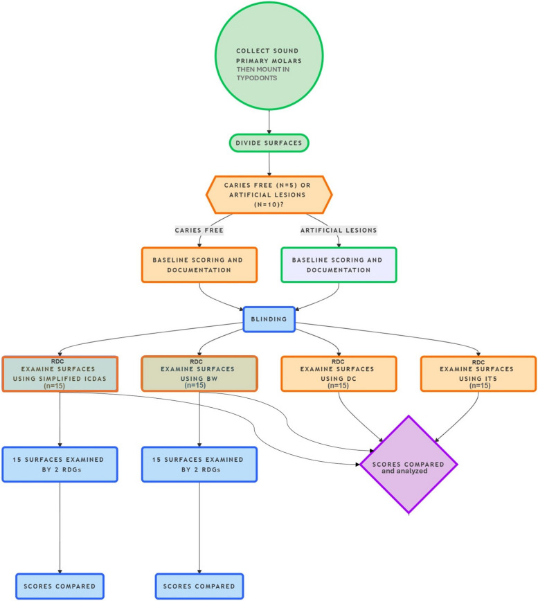
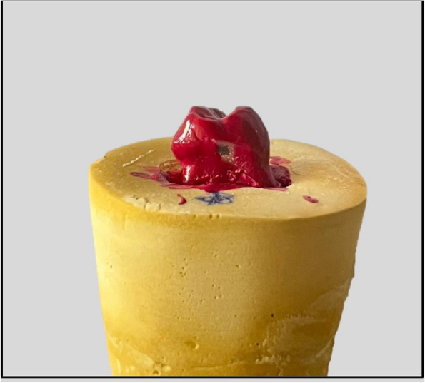
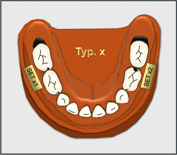
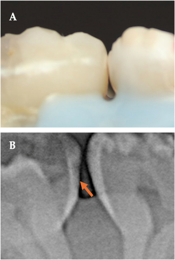
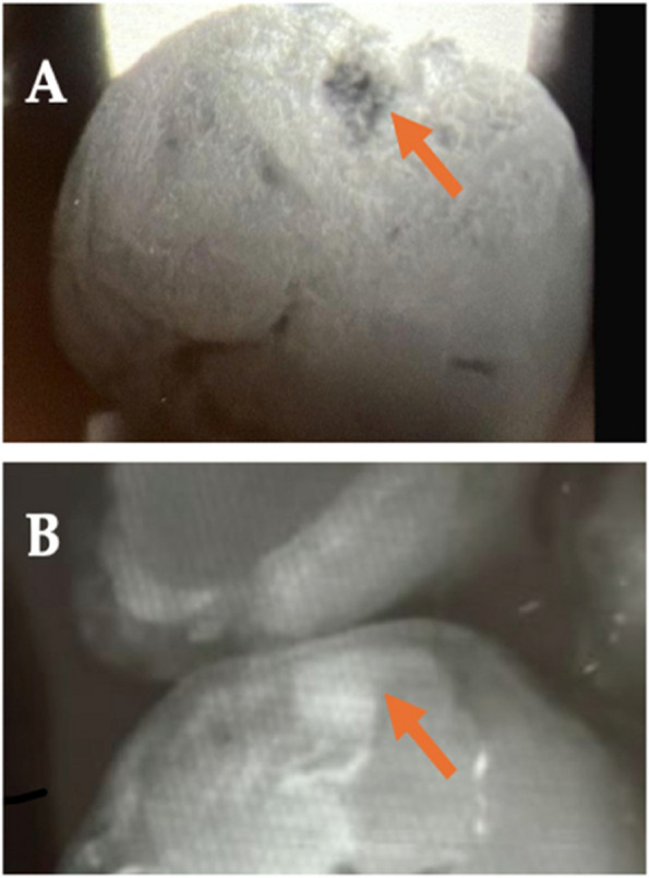
Methods: Fifteen examined tooth surfaces were used to produce a total of 60 scores by a restorative dentistry consultant (RDC). The tooth surfaces were categorized into caries-free (n = 5 × 4 exam methods) and carious (n = 10 × 4 exam methods) subgroups. Artificial caries lesions were created on specified surfaces and mounted on typodont in sets for evaluation using a simplified modified ICDAS visual and BW radiographic examination, DIAGNOcam device, and iTero Element 5D NIRI-assisted IOS. Reference surface scores were recorded. Investigators (RDCs and recent dental graduates (RDGs)) were trained and calibrated. Inter-examiner agreement, agreement with reference, specificity, and sensitivity were checked.
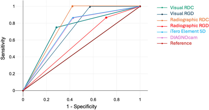
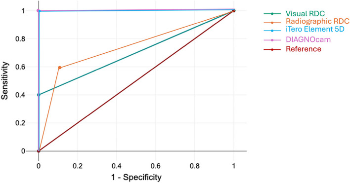
Results: The results showed that the sensitivity and specificity differed between the diagnostic tests. The best agreement of all investigated diagnostic methods with the reference was found using the DIAGNOcam device (ĸ = 0.87) and BW radiography (ĸ = 1.00). High agreement was found for visual examination (by the RDC and RDGs (ĸ ≈ 0.85)) and iTero 5D examination (ĸ = 0.87).
Conclusions: The iTero Element 5D IOS had lower sensitivity and specificity compared to other methods. The potential use of IOSs with NIRI as a substitute for conventional diagnostic methods in primary teeth shows promise but requires further investigation.
Keywords: Artificial caries; Caries detection; DIAGNOcam; Intra-oral scanners; Near infra-red irradiation; Near infra-red transillumination; Preventive; Primary teeth; Proximal lesions; Restorative dentistry; iTero Element 5D.
© 2025. The Author(s).
Conflict of interest statement
Declarations. Ethics approval and consent to participate: This study received approval from the ethical committee at the Faculty of Dentistry, King Abdulaziz University, Jeddah, Saudi Arabia (IRB# 192–11-23). The study adhered to the principles of the Declaration of Helsinki, and informed consent was obtained from all subjects and/ or their legal guardian(s). Consent for publication: Not applicable. Competing interests: The authors declare no competing interests.
Figures
Similar articles
-
Intraoral scanner featuring transillumination for proximal caries detection. An in vitro validation study on permanent posterior teeth.J Dent. 2022 Jan;116:103841. doi: 10.1016/j.jdent.2021.103841. Epub 2021 Oct 6. J Dent. 2022. PMID: 34624420
-
Near-infrared imaging in orthodontic intraoral scanners for early interproximal caries detection.Am J Orthod Dentofacial Orthop. 2024 Aug;166(2):138-147. doi: 10.1016/j.ajodo.2024.03.013. Epub 2024 May 9. Am J Orthod Dentofacial Orthop. 2024. PMID: 38727656
-
Accuracy of the DIAGNOcam and bitewing radiographs in the diagnosis of cavitated proximal carious lesions in primary molars.Niger J Clin Pract. 2019 Nov;22(11):1576-1582. doi: 10.4103/njcp.njcp_237_19. Niger J Clin Pract. 2019. PMID: 31719280
-
Performance of the caries diagnosis feature of intraoral scanners and near-infrared imaging technology-A narrative review.J Prosthodont. 2023 Dec;32(S2):114-124. doi: 10.1111/jopr.13770. Epub 2023 Sep 24. J Prosthodont. 2023. PMID: 37701946 Review.
-
Imaging modalities to inform the detection and diagnosis of early caries.Cochrane Database Syst Rev. 2021 Mar 15;3(3):CD014545. doi: 10.1002/14651858.CD014545. Cochrane Database Syst Rev. 2021. PMID: 33720395 Free PMC article.
References
MeSH terms
Grants and funding
LinkOut - more resources
Full Text Sources
Medical
