

The Usefulness of Concomitant Ultrasound Guidance With Surgery for Acute Achilles Tendon Rupture Using an Internal Brace
- PMID: 39980712
- PMCID: PMC11840447
- DOI: 10.7759/cureus.79340
The Usefulness of Concomitant Ultrasound Guidance With Surgery for Acute Achilles Tendon Rupture Using an Internal Brace
Abstract
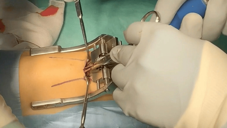
Background This study investigated the usefulness of intraoperative ultrasonography in the treatment of acute Achilles tendon rupture (ATR) using an internal brace (Achilles Midsubstance SpeedBridge, Arthrex Inc., Naples, FL), a technique that provides strong internal fixation. Methodology Forty-three patients were included and divided into two groups: Group A (n = 22), which received ultrasonography, and Group B (n = 21), which did not. In Group A, ultrasonography was used during suturing with a specialized jig to confirm the suture needle's position at the center of the proximal stump. Postoperative care in both groups involved initiating active dorsiflexion exercises on the day following surgery and permitting weight-bearing without orthosis once 0° dorsiflexion was achieved. The operative time, Japanese Society for Surgery of the Foot (JSSF) ankle/hindfoot scale, T2-weighted magnetic resonance imaging (MRI) findings at three months postoperatively, and complications were evaluated. Results Group A had a significantly shorter operative time (41.9 ± 7.5 minutes vs. 52.1 ± 6.5 minutes, P < 0.001) and a lower percentage of high-intensity areas on T2-weighted MRI (1.76% ± 2.68% vs. 8.74% ± 7.02%, P < 0.001) compared to Group B. No significant difference was observed in JSSF scale scores (P = 0.948). Additionally, no cases of re-rupture or wound infection were reported in either group. Conclusions Intraoperative ultrasonography in conjunction with this method may enable precise and reliable suturing, facilitating strong internal fixation and potentially enhancing clinical outcomes.
Keywords: acute achilles tendon rupture; early rehabilitation; internal brace; pars; ultrasonography.
Copyright © 2025, Chida et al.
Conflict of interest statement
Human subjects: Consent for treatment and open access publication was obtained or waived by all participants in this study. The Institutional Ethics Committee of Hiraka General Hospital issued approval 248. The original IRB approval document was issued in Japanese. For the convenience of the reviewers and editors, an English translation has been provided as a supplementary document. Animal subjects: All authors have confirmed that this study did not involve animal subjects or tissue. Conflicts of interest: In compliance with the ICMJE uniform disclosure form, all authors declare the following: Payment/services info: All authors have declared that no financial support was received from any organization for the submitted work. Financial relationships: All authors have declared that they have no financial relationships at present or within the previous three years with any organizations that might have an interest in the submitted work. Other relationships: All authors have declared that there are no other relationships or activities that could appear to have influenced the submitted work.
Figures


References
-
- Surgical treatment versus conservative management for acute Achilles tendon rupture: a systematic review and meta-analysis of randomized controlled trials. Deng S, Sun Z, Zhang C, Chen G, Li J. J Foot Ankle Surg. 2017;56:1236–1243. - PubMed
-
- Acute Achilles tendon ruptures: efficacy of conservative and surgical (percutaneous, open) treatment - a randomized, controlled, clinical trial. Manent A, López L, Corominas H, et al. J Foot Ankle Surg. 2019;58:1229–1234. - PubMed
-
- Outcome of percutaneous fixation of acute Achilles tendon ruptures. Rozis M, Benetos IS, Karampinas P, Polyzois V, Vlamis J, Pneumaticos SG. Foot Ankle Int. 2018;39:689–693. - PubMed
-
- Operative versus nonoperative treatment of acute Achilles tendon ruptures: a multicenter randomized trial using accelerated functional rehabilitation. Willits K, Amendola A, Bryant D, et al. J Bone Joint Surg Am. 2010;92:2767–2775. - PubMed
LinkOut - more resources
Full Text Sources
Miscellaneous