. 2025 Feb 19;27(Suppl 1):i73-i82.
doi: 10.1093/eurheartjsupp/suae108. eCollection 2025 Feb.

The 'Padua classification' of cardiomyopathies into three groups: hypertrophic/restrictive, dilated/hypokinetic, and scarring/arrhythmogenic

Affiliations

The 'Padua classification' of cardiomyopathies into three groups: hypertrophic/restrictive, dilated/hypokinetic, and scarring/arrhythmogenic

Domenico Corrado et al. Eur Heart J Suppl. .

Abstract

The newly proposed classification of cardiomyopathies, referred to as 'the Padua Classification', is based on both pathobiological basis (genetics, molecular biology, and pathology) and clinical features (morpho-functional and structural ventricular remodelling as evidenced by cardiac magnetic resonance). Cardiomyopathies are grouped into tree main categories and characterized by a designation combining both 'anatomical' and 'functional' features: hypertrophic/restrictive, dilated/hypokinetic, and scarring/arrhythmogenic; each cardiomyopathy group includes either genetic or non-genetic aetiologic variants. This novel approach aims to enhance the diagnostic accuracy and to support 'disease-specific' therapeutic strategies, with the objective to improve patient management and outcome.

Keywords: Cardiac magnetic resonance; Cardiomyopathy; Diagnosis.


Conflict of interest statement

Conflict of interest: None declared.

Figures

Figure 1
Figure 1
Evolution of Cardiomyopathy Classification. This flowchart illustrates the main steps of development of cardiomyopathy classification from the 1970s to 2024. The initial classification of Goodwin and Oakley in the 1970s was followed by those of the World Health Organization (WHO) and International Society and Federation of Cardiology in 1980 and 1996. In 1996, the classification endorsed by the WHO included: dilated, hypertrophic, restrictive, and arrhythmogenic right ventricular cardiomyopathies. In 2006, the American Heart Association task force proposed a classification of cardiomyopathies, which included ion channelopathies and distinguished conditions primarily involving the heart (genetic, acquired, or mixed aetiology) from conditions secondarily affecting the heart in the setting of systemic diseases. In 2008, the European Society of Cardiology proposed a classification of cardiomyopathies, which excluded ion channelopathies, separated familial vs. non-familial conditions, and sub-divided specific disease and idiopathic. The 2024 ‘Padua Classification’ grouped cardiomyopathies into three main categories: Hypertrophic/Restrictive, Dilated/Hypokinetic and Scarring/Arrhythmogenic, on the basis of distinctive pathobiology and morpho-functional ventricular remodelling. AHA, American Heart Association; CM, cardiomyopathy; ESC, European Society of Cardiology; ISFC; International Society and Federation of Cardiology; WHO, World Health Organization.
Figure 2
Figure 2
Cardiomyopathy phenotypes and patterns of cardiac remodelling. The ‘Padua classification’ of cardiomyopathy is in agreement with the original definition of ‘primary heart muscle disease of unknown aetiology’, whose phenotype reproduces the patterns of cardiac remodelling (i.e. changes in the size, shape, structure, and function) caused by conditions of known aetiology such as myocardial infarction, hypertensive heart disease, valvular diseases, and congenital heart diseases. The original phenotype of hypertrophic/restrictive reproduces the cardiac remodelling induced by systemic hypertension or aortic stenosis leading to haemodynamic pressure overload; the phenotype of dilated/hypokinetic the cardiac remodelling induced by mitral or aortic regurgitation leading to haemodynamic volume overload; and the phenotype of scarring/arrhythmogenic the cardiac remodelling of post-infarction myocardial scar leading to ventricular arrhythmias and ventricular systolic dysfunction/dilatation as a function of the amount of the post-necrotic reduction of myocardial mass. Adapted from Corrado et al..
Figure 3
Figure 3
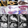
Pathologic and cardiac magnetic resonance (CMR) imaging features of hypertrophic/restrictive (H/RC) phenotypic variants. HCM and RCM are no longer considered two distinct entities on the basis of arbitrary cut offs in the left ventricle (LV) wall thickness (dotted vertical black line), but phenotypic variants of the same genetically determined sarcomeric heart muscle disease manifesting with variable degrees of LV hypertrophy and LV diastolic dysfunction. The spectrum of genetically determined H/RC, encompasses HCM with mild to moderate diastolic dysfunction (A, B), HCM with restrictive physiology (C, D), and RCM (E, F) phenotypes (continuous horizontal red line). Note that myocardial disarray and interstitial fibrosis are common histopathologic features to the different phenotypic variants (A, C, E). Note the increasing left atrial dilatation (from B to D and F) as an indirect sign of augmented diastolic filling pressure in the CMR four chamber view sequences of HCM (B), HCM with restrictive physiology (D) and RCM (F). Adapted from Corrado et al..
Figure 4
Figure 4
Pathologic and cardiac magnetic resonance (CMR) imaging features of scarring/arrhythmogenic phenotypes. Arrhythmogenic right ventricular cardiomyopathy phenotypic variant. Top left: Macroscopic longitudinal section of the heart showing apicobasal and transmural right ventricular myocardial scarring (A). Panoramic histologic view of the right ventricular (RV) free wall showing full-thickness myocardial loss with replacement by fibrous and fatty tissue, with residual myocardium confined to the endocardial trabeculae (trichrome stain) (B). Bottom left: Post-contrast CMR images showing late gadolinium enhancement (LGE)/fibrous replacement (solid arrows) of RV free wall in both long-axis 4-chamber view (A) and the sagittal view (B). ABVC phenotypic variant. Top mid: Macroscopic transverse section of the heart showing infundibular and inferior sub-tricuspidal aneurysms (C). Panoramic histological view of the inferior aneurysm showing wall thinning with transmural fibro-fatty myocardial replacement (D). Panoramic histological view showing sub-epicardial fibro-fatty scar of the left ventricular free wall (E). Bottom mid: Post-contrast images—long-axis 2-chamber view (C) and short-axis view (D) showing LGE/myocardial fibrosis from basal to apical anterolateral and inferolateral LV wall segments and RV free wall (solid arrows). ALVC phenotypic variant. Top right: Macroscopic transverse section of the heart showing a whitish, thin, linear discoloration in the LV posterior-lateral wall, involving the sub-epicardial and mid-mural myocardial layers (F). Histologic examination showing sub-epicardial/mid-myocardial fibrous (G) and fibro-fatty (H) myocardial replacement. Bottom right: Post-contrast long-axis 2-chamber view (E) showing LV sub-epicardial LGE (solid arrows), extending from basal-to-apical segments. Post-contrast short-axis view showing the involvement of the LV free wall and the septum (‘ring-like’ pattern) (open arrows) (F). ACM, arrhythmogenic cardiomyopathy; ALVC, arrhythmogenic left ventricular cardiomyopathy; CMR, cardiac magnetic resonance; LGE, late gadolinium enhancement. Adapted from Corrado et al..

References

    1. Goodwin JF, Oakley CM. The cardiomyopathies. Br Heart J 1972;34:545–552. - PMC - PubMed
    1. Braunwald E. Cardiomyopathies: an overview. Circ Res 2017;121:711–721. - PubMed
    1. McKenna WJ, Maron BJ, Thiene G. Classification, epidemiology, and global burden of cardiomyopathies. Circ Res 2017;121:722–730. - PubMed
    1. Thiene G, Corrado D, Basso C. Revisiting definition and classification of cardiomyopathies in the era of molecular medicine. Eur Heart J 2008;29:144–146. - PubMed
    1. Kramer CM. Role of cardiac MR imaging in cardiomyopathies. J Nucl Med 2015;56 Suppl 4:39S–45S. - PMC - PubMed

LinkOut - more resources