. 2025 Feb;121(4):e17243.
doi: 10.1111/tpj.17243.

Establishment and maintenance of embryogenic cell fate during microspore embryogenesis

Affiliations

Establishment and maintenance of embryogenic cell fate during microspore embryogenesis

Charlotte Siemons et al. Plant J. 2025 Feb.

Abstract

Microspore embryogenesis is a type of in vitro totipotency in which the immature male gametophyte (pollen) develops into a haploid embryo after an abiotic stress treatment. In Brassica napus, heat-stress treatment of male gametophytes induces the development of different types of multicellular embryogenic structures, each with different cellular characteristics and the capacity to form a differentiated embryo. The origin and early development of these different embryogenic structures have not been determined. We used two-photon excitation fluorescence microscopy and time-lapse imaging of cells expressing either a LEAFY COTYLEDON1 (LEC1) embryo identity reporter or a DR5v2 auxin response reporter to follow the development of embryogenic structures starting at the single- to few-cell stage. We show for the first time that the developmental fate of embryogenic structures is defined by the symmetry of the first embryogenic division and that the division plane also predicts the timing of subsequent pollen wall (exine) rupture: suspensorless embryos develop after a symmetric division and undergo late exine rupture, while suspensor-bearing embryos and embryogenic callus develop after an asymmetric division and undergo early exine rupture. Live imaging also captured previously unknown dynamic LEC1 and DR5v2 expression patterns that are associated with changes in exine integrity. This study highlights the developmental plasticity of cultured pollen and uncovers new roles for the first embryogenic cell division plane and the exine in defining and maintaining cell fate during microspore embryogenesis.

Keywords: Brassica napus; DR5v2; LEAFY COTYLEDON1; auxin; cell division plane; cell fate; microspore embryogenesis; totipotency.


Conflict of interest statement

The authors have not declared a conflict of interest.

Figures
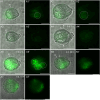
Figure 1
Figure 1
LEC1:LEC1‐GFP expression during suspensorless embryo development. The single‐cell microspore moved from a lateral position (A1–A3) to a central position (A4), where it divided symmetrically to form two equal‐sized cells (A4). A transient reduction in LEC1:LEC1‐GFP expression was observed at the one‐ and two‐cell stages (A3, A4). The exine started to burst around 4 days after the start of tracking (A6). A transmission image is shown next to the fluorescence image for each timepoint. The green signal corresponds to GFP fluorescence. All images were autoscaled to reduce the fluorescence intensity. White arrows indicate the first embryogenic cell division plane; white dashed circles indicate nuclear LEC1 nuclear expression; e, exine; *, site of exine rupture. Scale bar = 10 μm. The non‐autoscaled videos used for this figure can be found in Supporting Information.
Figure 2
Figure 2
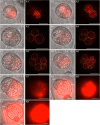
DR5v2:ntdTomato expression during suspensorless embryo development. The embryo developed after an initial symmetric division (A1), as inferred from the position of the nuclei in the two‐celled structure. DR5v2:ntdTomato expression transiently decreased during the two‐celled stage (A2, A3), followed by an increase in expression at the multicellular stage (A4, A5). Exine rupture started on day 4 of tracking (A6). For each time point, a transmission image is shown next to the fluorescence image. The red signal corresponds to tdTomato fluorescence. White arrows indicate the first embryogenic cell division plane; white dashed circles indicate DR5v2 nuclear expression; e, exine; asterisk (*), site of exine rupture. Scale bar = 10 μm. The videos used for this figure can be found in Supporting Information.
Figure 3
Figure 3
LEC1:LEC1‐GFP expression during suspensor‐bearing embryo development. The suspensor‐bearing embryo divided asymmetrically before the start of tracking (A1). Exine rupture started on day 3 of tracking (A3). The exine rupture plane was parallel to the first cell division plane. A multicellular structure developed with an embryo proper and a smaller suspensor that was subtended by the exine (A4, A5). Initially, relatively strong LEC1‐GFP expression was observed in the future suspensor and weaker expression in the future embryo proper (A2, A3). After exine rupture, LEC1:LEC1‐GFP expression gradually increased in the embryo proper, while LEC1:LEC1‐GFP expression in the suspensor gradually decreased (A4, A5). For each timepoint, a transmission image is shown next to the fluorescence image. The green signal corresponds to GFP fluorescence. White arrows indicate the first embryogenic cell division plane; white dashed circles indicate apical nuclear LEC1 expression; orange dashed circles indicate basal nuclear LEC1 expression; e, exine; *, site of exine rupture; sus, suspensor; ep, embryo proper. The videos used for this figure can be found in Supporting Information. Scale bar = 10 μm.
Figure 4
Figure 4
DR5v2:ntdTomato expression during suspensor embryo development. Suspensor embryos developed after an initial asymmetric division (A1, A3), as inferred from the cell division plane and position of the two nuclei within the dividing structure. DR5v2:ntdTomato expression was observed in the cells that contributed to both the future embryo proper and suspensor (A1, A2). In A4–A6, the nuclei in the two‐celled suspensor are indicated with orange dashed circles and the future embryo proper with white dashed circles. Exine rupture started on day 3 of tracking (A3) and occurred parallel to the first embryogenic division plane (A3). After exine rupture, DR5v2:ntdTomato expression was only observed in the embryo proper (A5–A9). For each timepoint, a transmission image is shown next to the fluorescence image with the same label. The relatively high tdTomato fluorescence intensity of the images in panel pair A9 was reduced by approximately 10%. The red signal corresponds to tdTomato fluorescence. sus, suspensor; ep, embryo proper; e, exine; asterisk, site of exine rupture; white arrows indicate the first embryogenic cell division plane. Scale bar = 10 μm. The videos used for this figure can be found in Supporting Information.
Figure 5
Figure 5
LEC1:LEC1‐GFP expression during embryogenic callus development. (A1–A7) Loose embryogenic callus development. Loose embryogenic callus development started with an asymmetric cell division that resulted in two large and equal‐sized nuclei (A1). Complete exine rupture occurred 4 h after the start of tracking (A2). The embryogenic structure lost LEC1:LEC1‐GFP expression in the uppermost cell that was no longer attached to the exine, while cells partially sheathed by the exine retained LEC1:LEC1‐GFP expression for a longer period of time (A3–A5). (B1–B4) Compact embryogenic callus development. The first embryogenic division was asymmetric (B2). The structure showed early (23 h) and partial exine rupture (B2). The partial exine rupture was followed by the gradual immediate loss of LEC1:LEC1‐GFP expression in the lower cell (B3) and gradually loss of LEC1:LEC1‐GFP expression in the upper two cells. For each timepoint, a transmission image is shown next to the fluorescence image labeled with the same letter. The green signal corresponds to GFP fluorescence. White arrows indicate the first embryogenic cell division plane; white dashed circle indicates nuclear LEC1:LEC1‐GFP expression; e, exine. The videos used for this figure can be found in Scale bar = 10 μm. Supporting Information.
Figure 6
Figure 6
DR5v2:ntdTomato expression during embryogenic callus development. (A1–A6) show compact embryogenic callus development. Compact and loose callus development begins with an asymmetric embryogenic division (A2, B1). The first embryogenic cell division was asymmetric (A1, A2). Exine rupture was initiated on day 3 of tracking (A4). DR5v2 expression was lost in both callus structures, including cells that remain attached to the exine (A5, A6). (B1–B4) show loose embryogenic callus development. The first embryogenic division is asymmetric (B1). Exine rupture was initiated on day 2 of tracking (B3). DR5v2:ntdTomato expression was lost in all cells, even those covered by the exine (B4). For each timepoint, a transmission image is shown next to the fluorescence image with the same label. The red signal corresponds to ntdTomato fluorescence. White arrows indicate the first embryogenic cell division plane; white dashed circles indicate nuclear DR5v2 expression; e, exine; asterisk, site of exine rupture. The videos used for this figure can be found in Scale bar = 10 μm. Supporting Information.
Figure 7
Figure 7
Schematic model of the main developmental pathways of embryogenic cells during Brassica napus microspore embryogenesis. Four different types of embryogenic structures can be recognized in culture: suspensorless embryos, suspensor‐bearing embryos, and compact and loose embryogenic callus. The four developmental routes can be distinguished based on the orientation of their first embryogenic division, the timing and extent of exine rupture, and the expression pattern of the LEC1:LEC1‐GFP (green) and DR5v2:ntdTomato (red) reporters. Suspensorless embryo development is initiated by a symmetric division of either the microspore or the vegetative cell of the bicellular pollen, followed by continued divisions of the two daughter cells inside the exine. Exine rupture occurs relatively late in the tracking period. A proportion of these exine‐enclosed structures develop into embryogenic callus. Suspensor‐bearing embryos and embryogenic callus are derived from an initial asymmetric division of either the microspore or the vegetative cell of the bicellular pollen. In embryogenic callus, the asymmetric division produces two cells that have two equal‐sized nuclei or nuclei that differ in size, resembling a smaller generative‐like (Gl) and a larger vegetative‐like (Vl) cell. Only the cells with vegetative‐like nuclei develop into the embryogenic callus. Compact and loose embryogenic callus show relatively early exine rupture, with compact callus showing partial rupture and loose callus showing complete rupture of the exine. These calli undergo limited cell divisions. In suspensor embryos, an asymmetric cell division produces two differently sized cells with two equal‐sized nuclei. Suspensor‐bearing embryos develop a larger apical multicellular embryo proper and a smaller basal few‐celled suspensor that is usually subtended by the original pollen wall. Suspensor‐bearing embryos can also develop into callus. The exine is indicated by a dotted line around the embryogenic structure.

References

    1. Camacho‐Fernández, C. , Seguí‐Simarro, J.M. , Mir, R. , Boutilier, K. & Corral‐Martínez, P. (2021) Cell wall composition and structure define the developmental fate of embryogenic microspores in Brassica napus . Frontiers in Plant Science, 12, 2260. - PMC - PubMed
    1. Castillo, A.M. , Valero‐Rubira, I. , Burrell, M.Á. , Allué, S. , Costar, M.A. & Vallés, M.P. (2020) Trichostatin A affects developmental reprogramming of bread wheat microspores towards n embryogenic route. Plants, 9, 1442. - PMC - PubMed
    1. Corral‐Martínez, P. , Parra‐Vega, V. & Seguí‐Simarro, J.M. (2013) Novel features of Brassica napus embryogenic microspores revealed by high pressure freezing and freeze substitution: evidence for massive autophagy and excretion‐based cytoplasmic cleaning. Journal of Experimental Botany, 64, 3061–3075. - PubMed
    1. Corral‐Martínez, P. , Siemons, C. , Horstman, A. , Angenent, G.C. , de Ruijter, N. & Boutilier, K. (2020) Live imaging of embryogenic structures in Brassica napus microspore embryo cultures highlights the developmental plasticity of induced totipotent cells. Plant Reproduction, 33, 143–158. - PMC - PubMed
    1. Custers, J. (2003) Microspore culture in rapeseed (Brassica napus L.). In: Maluszynski, M. , Kasha, K.J. , Forster, B.P. & Szarejko, I. (Eds.) Doubled haploid production in crop plants. Dordrecht: Kluwer Academic Publishers, pp. 185–193.

LinkOut - more resources