. 2025 Mar;34(3):e70072.
doi: 10.1002/pro.70072.

Taming interleukin-12: Engineering of bispecific antibody-based IL-12 mimetics with biased agonism capacities

Affiliations

Taming interleukin-12: Engineering of bispecific antibody-based IL-12 mimetics with biased agonism capacities

Britta Lipinski et al. Protein Sci. 2025 Mar.

Abstract

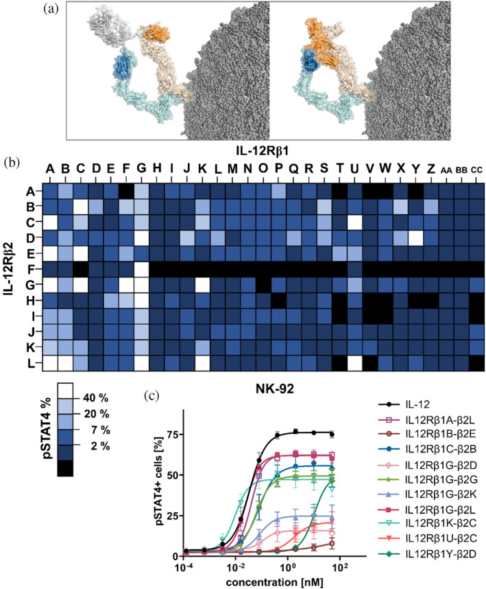
In this work, we have generated bispecific interleukin (IL)-12 surrogate agonists based on camelid-derived single-domain antibodies (sdAbs) targeting the IL-12 receptor (IL-12R) subunits IL-12Rβ1 and IL-12Rβ2. Following immunization and antibody display-based paratope isolation, respective sdAbs were combinatorially reformatted into a monovalent bispecific architecture by grafting resulting paratopes onto the hinge region of a heterodimeric Fc region. Functional characterization using NK-92 cells enabled the identification of multiple different sdAb-based bispecifics displaying divergent IL-12R agonism capacities as analyzed by STAT4 phosphorylation. Further investigations by harnessing peripheral blood mononuclear cells (PBMCs) from healthy donors revealed attenuated pSTAT4 activation compared to recombinant human (rh) wild-type IL-12 regarding both natural killer (NK)-cell and T-cell activation but robust IL-12R agonism on stimulated T cells. While several sdAb-based IL-12 mimetics were nearly inactive on NK cells as well as T cells obtained from PBMCs, they elicited significant STAT4 phosphorylation and interferon (IFN)-γ release on stimulated T cells as well as an IL-12-like transcriptional signature. Furthermore, we demonstrate that the activity of receptor agonism of generated bispecific IL-12 mimetics can also be biased towards stimulated T cells by changing the spatial orientation of the individual sdAbs within the molecular design architecture. Taken together, we present an alternative strategy to generate IL-12-like biologics with tailor-made characteristics.

Keywords: IL‐12; NK cell; T cell; VHH; antibody engineering; bispecific antibody; cytokine mimetic; single‐domain antibody; surrogate cytokine; yeast surface display.


Conflict of interest statement

BL, LU, PA, SB, SK, JH, AD, LH, CX, AM, AE, DE, LP, and SZ are employees at either Merck Healthcare KGaA or EMD Serono. Besides, this work was conducted in the absence of any further commercial interest.

Figures

FIGURE 1
FIGURE 1
Combinatorial reformatting of sdAbs targeting IL‐12Rβ1 and IL‐12Rβ2 into a bispecific antibody architecture (1 + 1) enables screening of IL‐12R surrogate agonists. (a) Left: bispecific single‐domain antibody binding to the heterodimeric IL‐12R by engaging IL‐12Rβ1 (light orange) and IL‐12Rβ2 (light blue) and eliciting downstream signaling. Right: heterodimeric IL‐12 initiates a complex with its own receptor through the subunits p35 (IL12A, blue) and p40 (IL12B, orange). Structural visualization was generated with PyMOL software version 2.3.0, based on PDB entry 8ODZ and structural modeling as described in section 4. (b) Heatmap of NK‐92 STAT4 phosphorylation capacity of IL‐12R agonists by combinatorial reformatting. Molecules failing expression are displayed in black, percentage of pSTAT4+ cells for active agonists are depicted in dark blue (<2%) to visualize functionally inactive bsAbs, while lighter blue shades (<7%, <20%, <40%) are given for moderately active surrogate agonists. The highest agonism is shown for constructs in white (>40%). Data from two independent experiments are depicted. (c) NK‐92 cells were incubated with IL‐12 and surrogate agonists in varying concentrations for 45 min. Prior to pSTAT4 staining, cells were fixed and permeabilized. Mean values ± SEM of three independent experiments are shown.
FIGURE 2
FIGURE 2
IL‐12 mimicking sdAb‐based surrogate agonists preferentially activate stimulated T cells with reduced activity on NK and T cells. (a–c) Intracellular staining of pSTAT4‐positive cells after 45 min incubation with IL‐12 or IL‐12 surrogate agonists (IL12Rβ1G‐β2K, IL12Rβ1G‐β2L, IL12Rβ1U‐β2C, or IL12Rβ1Y‐β2D). Graphs display mean values ± SEM of three (NK cells) to four independent experiments. (d) Absolute STAT4 phosphorylation in T cells displayed for each donor depending on their stimulation state. (e) Ratio of maximal pSTAT4 signaling of stimulated T cells vs. NK or vs. T cells. (f) IFN‐γ levels measured by HTRF in supernatant of stimulated T cells after 24 h stimulation with IL‐12 or IL‐12 surrogate agonists (IL12Rβ1G‐β2K, IL12Rβ1G‐β2l, IL12Rβ1U‐β2C, or IL12Rβ1Y‐β2D) in a dose–response curve. Mean values ± SEM of four independent experiments are shown.
FIGURE 3
FIGURE 3
Antibody engineering enables generation of IL‐12 mimetic with pronounced bias for activated T cells. (a) Two different bsAb architectures incorporating paratopes IL12Rβ1G and IL12Rβ2L were functionally compared. For the C‐terminal IgG‐VHH format, VHHs were grafted to the C‐terminus of the heavy chain of an isotype control antibody (aDIG) by incorporating a 15 amino acid linker (3xGly4Ser). (b–d) Intracellular staining of pSTAT4+ cells after 45 min incubation with IL‐12 or IL‐12 surrogate agonists at different concentrations (IL12Rβ1G‐β2L and aDIG_IL12Rβ1G‐β2L). Graphs display mean values ± SEM of three (NK cells) to four independent experiments. (e) Absolute pSTAT4 signaling in T cells displayed for each donor depending on their stimulation state. (f) Ratio of maximal pSTAT signaling of pre‐activated T cells vs. NK or vs. T cells. (f) IFN‐γ levels measured by HTRF in supernatant of stimulated T cells after 24 h stimulation with IL‐12 or IL‐12 surrogate agonists (IL12Rβ1G‐β2L and aDIG_IL12Rβ1G‐β2L) at varying concentrations. Mean values ± SEM of four independent experiments are given.
FIGURE 4
FIGURE 4
Hierarchical clustering of target DEGs by IL‐12 and antibody based surrogate agonists. Gene expression analysis in stimulated T cells from three donors treated with IL‐12 or surrogate agonists for 24 h. (a) Heatmap is generated based on FPKM values of differentially expressed genes with mainstream hierarchical clustering and normalized rows via z‐score (Gene expression value in sample of interest) − (Mean expression across all samples)/Standard Deviation). Genes are arrayed by row and agonists by column. IL‐12‐related genes that were aligned to the molecular signature database MSigDB (Liberzon et al. ; Subramanian et al. 2005) and significantly altered (p adj <0.05) by IL‐12 or surrogate agonist treatments are displayed.

References

    1. Airoldi I, Gri G, Marshall JD, Corcione A, Facchetti P, Guglielmino R, et al. Expression and function of IL‐12 and IL‐18 receptors on human tonsillar B cells. J Immunol. 2000;165:6880–6888. - PubMed
    1. Andrews LP, Butler SC, Cui J, Cillo AR, Cardello C, Liu C, et al. LAG‐3 and PD‐1 synergize on CD8(+) T cells to drive T cell exhaustion and hinder autocrine IFN‐γ‐dependent anti‐tumor immunity. Cell. 2024;187:4355–4372.e22. - PMC - PubMed
    1. Bauer C, Ciesielski E, Pekar L, Krah S, Toleikis L, Zielonka S, et al. Facile one‐step generation of camelid VHH and avian scFv libraries for phage display by golden gate cloning. Methods Mol Biol. 2023;2681:47–60. - PubMed
    1. Bloch Y,Felix J,Merceron R. et al. Structures of complete extracellular receptor assemblies mediated by IL‐12 and IL‐23. Nat Struct Mol Biol 2024; 31:591–597. 10.1038/s41594-023-01190-6 - DOI - PubMed
    1. Boje AS, Pekar L, Koep K, Lipinski B, Rabinovich B, Evers A, et al. Impact of antibody architecture and paratope valency on effector functions of bispecific NKp30xEGFR natural killer cell engagers. MAbs. 2024;16(2):315–640. - PMC - PubMed

MeSH terms

LinkOut - more resources