. 2025 Mar 4;19(8):7650-7660.
doi: 10.1021/acsnano.4c09661. Epub 2025 Feb 21.

Electronically Perturbed Vibrational Excitations of the Luminescing Stable Blatter Radical

Affiliations

Electronically Perturbed Vibrational Excitations of the Luminescing Stable Blatter Radical

Jonathan Bar-David et al. ACS Nano. .

Abstract

Stable radicals are spin-active species with a plethora of proposed applications in fields from energy storage and molecular electronics to quantum communications. However, their optical properties and vibrational modes are so far not well understood. Furthermore, it is not yet clear how these are affected by the radical oxidation state, which is key to understanding their electronic transport. Here, we identify the properties of 1,2,4-benzotriazin-4-yl, a stable doubly thiolated variant of the Blatter radical, using surface-enhanced Raman scattering (SERS). Embedding molecular monolayers in plasmonic nanocavities gives access to their vibrational modes, photoluminescence, and optical response during redox processes. We reveal the influence of the adjacent metallic surfaces and identify fluctuating SERS signals that suggest a coupling between the unpaired radical electron and a spatially overlapping vibrational mode. This can potentially be exploited for information-storage devices and chemically designed molecular qubits.

Keywords: Raman spectroscopy; SAM; SERS; electrochemistry; nanoparticles; nanophotonics; radicals.


Conflict of interest statement

The authors declare no competing financial interest.

Figures
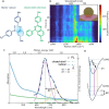
Figure 1
Figure 1
(A) Molecular structure of the Blatter radical and reference molecule with a closed shell (“closed-shell”). The standard acetyl [C(O)Me] protecting groups on the sulfur atoms are removed upon contact with gold. Delocalization of the radical electron over the conjugated core is shaded. (B) Time-resolved SERS spectra of 200 Blatter radicals in a plasmonic nanoparticle-on-mirror (NPoM) system (illustrated in the inset). Dotted lines follow the maxima of the 1065 and 1360 cm–1 SERS lines, showing their energy variation. (C) Absorption (α, solid) and photoluminescence (PL, dashed) spectra for the radical (blue) and closed-shell reference (green) molecules in ethanol. The radical has a 40 meV smaller band gap but PL 400× stronger. PL is decomposed into two Gaussians, from 0′ to 0 and 0′–1 decay to ground and excited vibrational levels, as in (D). Vibrational energy, ΔE ∼ 0.16 eV∼1360 cm–1, from the dominantly coupled molecular vibration.
Figure 2
Figure 2
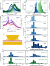
White-light scattering spectra of NPoMs. (A) Raw DF spectra for two NPoMs containing either the Blatter radical (blue) or the closed shell (green), constructed from focus-stacks to give a chromatically corrected scattering spectrum. (B) Histograms of the initial NPoM peak scattering wavelength showing 80 nm spectral shift between radical and closed-shell molecules. (C) Scattering spectra (dashed) from a single NPoM before (blue) and after (red) laser irradiation, overlaid on emission spectra [solid, from initial (blue) to final (red)], showing that both scattering and emission peaks redshift during irradiation. (D) Schematic of the bottom facet rearrangement (orange arrows) during irradiation. (E) Histograms of the scattering shift from before vs after laser irradiation for excitation powers from (i) 10 μW to (v) 100 μW. (F) Scattering shifts for radical vs closed-shell molecules for 50 μW irradiation at (i,iii) 633 nm and (ii,iv) 785 nm. Dashed line shows the average shift for each case (larger for the radical).
Figure 3
Figure 3
Average emission spectra of NPoMs. (A) Emission spectra of the Blatter radical (blue) composed of SERRS spectra (dashed) on a broad PL background (dotted), compared to the closed-shell molecule (green) without PL. (B) Average initial and final spectra of radical-filled NPoMs, showing PL bleaching over T = 30 s. For the closed-shell molecule, no bleaching is observed, and the initial and final spectra overlap exactly. (C) Comparison of SERRS peaks of the radical for 633 nm (blue) vs 785 nm (pink) excitation compared to DFT (black) calculation. The main vibrations shaded are 1065 (α), 1365 (β), and 1580 cm–1. (D) SERS spectrum of the closed-shell reference molecule (green) compared to the DFT calculation (black). (E,F) Illustrations of the α, β vibrational modes for the (E) Blatter radical and (F) closed-shell reference.
Figure 4
Figure 4
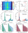
SERRS decay. (A) Time-dependent emission from an NPoM with the Blatter radical SAM, showing fast PL quenching in ∼2 s. (B) Time-dependent emission from NPoM with closed-shell SAM. (C) Average temporal evolution of 1065 (light) and 1365 cm–1 (dark) SERRS lines for the Blatter radical with 633 nm (blue) and 785 nm (red) pumps for closed-shell (green circles) and PL (purple). Fits are to a sum of fast exponential and slow linear decay: I(t) = Aexp{−te} – BtL + C (radical) or linear only (closed-shell) decay. (D,E) Evolution of radical (D) 1065 and (E) 1365 cm–1 SERS peaks at increasing powers (10–100 μW, 633 nm).
Figure 5
Figure 5
Time-jitter of Blatter radical SERS lines. (A) Evolving SERS of the Blatter radical in NPoM (785 nm pump), showing jitter of α and β peaks (dashed). (B) Extracted jittering of SERS peaks for three individual NPoMs, comparing radical and closed-shell SAMs at different laser λ indicated. (C,D) Histograms of SERS νpk for radical (blue) vs closed shell (green, C) and radical at 633 vs 785 nm (D). (E,F) Histograms of jitter rate, showing faster jitter for β vibration of the radical.
Figure 6
Figure 6
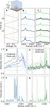
Measured SERS and PL for different potential steps. (A) Voltage steps applied to the Au mirror working electrode (inset). (B,C) Average spectra at each potential for radical and closed-shell SAMs, expanded to the range of interest in (D,E). (F,G) DFT-calculated SERS spectra for the (F) radical and (G) closed shell in oxidized and reduced states.

References

    1. Chen Z. X.; Li Y.; Huang F. Persistent and Stable Organic Radicals: Design, Synthesis, and Applications. Chem. 2021, 7 (2), 288–332. 10.1016/j.chempr.2020.09.024. - DOI
    1. Hicks R. G. What’s New in Stable Radical Chemistry?. Org. Biomol Chem. 2006, 5 (9), 1321–1338. 10.1039/b617142g. - DOI - PubMed
    1. Ratera I.; Veciana J. Playing with Organic Radicals as Building Blocks for Functional Molecular Materials. Chem. Soc. Rev. 2012, 41 (1), 303–349. 10.1039/C1CS15165G. - DOI - PubMed
    1. Casu M. B. Nanoscale Studies of Organic Radicals: Surface, Interface, and Spinterface. Acc. Chem. Res. 2018, 51 (3), 753–760. 10.1021/acs.accounts.7b00612. - DOI - PubMed
    1. Constantinides C. P.; Koutentis P. A. Stable N- and N/S-Rich Heterocyclic Radicals: Synthesis and Applications. Adv. Heterocycl. Chem. 2016, 119, 173–207. 10.1016/bs.aihch.2016.03.001. - DOI

LinkOut - more resources