. 2025 Apr;67(3):119-135.
doi: 10.1111/dgd.70000. Epub 2025 Feb 21.

The property of larval cells of the scleractinian coral, Acropora tenuis, deduced from in vitro cultured cells

Affiliations

The property of larval cells of the scleractinian coral, Acropora tenuis, deduced from in vitro cultured cells

Kaz Kawamura et al. Dev Growth Differ. 2025 Apr.

Abstract

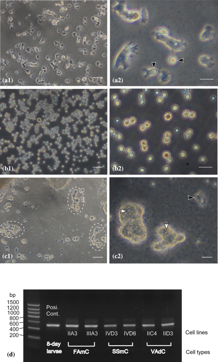
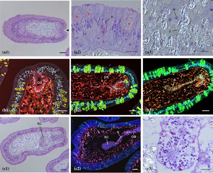
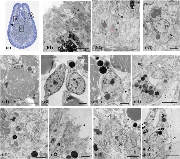
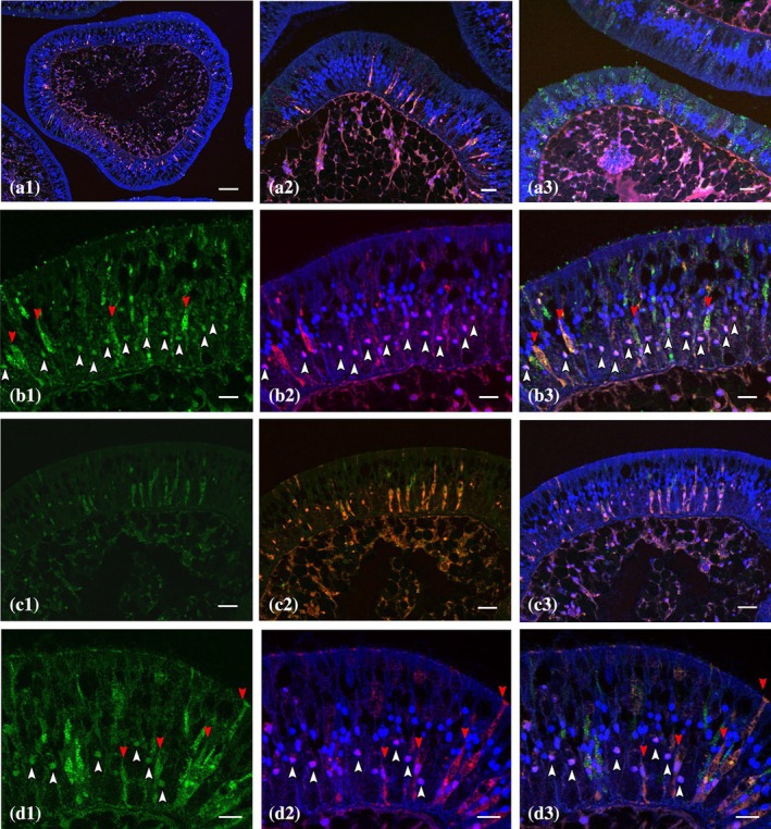
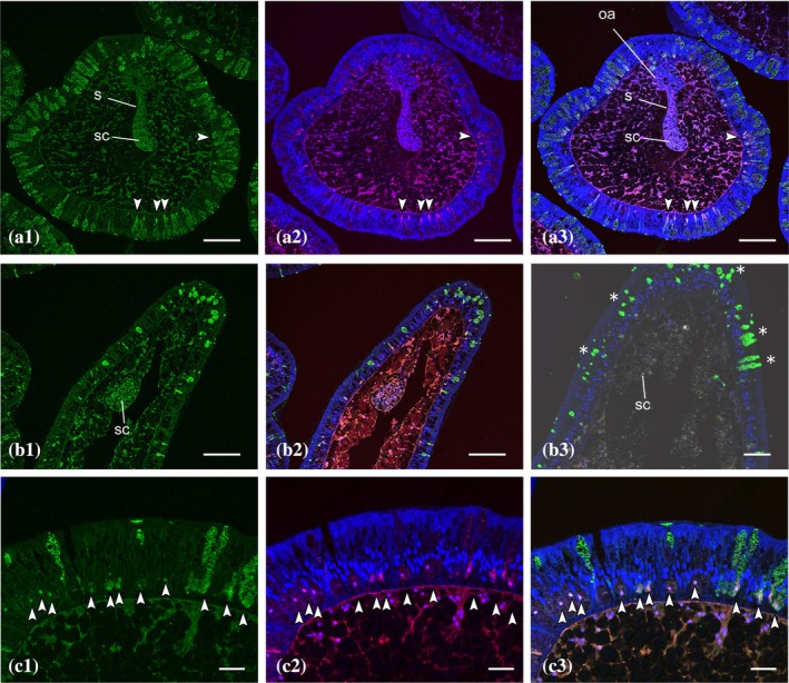
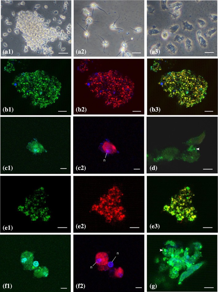
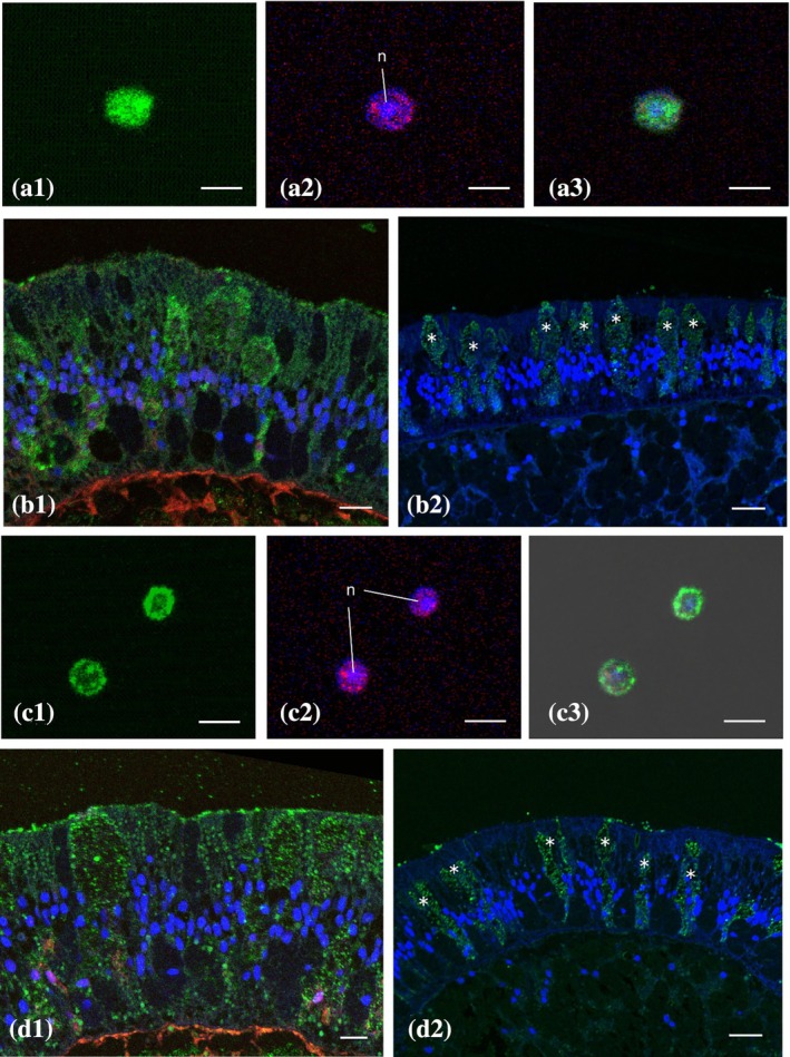
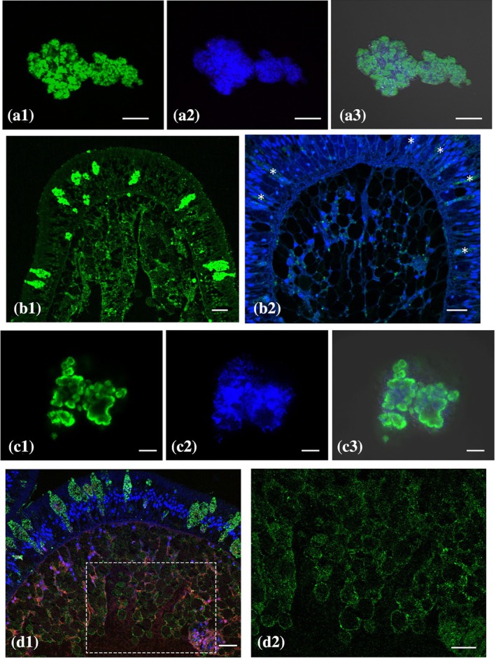
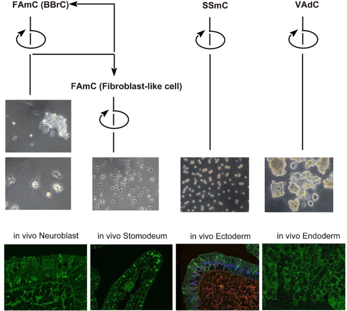
In previous studies, we have established approximately 15 cultured cell-lines derived from planula larvae of Acropora tenuis. Based on their morphology and behavior, these cells were classified into three types, flattened amorphous cells (FAmCs), vacuolated adherent cells (VAdCs), and small smooth cells (SSmCs). FAmCs include fibroblast-like cells and spherical, brilliant brown cells (BBrCs), which are transformable to each other. To examine the larval origin of the three cell types, we raised antibodies: anti-AtMLRP2 that appears to recognize FAmC, anti-AtAHNAK for BBrC, anti-AtSOMP5 and anti-AtEndoG for SSmC, and anti-AtGal and anti-AtFat4 for VAdC, respectively. Anti-AtMLRP2 antibody stained in vivo stomodeum and neuroblast-like cells embedded in larval ectoderm around the aboral pole. Anti-AtAHNAK antibody stained neuron-like and neuroblast-like cells, both of which were also stained with neuron-specific tubulin β-3 antibody. These results suggest that in vitro BBrCs and in vivo neuroblast-like cells share neuronal properties in common. Two antibodies for SSmCs, anti-AtSOMP5 and anti-AtEndoG, stained larval ectoderm cells, suggesting that SSmCs have larval ectoderm properties. Two antibodies for VAdCs, anti-AtGal and anti-AtFat4, stained larval endoderm cells, suggesting that VAdCs have larval endoderm properties. Therefore, the in vitro cell lines appear to retain properties of the stomodeum, neuroblast, ectoderm, or endoderm. Each of them may be used in future investigations to reveal cellular and molecular properties of cell types of coral larvae, such as the potential for symbiosis.

Keywords: Acropora coral; ectoderm; endoderm; in vitro cell system; larval cell property; neuroblast; stomodeum.


Conflict of interest statement

We declare no competing interests.

Figures

FIGURE 1
FIGURE 1
Three morphological types of cultured cells in Acropora tenuis. (a) Flattened amorphous cells (FAmCs). (a1) IIA3 cell line. (a2) Higher magnification of IIA3 showing characteristic, well‐developed lamellipodia and filopodia. FAmCs are interchangeable with brilliant brown cells (black arrowheads). (b) Small smooth cells (SSmCs). (b1) IVC3 cell line. (b2) Higher magnification of IVC3. These cells do not extend pseudopodia. (c) Vacuolated adherent cells (VAdCs). (c1) IVC1 cell line showing cell–cell adhesion (broken circles). (c2) Higher magnification of IVC1. Black arrowhead shows a single cell. White arrowheads show vacuoles in cell aggregates. Bars in a1, b1, c1, 50 μm. Bars in a2, b2, c2, 20 μm. (d) RT‐PCR amplification of RNAs isolated from 8‐day larvae, FAmCs (IIA3 and IIIA3 lines), SSmCs (IVD3 and IVD6 lines), and VAdCs (IIC4 and IID3 lines), using primers specific to an A. tenuis cytoplasmic Actin gene. Approximately 510‐bp‐long amplicons were amplified, of which sequences were completely matched to the A. tenuis gene.
FIGURE 2
FIGURE 2
Anatomy of Acropora tenuis larvae. (a) 6‐day larva. Toluidine blue staining. (a1) Longitudinal section of the whole body. The oral concavity (arrowhead) is visible. Bar, 50 μm. (a2) Ectodermal cell layer. A portion of cells (asterisks) have light brown color and contain vacuoles in the cytoplasm. n, nucleus. Bar, 20 μm. (a3) Endodermal cell layer. e, endodermal cell; y, yolk cell. Bar, 10 μm. (b) 8‐ and 11‐day larvae. Rhodamine‐phalloidin (red) and DAPI (blue) staining, confocal microscopy image. Non‐specific fluorescence colored green is observable in the ectoderm. (b1) Oral area of an 8‐day larva. oa, oral aperture. 50 μm. (b2) Developing stomodeum of an 8‐day larva. Cells seem to spread in the aboral direction in line. ps, primordial stomodeum. Bar, 50 μm. (b3) The stomodeal epithelium of an 11‐day larva in contact with the oral concavity. s, stomodeum. Bar, 50 μm. (c) 18‐day larvae. (c1) Toluidine blue staining. The stomodeum reaches the middle of the larva along the long (oral–aboral) axis. sc, stomodeum clump. Bars, 50 μm. (c2) Confocal microscopy of rhodamine‐phalloidin and DAPI staining. The leading tip of the stomodeum forms a clump. oa, oral aperture. Bars, 50 μm. (c3) Toluidine blue staining of stomodeal clump that consists of elongated cells. Bar, 20 μm.
FIGURE 3
FIGURE 3
Ultrastructure of major tissues of a 14‐day planula larva of Acropora tenuis. (a) Semi‐thin section of a larva along the oral–aboral axis. Bar, 50 μm. (b) Ectodermal cell layer. (b1) Apical region of the cell layer. Large vacuoles are prominent. m, mitochondrion; v, vacuole. (b2) Basal region of the cell layer. The nucleus occurs in the middle. Undifferentiated cells (asterisk) are scattered near the bottom. n, nucleus. (b3) An undifferentiated cell. (c) Endodermal cell layer. (c1) A yolk cell filled with oil droplets. (c2) Endodermal cells are embedded among yolk cells. (c3) Endodermal cells adhere to other endodermal cells and to yolk cells via adhesion plaques (arrowheads). (c4) Gastrodermis is associated with mesoglea. mg, mesoglea. (d) Stomodeum. (d1) The cell body is enriched with mitochondria. (d2) Flagella (black arrowheads) are located at the apical surface of the stomodeum. (d3) Growing extremity of the stomodeum. A white arrowhead shows pseudopodia. (d4) Pseudopodia (white arrowheads) are prominent at the extremity. Bars in b2 and d2, 5 μm. Other bars in b–d, 2 μm.
FIGURE 4
FIGURE 4
Immunostaining of 18‐day planulae stained with anti‐neuroblast differentiation‐associated protein AHNAK antibody (green), counterstained with DAPI. Sections were also stained with rhodamine‐phalloidin (red) in (a)–(c). White and red arrowheads show neuroblasts and neurons, respectively. (a) Specificity of fluorescent signals. (a1) A whole larva stained without antibodies. Bar, 50 μm. (a2) Oral region stained with the secondary antibody. Bar, 20 μm. (a3) Immunostaining of oral region in the presence of the preimmune rabbit serum. Bar, 20 μm. (b) Immunostaining at the aboral pole. Bars, 10 μm. (b1) FITC image of anti‐AHNAK antibody staining. (b2) Rhodamine‐phalloidin staining merged with DAPI image. (b3) Triple images of FITC, rhodamine‐phalloidin, and DAPI. (c) Negative control of aboral region stained by anti‐AtAHNAK antibody preabsorbed by AHNAK peptide. Bars, 20 μm. (c1) FITC image. (c2) Rhodamine‐phalloidin staining merged with DAPI image. (c3) Triple images of FITC, rhodamine‐phalloidin, and DAPI. (d) Double immunostaining of aboral region with anti‐AtAHNAK antibody (green) and anti‐neuron‐specific tubulin β‐3 TUBB3 antibody (red). Bars, 10 μm. (d1) AHNAK‐FITC image. (d2) TUBB3‐rhodamine B image merged with DAPI image. (d3) Triple image of FITC, rhodamine B, and DAPI.
FIGURE 5
FIGURE 5
Immunostaining of planulae with anti‐AtMLRP2 (a1–c1) and anti‐TUBB3 antibodies (c2). Specimens were counterstained with rhodamine‐phalloidin and DAPI (a2, b2). Merged images are shown in a3 and c3. Absorption test by peptide antigen is shown in b3. Asterisks show non‐specific signals. (a) 8‐day planula. Arrowheads show immuno‐responsive cells embedded in the ectoderm. oa, oral aperture; s, stomodeum. Bars, 100 μm. (b) 18‐day planula. sc, stomodeum clump. Bars in b1 and b2, 100 μm. Bar in B3, 50 μm. (c) Double immunostaining of 18‐day planula using anti‐AtMLRP2 (green) and anti‐TUBB3 (red) antibodies. Arrowheads show double‐positive cells. Bars, 20 μm.
FIGURE 6
FIGURE 6
Fluorescent immunocytochemistry of the BBrC subpopulation. (a) In vitro behavior of BBrCs. (a1) Cell aggregates. Bar, 50 μm. (a2) Dissociation of floating aggregates. Note that most dissociated cells look brilliant brown, and they possess elongated filopodia. Bar, 20 μm. (a3) One day after cell dissociation, the majority of cells assume a fibroblast‐like configuration. Bar, 20 μm. (b) Anti‐AtAHNAK immunostaining (b1) and anti‐TUBB3 immunostaining (b2) of aggregated cells. (b3) Merged image. Bars, 20 μm. (c) A single BBrC stained with anti‐AtAHNAK antibodies (c1) or with anti‐TUBB3 antibodies (c2). Cells are counterstained with DAPI. n, nucleus. Bars, 5 μm. (d) Fibroblast‐like cells not stained with anti‐AtAHNAK antibody. Arrowhead shows dot‐like weak signal. Bar, 5 μm. (e) Anti‐AtMLRP2 (e1) and anti‐TUBB3 immunostaining (e2) of aggregated cells. (e3) Merged image. Bars, 20 μm. (f) A few BBrCs stained with anti‐AtMLRP2 antibodies (f1) or with anti‐TUBB3 antibodies (f2). Cells are counterstained with DAPI. Bars, 5 μm. (g) Fibroblast‐like cells stained with anti‐AtMLRP2 antibodies. Arrowhead shows dot‐like strong signal. Bar, 5 μm.
FIGURE 7
FIGURE 7
Fluorescent immunohistochemistry of AtSOMP5 and AtEndoG, confocal microscopy. (a, b) Anti‐AtSOMP5 immunocytochemistry. (a) The SSmC cell line, IVC3. Bars, 5 μm. (a1) FITC image of AtSOMP5 (green). (a2) DAPI and rhodamine‐phalloidin (red) images. n, nucleus. (a3) Merged image. (b) Ectodermal cells in a 10‐day larva. (b1) FITC image merged with DAPI and rhodamine‐phalloidin images. Bar, 10 μm. (b2) Negative control absorbed by peptide antigen. Asterisks show non‐specific signals. Bar, 20 μm. (c, d) Anti‐AtEndoG immunocytochemistry. (c) The SSmC cell line, IVC3. Bars, 5 μm. (c1) FITC image of AtEndoG. (c2) DAPI and rhodamine‐phalloidin images. (c3) Merged image. (d) 6‐day larva. (d1) FITC image of AtEndoG in ectodermal cell layer merged with DAPI and rhodamine‐phalloidin images. Bar, 10 μm. (d2) Negative control absorbed by peptide antigen. Asterisks show non‐specific signals. Bar, 20 μm.
FIGURE 8
FIGURE 8
Fluorescence immunohistochemistry of AtGal and AtFAT4, confocal microscopy. (a) Anti‐AtGal immunocytochemistry of the VAdC cell line, IVC1. Bars, 50 μm. (a1) FITC image of AtGal. (a2) DAPI image. (a3) Merged image. (b) An 18‐day larva. Bars, 20 μm. (b1) FITC image. (b2) Negative control absorbed by peptide antigen. Asterisks show non‐specific signals. (c) Anti‐AtFAT4 immunocytochemistry of the VAdC cell line, IVC1. Bars, 20 μm. (c1) FITC image of AtFAT4. (c2) DAPI image. (c3) Merged image. (d) An 8‐day larva. 20 μm. (d1) FITC image merged with DAPI and rhodamine‐phalloidin images. (d2) FITC image of the square shown in (d1).
FIGURE 9
FIGURE 9
Summary of results. Three lines of in vitro cultured cells have been established: flattened amorphous cells (FAmCs), small smooth cells (SSmCs), and vacuolated adherent cells (VAdCs). They correspond immunohistochemically to in vivo larval cells and tissues by the aid of six gene products deduced from differential transcriptome assay. FAmCs include two subpopulations, brilliant brown cells (BBrCs) and fibroblast‐like cells that renew by themselves and are interchangeable. BBrCs share some molecular features with in vivo neuroblasts. Fibroblast‐like cells are similar to in vivo stomodeum, based upon antigenic expression, cell morphology, and the presence of pseudopodia. On the other hand, SSmCs share antigens with ectodermal cells. VAdCs adhere to the same types of cells and share antigens with endodermal cells.

Similar articles

References

    1. Attenborough, R. M. F. , Hayward, D. C. , Wiedemann, U. , Forêt, S. , Miller, D. J. , & Ball, E. E. (2019). Expression of the neuropeptides RFamide and LWamide during development of the coral *Acropora millepora* in relation to settlement and metamorphosis. Developmental Biology, 446, 56–67. - PubMed
    1. Bernasconi, R. , Stat, M. , Koenders, A. , Paparini, A. , Bunce, M. , & Huggett, M. J. (2019). Establishment of coral‐bacteria symbioses reveal changes in the core bacterialcommunity with host ontogeny. Frontiers in Microbiology, 10, 1529. - PMC - PubMed
    1. Bode, H. R. , Heimfeld, S. , Chow, M. A. , & Huang, L. W. (1987). Gland cells arise by differentiation from interstitial cells in Hydra attenuata . Developmental Biology, 122, 577–585. - PubMed
    1. Chapman, D. M. (1999). Microanatomy of the bell rim of Aurelia aurita (cnidaria: Scyphozoa). Canadian Journal of Zoology, 77, 34–46.
    1. Coffroth, M. A. , & Santos, S. R. (2005). Genetic diversity of symbiotic dinoflagellates in the genus Symbiodinium . Protist, P156, 19–34. 10.1016/j.protis.2005.02.004 - DOI - PubMed

Grants and funding