. 2025 Aug;603(15):4345-4366.
doi: 10.1113/JP285194. Epub 2025 Feb 21.

Endothelial TRPV4-Cx43 signalling complex regulates vasomotor tone in resistance arteries

Affiliations

Endothelial TRPV4-Cx43 signalling complex regulates vasomotor tone in resistance arteries

Pía C Burboa et al. J Physiol. 2025 Aug.

Abstract

S-nitrosylation of Cx43 gap junction channels critically regulates communication between smooth muscle cells and endothelial cells. This post-translational modification also induces the opening of undocked Cx43 hemichannels. However, its specific impact on vasomotor regulation remains unclear. Considering the role of endothelial TRPV4 channel activation in promoting vasodilatation through nitric oxide (NO) production, we investigated the direct modulation of endothelial Cx43 hemichannels by TRPV4 channel activation. Using the proximity ligation assay, we identified that Cx43 and TRPV4 are found in close proximity in the endothelium of resistance arteries. In primary endothelial cell (EC) cultures from resistance arteries, GSK 1016790A-induced TRPV4 activation enhances eNOS activity, increases NO production, and opens Cx43 hemichannels via direct S-nitrosylation. Notably, the elevated intracellular Ca2+ levels caused by TRPV4 activation were reduced by blocking Cx43 hemichannels. In ex vivo mesenteric arteries, inhibiting Cx43 hemichannels reduced endothelial hyperpolarization without affecting NO production in ECs, underscoring a critical role of TRPV4-Cx43 signalling in endothelial electrical behaviour. We perturbed the proximity of Cx43/TRPV4 by disrupting lipid rafts in ECs using β-cyclodextrin. Under these conditions, hemichannel activity, Ca2+ influx and endothelial hyperpolarization were blunted upon GSK stimulation. Intravital microscopy of mesenteric arterioles in vivo further demonstrated that inhibiting Cx43 hemichannel activity, NO production and disrupting endothelial integrity reduce TRPV4-induced relaxation. These findings underscore a new pivotal role of the Cx43 hemichannel associated with the TRPV4 signalling pathway in modulating endothelial electrical behaviour and vasomotor tone regulation. KEY POINTS: TRPV4-Cx43 interaction in endothelial cells: the study reveals a close proximity between Cx43 proteins and TRPV4 channels in endothelial cells of resistance arteries, establishing a functional interaction that is critical for vascular regulation. S-nitrosylation of Cx43 hemichannels: TRPV4 activation via GSK treatment induces S-nitrosylation of Cx43, facilitating the opening of Cx43 hemichannels. TRPV4-mediated calcium signalling: activation of TRPV4 leads to increased intracellular Ca2+ levels in endothelial cells, an effect that is mitigated by the inhibition of Cx43 hemichannels, indicating a regulatory feedback mechanism between these two channels. Endothelial hyperpolarization and vasomotor regulation: Blocking Cx43 hemichannels impairs endothelial hyperpolarization in mesenteric arteries, without affecting NO production, suggesting a role for Cx43 in modulating endothelial electrical behaviour and contributing to vasodilatation. In vivo role of Cx43 hemichannels in vasodilatation: intravital microscopy of mouse mesenteric arterioles demonstrated that inhibiting Cx43 hemichannel activity and disrupting endothelial integrity significantly impair TRPV4-induced vasodilatation, highlighting the crucial role of Cx43 in regulating endothelial function and vascular relaxation.

Keywords: ECs; caveolae; hyperpolarization; resting membrane potential.


Conflict of interest statement

No competing interests declared.

Figures

Figure 1
Figure 1. TRPV4–Cx43 complex is primarily observed within the endothelial cell layer of resistance arteries
Proximity ligation assay (PLA) was utilized to illustrate the spatial co‐localization between TRPV4 and Cx43. Notably, TRPV4 and Cx43 exhibit predominant distribution within the endothelial layer (EC) compared to the smooth muscle cells (SMC). Controls were conducted to validate the specificity of PLA analysis, wherein primary antibodies were omitted as a negative control. Asterisks indicate the luminal region of the vessels, while the green, fluorescence signal corresponds to the internal elastic lamina (IEL). BF, brightfield. Scale bar: 80 µm.
Figure 2
Figure 2. TRPV4 activation promote S‐nitrosylated Cx43 hemichannel activity by NO production
A, representative immunofluorescence microscopy images illustrating the detection of phosphorylation status at serine 177 and threonine 495 residues of endothelial nitric oxide synthase (eNOS), alongside the assessment of protein levels of S‐nitrosylated proteins, in vehicle conditions and upon stimulation with 10 nM GSK 1016790A on primary cultures of ECs. B, representative images from proximity ligation assay demonstrating Cx43 S‐nitrosylation levels in both control and 10 nM GSK 1016790A‐stimulated conditions. C, quantification of Cx43‐SNO dot signals per cell. Comparisons between groups were made using Student's t test. D, percentage of positive cells displaying Cx43‐SNO signal. Comparisons between groups were made using Student's t test. E, western blot analysis (left) and quantification (right) of S‐nitrosylated Cx43. The S‐nitrosylated Cx43 levels were expressed as fold change relative to total Cx43 protein levels per sample. Comparisons between groups were made using Student's t test. F, time course analysis to measure the uptake of 5 µM ethidium, assessing hemichannel activity upon 10 nM GSK 1016790A and under pre‐treatment of 100 µM L‐NAME, 50 µM glycyrrhetinic acid (β‐GA) and 50 µM Gap19. Statistical comparisons between the time course curves were performed using one‐way ANOVA followed by Tukey's post hoc test. The total number of mice used in the experiments is indicated in parentheses. Scale bar: 80 µm.
Figure 3
Figure 3. TRPV4 controls Cx43 hemichannel activity in a heterologous expression system
Activation of Cx43 hemichannels was evaluated by assessing ethidium uptake. Connexin‐free HeLa cells were transfected with human EGFP‐tagged Cx43, either alone or in combination with human TRPV4. Cells transfected with EGFP alone were used as controls. Dye uptake was evaluated in basal conditions and in the presence of 1 µM GSK under constant superfusion. A, representative time courses of ethidium uptake. Each group corresponds to the average of several GFP‐positive cells. B, quantification of the ethidium uptake rate. Statistical comparisons between groups were performed using one‐way ANOVA and Tukey's post hoc test.
Figure 4
Figure 4. TRPV4 decreases Cx43 expression
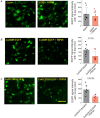
A, representative images of the EGFP signal in cells transfected with EGFP alone or EGFP + TRPV4. Images were taken with the same exposure time (500 ms) for comparison. The quantification of EGFP signal intensity with 1 s exposure is shown in the bar graph. B, representative images of the EGFP signal in cells transfected with Cx43WT‐EGFP alone or Cx43WT‐EGFP + TRPV4. Images were taken with the same exposure time (1 s). The quantification of EGFP signal intensity is shown in the bar graph. C, representative images of the EGFP signal in cells transfected with Cx43C271S‐EGFP alone or Cx43C271S‐EGFP + TRPV4. Images were taken with the same exposure time (1 s). The quantification of EGFP signal intensity is shown in the bar graph. Scale bar: 80 µm.
Figure 5
Figure 5. TRPV4–Cx43 hemichannels mediate Ca2+ increases in endothelial cells in response to TRPV4 activation
A, left: representative imaging captures the endothelial cell responses before and after exposure to 10 nM GSK 1016790A stimulation, both under control conditions and in the presence of 50 µM Gap19. Right: time course analysis illustrating the fluctuations in Fluo‐4 signalling subsequent to GSK stimulation. Scale bar: 80 µm. B, peak Ca2+ increases observed in A. Comparisons between groups were made using Student's t test. C, analysis conducted to assess the time required to detect the Ca2+ peak under control conditions and in the presence of 50 µM Gap19 upon GSK simulation. Comparisons between groups were made using Student's t test. The total number of mice used in the experiments is indicated in parentheses. D, time course analysis illustrating the fluctuations in Fluo‐4 signalling subsequent to 10 µM DEANO under 50 µM Gap19 and 1 µM HC‐067047. Comparisons between groups were made using two‐way ANOVA. E, peak Ca2+ increases observed in D. Comparisons between groups were made using nested one‐way ANOVA. F, analysis conducted to assess the time required to detect the Ca2+ peak under control conditions, Ca2+‐free medium, 50 µM Gap19 (I130A) and 1 µM HC‐067047 upon 10 nM GSK 1016790A. G, peak Ca2+ increases observed in F. Comparisons between groups were made using nested one‐way ANOVA.
Figure 6
Figure 6. Cx43 hemichannels do not contribute to NO production in endothelial cells but mediate endothelium hyperpolarization upon TRPV4 activation in intact endothelium
A, NO production was detected using DAF‐2DA in primary endothelial cell cultures after stimulation with 10 nM GSK 1016790A, preceded by pretreatment with 50 µM Gap19 and 100 µM L‐NAME (a NOS blocker). The bars represent the duration of stimulation. B, left: representative en face imaging of an arteriole showing ECs detected using FITC–Dextran 70 KDa in the pipette solution to measure resting membrane potential changes. Scale bar: 20 µm. Right: hyperpolarization peaks were induced by TRPV4 activation in intact ECs from mesenteric arteries under control conditions, 50 µM Gap19 and KCa channel blockers (300 nM charybdotoxin and 200 nM apamin). In another set of experiments, ECs were treated with 10 µM SKA‐31 and Gap19. Group comparisons were made using nested ANOVA. C, time to detect hyperpolarization peaks in ECs treated under control and 50 µM Gap19 conditions following stimulation with 10 nM GSK 1016790A. Group comparisons were made using Student's t test. D, resting membrane potential of arteries under control conditions, with 50 µM Gap19, and with 300 nM charybdotoxin and 200 nM apamin. Group comparisons were made using nested ANOVA. The total number of mice used in the experiments is indicated in parentheses.
Figure 7
Figure 7. Critical role of eNOS/Cx43/TRPV4 proximity in regulating endothelial Ca2+ increases and electrical behaviour
A, representative imaging of proximity ligation assay (PLA) in control conditions and in ECs treated with 5 mM β‐cyclodextrin to assess the spatial proximity between eNOS–TRPV4, Cx43–TRPV4 and Cx43–eNOS. Strong interactions between these proteins are indicated by red dots, while complete disruption of the PLA signal is observed with 5 mM β‐cyclodextrin treatment. Scale bar: 80 µm. B, ethidium bromide uptake rate of ECs following stimulation with 10 nM GSK 1016790A under control conditions, extracellular zero Ca2+ and β‐cyclodextrin treatment. Group comparisons were conducted using nested ANOVA. C, changes in intracellular calcium peak increases under control and cyclodextrin conditions in cells treated with 10 nM GSK 1016790A. D, hyperpolarization peak induced by 10 nM GSK 1016790A under control and cyclodextrin conditions. To assess the activity of KCa channels under β‐cyclodextrin conditions, we used 10 µM SKA‐3. Group comparisons were made using nested ANOVA. The total number of mice used in the experiments is indicated in parentheses.
Figure 8
Figure 8. Endothelial Cx43 hemichannels mediate vasomotor responses in mesenteric arteries in vivo
A, schematic illustration of intravital microscopy approaches. To enhance vessel visualization, 50 µg/kg FITC–Dextran (70 kDa) and 10 µg/kg Gap19, to block Cx43 hemichannel activity, were administered via retroorbital injection. B, representative imaging of the mesenteric bed at baseline, during the phenylephrine (PE) peak, and at the dilatation peak in control and 50 µM Gap19 conditions. Scale bar: 500 µm. C, representative traces of vasomotor responses induced by 100 nM GSK 1016790A, 10 µM PE, 10 µM ACh and 10 µM NO donor DEANO in control, 50 µM Gap19 and L‐NAME conditions. D, percentage of relaxation in vessels pre‐contracted with 10 µM PE upon GSK, 10 µM ACh. Group comparisons were made using Student's t test. The total number of mice used in the experiments is indicated in parentheses. E, percentage of relaxation in vessels precontracted with 10 µM PE upon 10 µM DEANO in control and Gap19‐treated conditions. Group comparisons were made using Student's t test. The total number of mice used in the experiments is indicated in parentheses. F, representative traces depicting vasomotor responses induced by 10 nM GSK 1016790A, 10 µM PE, 10 µM ACh and 10 µM NO donor DEANO in control conditions, alongside conditions where endothelium damage (ED) is present. G, percentage of relaxation in vessels pre‐contracted with 10 µM PE upon GSK, 10 µM ACh and 10 µM DEANO stimulation in both control and ED conditions. Group comparisons were conducted using Student's t test. The total number of mice used in the experiments is indicated in parentheses.
Figure 9
Figure 9. Inhibition of Cx43 hemichannels by Gap19 did not affect smooth muscle function in resistance arteries
A, contraction curve of phenylephrine (PE) with and without 50 µM Gap19. B, smooth muscle membrane potential changes (green) observed in intact isolated arteries under control conditions and in the presence of 10 µM phenylephrine. Group comparisons were conducted using Student's t test. The total number of mice used in the experiments is indicated in parentheses.
Figure 10
Figure 10. NO production is critical for the endothelium hyperpolarization upon TRPV4 activation
A, rate of NO production in ECs under different minutes of incubation time with 100 µM L‐NAME. B, endothelium hyperpolarization peak displayed in control conditions as well as at different minutes of incubation time with 100 µM L‐NAME. Group comparisons were made using nested ANOVA. The total number of mice used in the experiments is indicated in parentheses.
Figure 11
Figure 11. Validation of Cx45 KO cell line
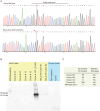
A, Sanger sequencing of Cx45 from wild‐type HeLa cells (top) and the clone no. 18 used in this work. The mutation induced by CRISPR (Synthego) induced the deletion of two nucleotides causing a frameshift in the Cx45 sequence (asterisk). The guide sequence used for guide RNA synthesis is shown as a horizontal black line. The vertical dotted line represents the cut site. B, western blot of HeLa lysates from four different monoclonal populations and two different batches of wild‐type HeLa cells. Clone 18 was transiently transfected with rat Cx45 as a positive control for the Cx45 antibody and to demonstrate transfection efficiency in this clone. Lysates from Xenopus laevis oocytes were also used as control. Oocytes were injected with RNA for Cx45 and cell lysates were obtained 2 days after RNA injection. Non‐injected oocytes were used as the negative control. C, summary of ICE analysis. The predicted KO scores correlates with the signal for Cx45 detected by western blot.

Update of

References

    1. Adapala, R. K. , Talasila, P. K. , Bratz, I. N. , Zhang, D. X. , Suzuki, M. , Meszaros, J. G. , & Thodeti, C. K. (2011). PKCalpha mediates acetylcholine‐induced activation of TRPV4‐dependent calcium influx in endothelial cells. American Journal of Physiology‐Heart and Circulatory Physiology, 301(3), H757–H765. - PMC - PubMed
    1. Archer, S. L. , Gragasin, F. S. , Wu, X. , Wang, S. , McMurtry, S. , Kim, D. H. , Platonov, M. , Koshal, A. , Hashimoto, K. , Campbell, W. B. , Falck, J. R. , & Michelakis, E. D. (2003). Endothelium‐derived hyperpolarizing factor in human internal mammary artery is 11,12‐epoxyeicosatrienoic acid and causes relaxation by activating smooth muscle BK(Ca) channels. Circulation, 107(5), 769–776. - PubMed
    1. Berridge, M. J. (2009). Inositol trisphosphate and calcium signalling mechanisms. Biochimica et Biophysica Acta, 1793(6), 933–940. - PubMed
    1. Billaud, M. , Lohman, A. W. , Straub, A. C. , Looft‐Wilson, R. , Johnstone, S. R. , Araj, C. A. , Best, A. K. , Chekeni, F. B. , Ravichandran, K. S. , Penuela, S. , Laird, D. W. , & Isakson, B. E. (2011). Pannexin1 regulates alpha1‐adrenergic receptor‐ mediated vasoconstriction. Circulation Research, 109(1), 80–85. - PMC - PubMed
    1. Bray, M. A. , Sartain, S. E. , Gollamudi, J. , & Rumbaut, R. E. (2020). Microvascular thrombosis: Experimental and clinical implications. Translational Research, 225, 105–130. - PMC - PubMed

LinkOut - more resources