. 2025 Feb 25;122(8):e2418481122.
doi: 10.1073/pnas.2418481122. Epub 2025 Feb 21.

A hormone-to-neuropeptide pathway inhibits sexual receptivity in immature Drosophila females

Affiliations

A hormone-to-neuropeptide pathway inhibits sexual receptivity in immature Drosophila females

Jie Chen et al. Proc Natl Acad Sci U S A. .

Abstract

Newborns, typically asexual, undergo a process of sexual transition to reach sexual maturity, but the regulatory mechanism underlying this transition is not clear. Here, we studied how female sexual behavior is modulated during sexual transition by hormones and neuromodulators in Drosophila. We found that neuropeptide Leucokinin (LK) inhibits female receptivity specifically during a sexual transition period in immature females, but not in younger or mature females. Moreover, the steroid hormone ecdysone, which is mainly synthesized in the female ovary during sexual maturation, acts on LK neurons via the ecdysone receptor to suppress sexual receptivity. We further found that LK suppresses female receptivity through its receptor LKR in central pC1 neurons, a decision center for female sexual behavior. These findings reveal a hormone-to-neuropeptide pathway that specifically inhibits sexual behavior during sexual maturation in female Drosophila, shedding light on how hormones and neuromodulators coordinate sexual development and behaviors.

Keywords: ecdysone; female receptivity; juvenile hormone; neuropeptide; sexual transition.


Conflict of interest statement

Competing interests statement:The authors declare no competing interest.

Figures
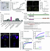
Fig. 1.
Fig. 1.
Neuropeptide LK inhibits female receptivity during sexual transition. (A) A video still in female receptivity assay. (B) The copulation rate of wild-type Canton-S or w1118 virgin female flies from 6 h to 7 d AE. n = 52 to 81. (C) Representative images showing the eggs in virgin female ovaries at the indicated time points AE. Yellow asterisks indicate mature eggs. (Scale bar, 500 μm.) (D) Quantification of the mature eggs retained in each pair of Canton-S ovaries. n = 11 for each. (E) Screening for neurotransmission-related genes required to inhibit the sexual receptivity of 36-h-old females. n = 58 to 89. (F) Top: Schematic diagram of the Lk gene loci and the locations of the deletion mutants, ∆Lk1and ∆Lk2. Bottom: Predicted amino acid sequences of the Lk gene in wild-type, ΔLk1, and ΔLk2 flies. The wild-type bioactive LK peptide sequence is marked in red and underlined. (G and H) Validation of ΔLk mutants by anti-LK staining (green) and anti-nc82 staining (magenta). Arrowheads and dashed boxes indicate anti-LK signals in the brains and ventral nerve cords of wild-type females and Lk mutant females. (Scale bars, 50 μm.) (IK) Cumulative mating percentages of Canton-S, ∆Lk1/+, ∆Lk2/+, and ∆Lk1/∆Lk2 females. (I) 18-h-old virgin females. n = 62, 53, 61, and 79, respectively. (J) 36-h-old virgin females. n = 104, 63, 66, and 112, respectively. (K) 7-d-old virgin females. n = 71, 89, 84, and 77, respectively. (L) Number of mature eggs in the ovaries of 36-h-old Lk mutant and control females. n =10 for each. One-way ANOVA with Tukey’s post hoc tests was performed in (D and L). Variables with different letters are significantly different (P < 0.05), while variables with the same letter or marked “n.s.” are not significantly different. Chi-square tests were performed in (IK), comparing the 10- and 30-min time points. Error bars indicate SEM.
Fig. 2.
Fig. 2.
LK neurons inhibit female receptivity. (A) Expression patterns of Lk-GAL4 in the CNS. Left: Lk-GAL4 > UAS-Stinger; Middle: Lk-GAL4 > UAS-myrGFP; Right: Lk-GAL4 > UAS-Denmark, UAS-sytGFP. White arrowheads indicate cell bodies of LHLKs, yellow arrowheads indicate cell bodies of SELKs, the red arrowhead indicates a cell body of ALK, and the dashed box indicates cell bodies of ABLKs. (Scale bars, 50 μm.) (B and C) Thermogenetic activation of LK neurons inhibits sexual receptivity of 36-h-old females. n = 65, 67, and 60, respectively (B, 22 °C); n = 65, 58, and 67, respectively (C, 30 °C). (D and E) Thermogenetic activation of LK neurons inhibits sexual receptivity of 7-d-old females. n = 66, 77, and 96, respectively (D, 22 °C); n = 48, 65, and 77, respectively (E, 30 °C). (F) Left: Otd-Flp/Lk-GAL4; Tub>GAL80>/UAS-myrGFP, restricted Lk-GAL4 expression in LHLKs. Right: Otd-Flp/Lk-GAL4; Tub>stop>GAL80/UAS-myrGFP, restricted Lk-GAL4 expression in SELKs and ABLKs. (G) Thermogenetic activation of LHLKs does not affect female receptivity. n = 72, 70, 82, 72, 95, and 89, respectively. (H) Thermogenetic activation of SELKs and ABLKs partially inhibit female receptivity. n = 84, 89, 98, 89, 100, and 102, respectively. (I) Stochastic labeling of subsets of LK neurons by the SPARC technique. The red dashed circle indicates a single LHLK neuron. (J) Thermogenetic activation of LK neurons in Lk mutant background does not affect female receptivity. n = 100, 86, 82, 96, 84, 78, 107, 100, 66, 96, 89, and 72, respectively. Chi-square tests were performed in (BE) and (G, H, and J), comparing the 10- and 30-min time points for (BE) and the 30-min time point for (G, H, and J). n.s., not significant; *P < 0.05, and ***P < 0.001.
Fig. 3.
Fig. 3.
20E signaling inhibits female receptivity during sexual transition through LK neurons. (AI) Cumulative mating percentages in females of indicated genotypes. (AC) Lk-GAL4>UAS-Met::Gce RNAi females of 18-h (A), 36-h (B), or 7-d-old (C) females. n = 60, 73, and 64 (A); n = 71, 74, and 68 (B); n = 60, 65, and 66 (C), respectively. (DF) Lk-GAL4>UAS-EcR-A RNAi females of 18-h (D), 36-h (E), or 7-d-old (F) females. n = 82, 79, and 50 (D); n = 68, 68, and 64 (E); n = 66, 66, and 60 (F), respectively. (GI) Lk-GAL4>UAS-EcR-B1 RNAi females of 18-h (G), 36-h (H), or 7-d-old (I) females. n = 75, 80, and 50 (G); n = 63, 68, and 81 (H); n = 66, 60, and 66 (I), respectively. (J) Number of mature eggs in the ovaries of 36-h-old females with downregulated EcR-A or EcR-B1 expression in LK neurons. n = 10 for each. (K) GCaMP signals in ABLK neurons upon 20E application. n = 9 for each. Chi-square tests were performed in (AI), comparing the 10- and 30-min time points. A one-way ANOVA with Tukey’s post hoc test was performed in (J). An unpaired t test was performed in (K). n.s., not significant; *P < 0.05, and ***P < 0.001. Error bars indicate SEM.
Fig. 4.
Fig. 4.
LK suppresses female receptivity via its receptor LKR. (A) Expression pattern of LkrGAL4 driving UAS-myrGFP in female CNS, stained with anti-GFP (green) and anti-nc82 (magenta). (Scale bars, 50 μm.) (BD) Cumulative mating percentages of 18-h (B), 36-h (C), or 7-d-old (D) LkrGAL4/UAS-Lkr RNAi and control females. n = 57, 54, and 54 (B); n = 75, 67, and 70 (C); n = 90, 73, and 84 (D), respectively. (E) Number of mature eggs in the ovaries of 36-h-old LkrGAL4/UAS-Lkr RNAi and control females. n = 10 for each. (F) Schematic view of Lkr gene loci and the locations of the deletion mutants, ∆Lkr1 and ∆Lkr2, which was confirmed by PCR. (GI) Cumulative mating percentages of 18-h (G), 36-h (H), or 7-d-old (I) Lkr mutant and control females. n = 53, 51, and 53 (G); n = 73, 78, and 88 (H); n = 90, 73, and 84 (I), respectively. (J) Number of mature eggs in the ovaries of 36-h-old Lkr mutant and control females. n =10 for each. (K) Cumulative mating percentages of 7-d-old females with LK neurons activated in the Lkr mutant background. The inhibition in female receptivity by activating LK neurons depends on LKR. n = 75, 48, 47, 84, 85, 100, 96, 104, 111, 101, 57, and 93 from left to right. Chi-square tests were performed in (AD), (GI), and (K), comparing the 10- and 30-min time points for (BD) and (GI), and the 30-min time point for (K). Unpaired t tests were performed in (E) and (J). n.s., not significant; *P < 0.05, **P < 0.01, and ***P < 0.001. Error bars indicate SEM.
Fig. 5.
Fig. 5.
LK inhibits female receptivity during sexual transition in pC1 neurons through LKR. (A) Intersectional expression between LkrGAL4 and dsxLexA in the female CNS, labeled with anti-GFP (green) and anti-nc82 (magenta). Genotype: 8XLexAop2-FlpL, UAS>stop>UAS-CsChrimson-mVenus/+;dsxLexA/LkrGAL4. The white arrowhead indicates the pC1 neurons. (B) Activation of LK neurons with dTrpA1 reduced Ca2+ signals in pC1 dsx neurons. The left representative pseudocolored images show Ca2+ imaging of pC1 neurons from 22 to 30 °C and back to 22 °C. Genotype: Lk-GAL4/UAS-dTrpA1;dsxLexA/13XLexAop2-IVS-GCaMP6m. n = 8 and 10 from left to right. (C) Expression of pC1-SS1 in the female CNS, labeled with anti-GFP (green) and anti-nc82 (magenta). The white arrowhead indicates the pC1 neurons. (D) Incubating dissected brains with LK but not scrambled control peptides decreases Ca2+ signals in pC1 neurons. n = 9 and 10 from left to right. (EG) Cumulative mating percentages of 18-h (E), 36-h (F), or 7-d-old (G) pC1-SS1>Lkr RNAi and control females. n = 58, 55, and 61 (E); n = 60, 65, and 66 (F); n = 66, 63, and 69 (G), respectively. (H) Number of mature eggs in the ovaries of 36-h-old pC1-SS1>Lkr RNAi and control females. n = 9 and 13 from left to right. Unpaired t tests were performed in (B) and (H). A Mann–Whitney U test was performed in (D). Chi-square tests were performed in (EG), comparing the 10- and 30-min time points. n.s., not significant; *P < 0.05, and **P < 0.01. Error bars indicate SEM. (Scale bars, 50 μm.)
Fig. 6.
Fig. 6.
A working model of a hormone-to-neuropeptide pathway that inhibits female receptivity during sexual transition. Virgin females are not receptive within 18 h AE. Their receptivity gradually increases, peaking at 3 d posteclosion. The regulation of this transition involves 20E, whose synthesis correlates positively with ovary maturation during sexual transition. 20E increases the activity of LK neurons through its receptor EcR, with LK neurons suppressing female receptivity via LKR in pC1 neurons. This mechanism prevents premature mating during sexual transition, though asexuality in young virgin females within the first 18 h posteclosion is independent of this regulation.

Similar articles

Cited by

  • Distributed control circuits across a brain-and-cord connectome.
    Bates AS, Phelps JS, Kim M, Yang HH, Matsliah A, Ajabi Z, Perlman E, Delgado KM, Osman MAM, Salmon CK, Gager J, Silverman B, Renauld S, Collie MF, Fan J, Pacheco DA, Zhao Y, Patel J, Zhang W, Serratosa Capdevilla L, Roberts RJV, Munnelly EJ, Griggs N, Langley H, Moya-Llamas B, Maloney RT, Yu SC, Sterling AR, Sorek M, Kruk K, Serafetinidis N, Dhawan S, Stürner T, Klemm F, Brooks P, Lesser E, Jones JM, Pierce-Lundgren SE, Lee SY, Luo Y, Cook AP, McKim TH, Kophs EC, Falt T, Negrón Morales AM, Burke A, Hebditch J, Willie KP, Willie R, Popovych S, Kemnitz N, Ih D, Lee K, Lu R, Halageri A, Bae JA, Jourdan B, Schwartzman G, Demarest DD, Behnke E, Bland D, Kristiansen A, Skelton J, Stocks T, Garner D, Salman F, Daly KC, Hernandez A, Kumar S; BANC-FlyWire Consortium; Dorkenwald S, Collman F, Suver MP, Fenk LM, Pankratz MJ, Jefferis GSXE, Eichler K, Seeds AM, Hampel S, Agrawal S, Zandawala M, Macrina T, Adjavon DY, Funke J, Tuthill JC, Azevedo A, Seung HS, de Bivort BL, Murthy M, Drugowitsch J, Wilson RI, Lee WA. Bates AS, et al. bioRxiv [Preprint]. 2025 Aug 2:2025.07.31.667571. doi: 10.1101/2025.07.31.667571. bioRxiv. 2025. PMID: 40766407 Free PMC article. Preprint.
  • Behavior choices amongst grooming, feeding, and courting in Drosophila show contextual flexibility, not an absolute hierarchy of needs.
    Ladd CE, Simpson JH. Ladd CE, et al. bioRxiv [Preprint]. 2025 May 13:2025.05.09.653186. doi: 10.1101/2025.05.09.653186. bioRxiv. 2025. PMID: 40463182 Free PMC article. Preprint.
  • Mating status-dependent dopaminergic modulation of auditory sensory neurons in Drosophila.
    Yamakoshi H, Horigome M, Yamamoto S, Iwanami S, Iwami S, Tanaka R, Ishikawa Y, Kamikouchi A. Yamakoshi H, et al. iScience. 2025 Jul 29;28(9):113232. doi: 10.1016/j.isci.2025.113232. eCollection 2025 Sep 19. iScience. 2025. PMID: 40831745 Free PMC article.

References

    1. Plant T. M., Neuroendocrine control of the onset of puberty. Front. Neuroendocrinol 38, 73–88 (2015). - PMC - PubMed
    1. Barredo C. G., Gil-Marti B., Deveci D., Romero N. M., Martin F. A., Timing the juvenile-adult neurohormonal transition: Functions and evolution. Front. Endocrinol. (Lausanne) 11, 602285 (2020). - PMC - PubMed
    1. Ellison P. T., et al. , Puberty as a life history transition. Ann. Hum. Biol. 39, 352–360 (2012). - PubMed
    1. Phoenix C. H., Goy R. W., Gerall A. A., Young W. C., Organizing action of prenatally administered testosterone propionate on the tissues mediating mating behavior in the female guinea pig. Endocrinology 65, 369–382 (1959). - PubMed
    1. McCarthy M. M., Arnold A. P., Reframing sexual differentiation of the brain. Nat. Neurosci. 14, 677–683 (2011). - PMC - PubMed

LinkOut - more resources