. 2025 Apr:81:103545.
doi: 10.1016/j.redox.2025.103545. Epub 2025 Feb 11.

Supersulfides contribute to joint homeostasis and bone regeneration

Affiliations

Supersulfides contribute to joint homeostasis and bone regeneration

Miki Maemura et al. Redox Biol. 2025 Apr.

Abstract

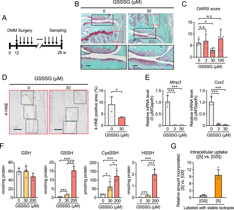
The physiological functions of supersulfides, inorganic and organic sulfides with sulfur catenation, have been extensively studied. Their synthesis is mainly mediated by mitochondrial cysteinyl-tRNA synthetase (CARS2) that functions as a principal cysteine persulfide synthase. This study aimed to investigate the role of supersulfides in joint homeostasis and bone regeneration. Using Cars2AINK/+ mutant mice, in which the KIIK motif of CARS2 essential for supersulfide production was replaced with AINK, we evaluated the role of supersulfides in fracture healing and cartilage homeostasis during osteoarthritis (OA). Tibial fracture surgery was performed on the wild-type (Cars2+/+) and Cars2AINK/+ mice littermates. Bulk RNA-seq analysis for the osteochondral regeneration in the fracture model showed increased inflammatory markers and reduced osteogenic factors, indicative of impaired bone regeneration, in Cars2AINK/+ mice. Destabilization of the medial meniscus (DMM) surgery was performed to produce the mouse OA model. Histological analyses with Osteoarthritis Research Society International and synovitis scores revealed accelerated OA progression in Cars2AINK/+ mice compared with that in Cars2+/+ mice. To assess the effects of supersulfides on OA progression, glutathione trisulfide (GSSSG) or saline was periodically injected into the mouse knee joints after the DMM surgery. Thus, supersulfides derived from CARS2 and GSSSG exogenously administered significantly inhibited inflammation and lipid peroxidation of the joint cartilage, possibly through suppression of ferroptosis, during OA development. This study represents a significant advancement in understanding anti-inflammatory and anti-oxidant functions of supersulfides in skeletal tissues and may have a clinical relevance for the bone healing and OA therapeutics.

Keywords: Bone regeneration; Cysteinyl-tRNA synthetase; Ferroptosis; Glutathione trisulfide; Osteoarthritis; Supersulfides.


Conflict of interest statement

Declaration of competing interest The authors declare that they have no competing interests.

Figures

Image 1
Graphical abstract
Fig. 1
Fig. 1
Comparison of bone fracture healing between Cars2+/+ and Cars2AINK/+ mice. (A) Representative soft X-ray images of fracture sites in male Cars2+/+ and Cars2AINK/+ mice at 2 weeks post-fracture. (B) Safranin-O staining of calluses 2 weeks after fracture. Scale bars, 500 μm (C) Semi-quantification of the areas of safranin-O-stained calluses in the tibias of Cars2+/+ and Cars2AINK/+ mice. Data are means ± SD. ∗P < 0.05. Symbols represent individual mice (n = 4 per group). (D) In vivo formation of supersulfides in Cars2+/+ and Cars2AINK/+ mice. Endogenous production of CysSH, GSH, CysSSH, and other related supersulfide metabolites in chondrocytes and chondral tissues obtained from Cars2+/+ and Cars2AINK/+ mice littermates were quantified via LC-MS/MS analysis with HPE-IAM labeling. Data are means ± SD. ∗P < 0.05.
Fig. 2
Fig. 2
Comprehensive gene expression in the calluses of Cars2+/+ and Cars2AINK/+ mice. Bulk RNA-seq analysis of calluses from the tibias of Cars2+/+ and Cars2AINK/+ mice (n = 2 each) at 2 weeks post-fracture. (A) Heatmap showing the top 20 downregulated differentially expressed genes (DEGs) in calluses of Cars2AINK/+ mice compared to those of Cars2+/+ mice. (B) mRNA levels of markers for endochondral ossification in calluses from the tibias of Cars2+/+ and Cars2AINK/+ mice at 2 weeks post-fracture. (C) The top 20 upregulated DEGs in calluses of Cars2AINK/+ mice, as compared with calluses of Cars2+/+ mice. (D) mRNA levels of inflammatory response markers in calluses from the tibias of Cars2+/+ and Cars2AINK/+ mice at 2 weeks post-fracture. Data are presented as dot plots and means ± SD (n = 3 per group). ∗P < 0.05, ∗∗P < 0.005, ∗∗∗P < 0.0005.
Fig. 3
Fig. 3
Development of osteoarthritis (OA) in Cars2+/+ and Cars2AINK/+ mice. (A) At 16 weeks post-DMM surgery, knee joints were stained with safranin-O (left panels). Boxed areas in the left panel are shown at higher magnification, articular cartilage (upper right panels, outlined in black), and synovial lesions (lower right panels, outlined in red). Representative images are shown. Scale bars, 100 μm. (B) Quantification of OA development using Osteoarthritis Research Society International (OARSI) histologic scoring for Cars2+/+ [n = 7] and Cars2AINK/+ [n = 4] mice. Severity of synovitis assessed using synovitis scores (Cars2+/+ [n = 7] and Cars2AINK/+ [n = 4] mice). Data are means ± SD. ∗P < 0.05, ∗∗P < 0.005. (C) Immunohistochemical assessment of 4-hydroxy-2-nonenal (4-HNE) expression in the synovium of knee joints that correspond to the boxed area in (A). The rates of 4-HNE-positive areas in the boxes are shown in the panels in right panel. Data are means ± SD. ∗∗∗P < 0.0005.
Fig. 4
Fig. 4
Effects of intra-articular administration of GSSSG on mouse OA model induced surgically and intracellular uptake of supersulfides in ATDC5 cells. (A) A scheme of GSSSG administration to the OA model used in this study. The intra-articular injections were initiated at 12 weeks post-DMM surgery and repeated 16 times once a week for consecutive weeks. (B) At 16 weeks post-surgery and GSSSG treatment, knee joints were stained with safranin-O. Areas within the articular cartilage outlined in black in the upper panels are shown in the lower panels. Scale bar, 100 μm. (C) Semi-quantification of OA development using OARSI histologic scoring in the GSSSG-treated OA model. Data are means ± SD. ∗P < 0.05 vs. 0 μM; N.S., not significant. Each group (0, 3, 30, 100 μM treatment) includes 4 mice. (D) Immunohistochemical assessment of 4-HNE formation in the synovium of knee joints of GSSSG-treated mice at 16 weeks after OA induction. High-magnification images of the areas outlined in red in (B) are shown. The two left panels show representative images; the right panel indicates the rates of 4-HNE-positive areas in the box regions. Data are means ± SD. ∗P < 0.05, vs. 0 μM; Student's t-test. (E) mRNA levels of inflammatory markers (Mmp3 and Cox2) in mouse ASF treated with different concentrations of GSSSG (0, 3, 30 μM) and exposed to 1 ng/ml IL-1β for 24 h Data are means ± SD (n = 3). ∗∗∗P < 0.0005. (F) Supersulfide metabolome analysis with ATDC5 cells treated with various doses of GSSSG (0, 30, 200 μM) for 3 h. Data are means ± SD (n = 4). ∗P < 0.05, ∗∗∗P < 0.001. (G) Intracellular uptake of stable isotope-labelled GS ([GS]) and S ([S]) with ATDC5 cells treated with stable isotope-labelled GSSSG ([13C2, 15N]GS-[34S]-SG[13C2, 15N], 200 μM) for 3 h. Amounts of intracellular [GS] and [S] were quantified (Fig. S9A) and their relative ratios ([S] vs. [GS]) are shown. Data are means ± SD (n = 4).

References

    1. Akaike T., Ida T., Wei F.Y., Nishida M., Kumagai Y., Alam M.M., Ihara H., Sawa T., Matsunaga T., Kasamatsu S., Nishimura A., Morita M., Tomizawa K., Nishimura A., Watanabe S., Inaba K., Shima H., Tanuma N., Jung M., Fujii S., Watanabe Y., Ohmuraya M., Nagy P., Feelisch M., Fukuto J.M., Motohashi H. Cysteinyl-tRNA synthetase governs cysteine polysulfidation and mitochondrial bioenergetics. Nat. Commun. 2017;8:1177. doi: 10.1038/s41467-017-01311-y. - DOI - PMC - PubMed
    1. Barayeu U., Sawa T., Nishida M., Wei F.Y., Motohashi H., Akaike T. Supersulfide biology and translational medicine for disease control. Br. J. Pharmacol. 2023 doi: 10.1111/bph.16271. - DOI - PubMed
    1. Ogata S., Matsunaga T., Jung M., Barayeu U., Morita M., Akaike T. Persulfide biosynthesis conserved evolutionarily in all organisms. Antioxidants Redox Signal. 2023;39:983–999. doi: 10.1089/ars.2023.0405. - DOI - PMC - PubMed
    1. Ida T., Sawa T., Ihara H., Tsuchiya Y., Watanabe Y., Kumagai Y., Suematsu M., Motohashi H., Fujii S., Matsunaga T., Yamamoto M., Ono K., Devarie-Baez N.O., Xian M., Fukuto J.M., Akaike T. Reactive cysteine persulfides and S-polythiolation regulate oxidative stress and redox signaling. Proc. Natl. Acad. Sci. U.S.A. 2014;111:7606–7611. doi: 10.1073/pnas.1321232111. - DOI - PMC - PubMed
    1. Fukuto J.M., Ignarro L.J., Nagy P., Wink D.A., Kevil C.G., Feelisch M., Cortese-Krott M.M., Bianco C.L., Kumagai Y., Hobbs A.J., Lin J., Ida T., Akaike T. Biological hydropersulfides and related polysulfides - a new concept and perspective in redox biology. FEBS Lett. 2018;592:2140–2152. doi: 10.1002/1873-3468.13090. - DOI - PMC - PubMed

LinkOut - more resources