. 2025 Apr;301(4):108336.
doi: 10.1016/j.jbc.2025.108336. Epub 2025 Feb 19.

Structural and functional characterization of the extended-diKH domain from the antiviral endoribonuclease KHNYN

Affiliations

Structural and functional characterization of the extended-diKH domain from the antiviral endoribonuclease KHNYN

Rebecca L Youle et al. J Biol Chem. 2025 Apr.

Abstract

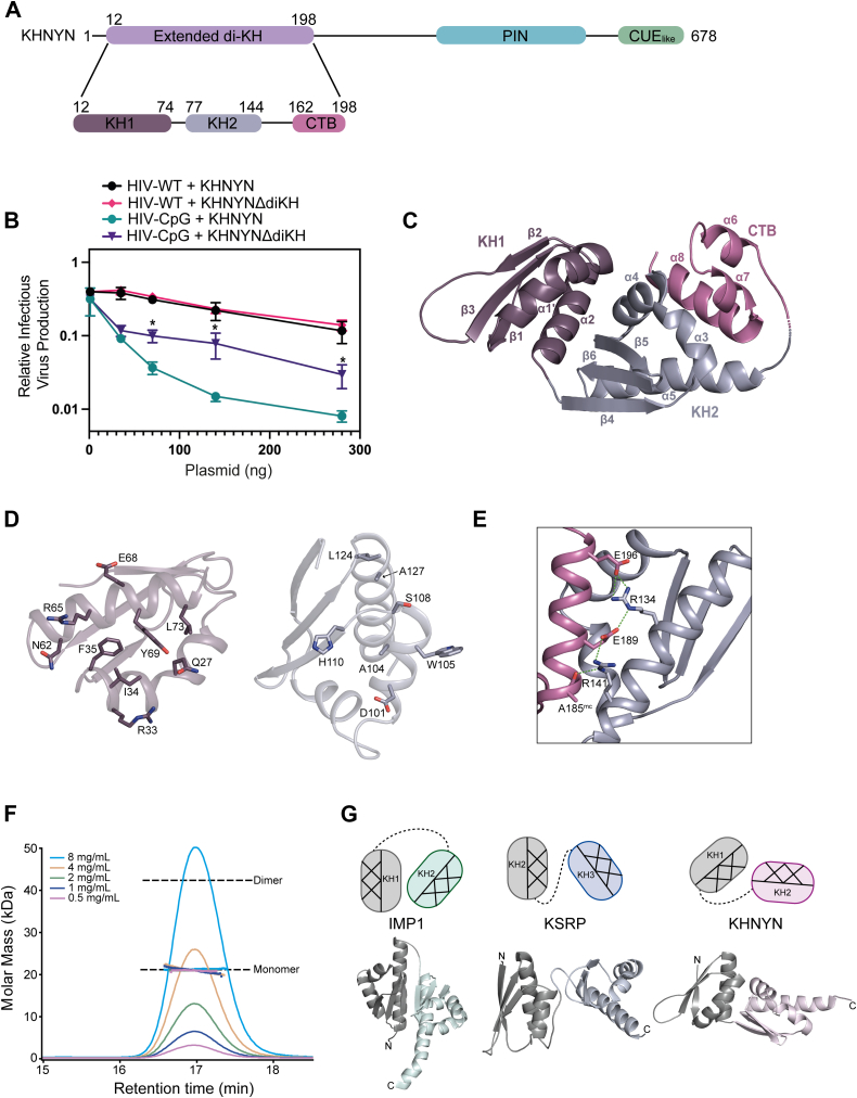
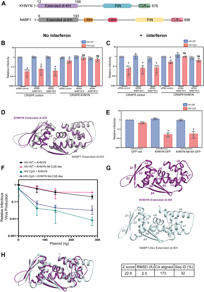
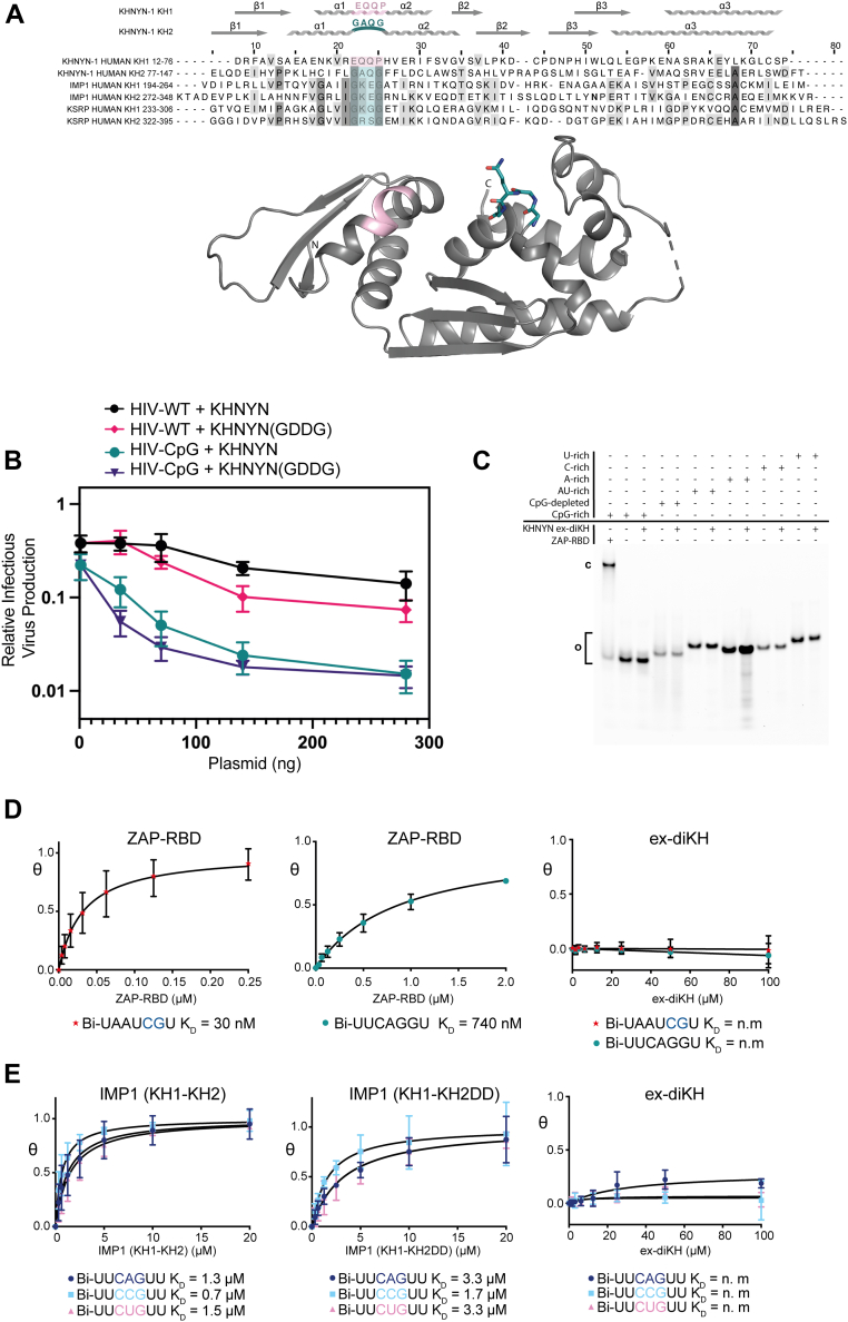
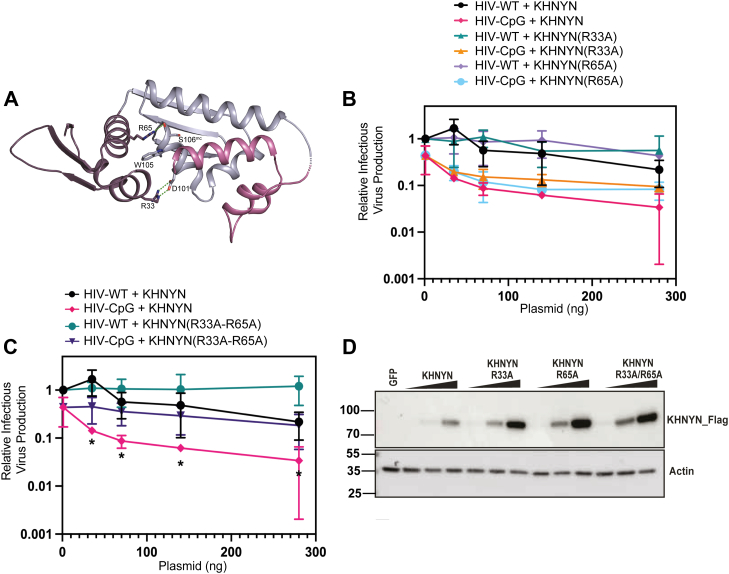
Zinc finger antiviral protein (ZAP) binds CpG dinucleotides in viral RNA and targets them for decay. ZAP interacts with several cofactors to form the ZAP antiviral system, including KHNYN, a multidomain endoribonuclease required for ZAP-mediated RNA decay. However, it is unclear how the individual domains in KHNYN contribute to its activity. Here, we demonstrate that the KHNYN amino-terminal extended-diKH (ex-diKH) domain is required for antiviral activity and present its crystal structure. The structure belongs to a rare group of KH-containing domains, characterized by a noncanonical arrangement between two type 1 KH modules, with an additional helical bundle. N4BP1 is a KHNYN paralog with an ex-diKH domain that functionally complements the KHNYN ex-diKH domain. Interestingly, the ex-diKH domain structure is present in N4BP1-like proteins in lancelets, which are basal chordates, indicating that it is evolutionarily ancient. While many KH domains demonstrate RNA binding activity, biolayer interferometry and electrophoretic mobility shift assays indicate that the KHNYN ex-diKH domain does not bind RNA. Furthermore, residues required for canonical KH domains to bind RNA are not required for KHNYN antiviral activity. By contrast, an inter-KH domain cleft in KHNYN is a potential protein-protein interaction site, and mutations that eliminate arginine salt bridges at the edge of this cleft decrease KHNYN antiviral activity. This suggests that this domain could be a binding site for an unknown KHNYN cofactor.

Keywords: CpG; HIV; KHNYN; RNA turnover; RNA virus; ZAP; ZC3HAV1; innate immunity; structure-function; viral replication.


Conflict of interest statement

Conflict of interest The authors declare that they have no conflicts of interest with the contents of this article.

Figures

Figure 1
Figure 1
TheKHNYN ex-diKH domain is required for antiviral activity and is a member of a novel group of diKH domain structures.A, (upper) schematic representation of KHNYN. The positions of structural domains are highlighted; lilac (ex-diKH, residues 12–198), cyan (PIN), and green CUElike. (Lower) schematic of the ex-diKH domain, purple (KH1, residues 12–74), blue gray (KH2, residues 77–144), and magenta (CTB, residues 162–198). B, infectious virus production from KHNYN CRISPR HeLa cells cotransfected with pHIV-WT or pHIV-CpG and increasing amount of CRISPR-resistant FLAG-tagged pKHNYN or pKHNYNΔdiKH expressing plasmids. Each point shows the average value of three independent experiments normalized to the value obtained for HIV-WT in the absence of KHNYN. Error bars represent standard deviation. ∗ indicates that HIV-CpG + KHNYN is significantly different compared to HIV-CpG + KHNYNΔdiKH, unpaired two-tailed t test p < 0.05. C, crystal structure of ex-diKH. The protein backbone is shown as a cartoon representation, domains are colored as in (A), secondary structure elements are labeled from N to Ctermini. D, details of the KH1-KH2 interface. Cartoon representations of the protein backbone of KH1 (left) and KH2 (right) are shown, colored as in (C). The view is from one domain looking into the interface of the other. Residues with side chains that make hydrogen bonding and hydrophobic interactions that contribute to interdomain packing (KH1: Q27, R33, I34, F35, N62, R65, E68, Y69, and L73) and (KH2: D101, A104, W105, S108, H110, A127, and L124) are shown as sticks, colored by atom type. E, the KH2-CTB interdomain interface. KH2 and the CTB are shown in the cartoon representation, colored as in (C). Residues R134, A185, E189, and E196 that make hydrogen bonds, represented by the green dashed lines, are shown as sticks, mc = mainchain. F, SEC-MALLS analysis of ex-diKH. The sample loading concentrations were 8 mg/ml (cyan), 4 mg/ml (wheat), 2 mg/ml (green), 1 mg/ml (blue), and 0.5 mg/ml (magenta). The differential refractive index (dRI) is plotted against column retention time, and the molar mass determined at 1 s intervals throughout the elution of each peak, is plotted as points. The monomer and dimer molecular mass for ex-diKH are indicated by the dashed lines. G, diKH domain arrangements. diKH domains from IMP1, KSRP, and KHNYN are shown in cartoon colored gray and mint, gray and light blue, and gray and pink, respectively, N and C termini are labeled. The relative orientation of KH domains in each structure is shown above schematically, color coded to the corresponding domain. The helical and β-sheet faces of each KH domain are shown hatched and plain, respectively. SEC-MALLS, size-exclusion chromatography coupled to multiangle laser light scattering; CTB, C-terminal bundle; ex-diKH domain, extended-diKH domain; KHNYN, KH and NYN containing protein; PIN, PilT N-terminal.
Figure 2
Figure 2
The KHNYN and N4BP1 ex-diKH domains are functionally equivalent.A, schematic representation of KHNYN and N4BP1. The position of structural/functional domains are highlighted; lilac (KHNYN ex-diKH domain residues 12–198), cyan (KHNYN PIN domain), green (KHNYN CUElike domain), gray (N4BP1 ex-diKH domain residues 6–193), orange (N4BP1 UBM domain), deep pink (N4BP1 UBA domain), yellow (N4BP1 PIN domain), and pink (N4BP1 CUElike domain). B and C, infectious virus production from HeLa CRISPR control or CRISPR KHNYN cells that were either untreated (B) or treated with 500 U of IFN-β (C) and transfected with either siRNA control or siRNAs against N4BP1 and infected with HIV-WT or HIV-CpG at MOI = 3. Each bar shows the average value of three independent experiments normalized to the value obtained for HIV-WT in the CRISPR control cells transfected with siRNA control. Error bars represent standard deviation. ∗ indicates that HIV-CpG is significantly different compared to HIV-WT, unpaired two-tailed t test p < 0.05. D, 3D DALI structural superimposition of KHNYN and N4BP1 (PDB: 6Q3V) ex-diKH domains. Structures, shown in cartoon representation, KHNYN in purple and N4BP1 in gray, were aligned over all backbone Cα atoms (Z score 23.9, RSMD = 1.8 Å over 174 Cα). E, infectious virus production from stable HeLa CRISPR KHNYN GFP control, KHNYN WT-GFP, or KHNYN N4-diKH-GFP infected with either HIV-1 WT or HIV-1 CpG at a MOI = 3. Bar plots represent the average value of three independent experiments normalized to the value obtained for HIV-WT in the absence of KHNYN. Error bars represent standard deviation. ∗ indicates that HIV-CpG is significantly different compared to HIV-WT, unpaired two-tailed t test p < 0.05. F, infectious virus production from KHNYN CRISPR HeLa cells cotransfected with pHIV-WT or pHIV-CpG and increasing amount of CRISPR-resistant FLAG-tagged pKHNYN or pKHNYN N4-CUE-like expressing plasmids. Each point shows the average value of three independent experiments normalized to the value obtained for HIV-WT in the absence of KHNYN. Error bars represent standard deviation. ∗ indicates that HIV-CpG + KHNYN is significantly different compared to HIV-CpG + KHNYN N4-CUE-like, unpaired two-tailed t test p < 0.05. G, structure of KHNYN ex-diKH domain (top) and AlphaFold model of the ex-diKH domain from Branchiostoma belcheri NEDD4-binding protein 1-like (NBP1-like) (bottom). Protein backbones are shown in cartoon representations, secondary structure elements are labeled from N to C termini. H, 3D DALI structural superimposition of KHNYN and N4BP1-like ex-diKH domains. Protein backbones are shown in cartoon representation, KHNYN in purple and N4BP1-like in pale cyan. Structures were aligned over all backbone Cα atoms. Alignment data are shown on the right (Z score = 22.6, rmsd = 2.5 Å over 173 Cα, 32% Seq ID). ex-diKH domain, extended-diKH domain; IFN-β, interferon-β; KHNYN, KH and NYN containing protein; MOI, multiplicity of infection; PDB, Protein Data Bank; PIN, PilT N-terminal.
Figure 3
Figure 3
The KHNYN ex-diKH domain has no detectable RNA binding activity.A, (Upper) multiple sequence alignment of KH domains from human KHNYN (Uniprot: O15037), Insulin-like growth factor 2 mRNA binding protein 1 (IMP1) (Uniprot: Q9NZI8) and KH type-splicing regulatory protein (KSRP) (Uniprot Q92945). Conserved residues are highlighted with increasing shades of gray. GxxG motifs are highlighted in cyan and the equivalent residues in pink for KHNYN KH1. KHNYN KH1 and KH2 secondary structure elements aligned with the sequence are shown above. (Lower) KHNYN ex-diKH domain is shown in the cartoon representation. The position of the proline-kinked helix in KH1 and GxxG motif in KH2 are colored according to the scheme above with the GxxG motif (94GAQG97) and also highlighted as sticks. B, infectious virus production from KHNYN CRISPR HeLa cells cotransfected with pHIV-WT or pHIV-CpG and increasing amount of CRISPR-resistant FLAG-tagged pKHNYN or pKHNYN (GDDG) expressing plasmids. Each point shows the average value of three independent experiments normalized to the value obtained for pHIV-WT in the absence of KHNYN. C, EMSA analysis of ex-diKH RNA-binding. U-rich, C-rich, A-rich, AU-rich, CpG-depleted, and CpG-rich 33mer 5′-FAM-labeled RNAs (0.2 μM) were incubated with ZAP RBD (2 μM) and ex-diKH (20 μM). Free (o) and bound (c) components were separated on a 10% acrylamide native TBE gel and visualized by fluorescence imaging. D and E, BLI analysis for (D) KHNYN ex-diKH and ZAP-RBD binding to CpG (Bi-UAAUCGU) and non-CpG (Bi-UUCAGGU) RNAs and (E) KHNYN ex-diKH, IMP1(KH1-KH2) and IMP1(KH1-KH2DD) RNA-binding to 5′-biotinylated CNG RNAs. Nonlinear fits of fraction bound plotted against protein concentration are shown. Data are the mean of triplicate measurements. Error bars represent the standard deviation of the mean for each triplicate measurement. The derived KD values are shown beneath plots and presented with 95% CI values in Table S4. BLI signal response for ex-diKH interaction with CpG, non-CpG, and CNG RNAs is unobservable. BLI, biolayer interferometry; CI, confidence interval; ex-diKH domain, extended-diKH domain; FAM, fluorescein amidite; KHNYN, KH and NYN containing protein; RBD, RNA binding domain; TBE, tris-borate-EDTA; ZAP, zinc finger antiviral protein.
Figure 4
Figure 4
The KHNYN KH1-KH2 interdomain cleft is required for antiviral activity.A, the KHNYN ex-diKH domain is shown in the cartoon representation. KH1, KH2, and CTB are colored purple, blue gray, and magenta respectively. Residues R33 and R65 together with D101, W105, and S106 that make salt bridge, hydrogen bond, and cation-π interactions across the KH1-KH2 interdomain cleft are shown in sticks. Salt bridge and hydrogen bonds are represented by the green dashes, mc = mainchain. B and C, infectious virus production from KHNYN CRISPR HeLa cells cotransfected with pHIV-WT or pHIV-CpG and (B) increasing amount of CRISPR-resistant FLAG-tagged pKHNYN, pKHNYN (R33A), or pKHNYN (R65A) and (C) pKHNYN or pKHNYN(R33A/R65A) expressing plasmids. Each point shows the average value of three independent experiments normalized to the value obtained for pHIV-WT in the absence of KHNYN. HIV-WT + KHNYN and HIV-CpG + KHNYN are the same data for panel B and C. ∗ indicates that HIV-CpG + KHNYN is significantly different compared to HIV-CpG + KHNYN(R33A/R65A), unpaired two-tailed t test p < 0.05. D, representative Western blotting corresponding to the expression level of FLAG-tagged KHNYN KHNYN(R33A), KHNYN(R65A), or KHNYN(R33A-R65A) employed in the infection experiments is shown in the panels B and C. CTB, C-terminal bundle; ex-diKH domain, extended-diKH domain; KHNYN, KH and NYN containing protein.

Similar articles

References

    1. Gao G., Guo X., Goff S.P. Inhibition of retroviral RNA production by ZAP, a CCCH-type zinc finger protein. Science. 2002;297:1703–1706. - PubMed
    1. Guo X., Carroll J.W., Macdonald M.R., Goff S.P., Gao G. The zinc finger antiviral protein directly binds to specific viral mRNAs through the CCCH zinc finger motifs. J. Virol. 2004;78:12781–12787. - PMC - PubMed
    1. Ficarelli M., Neil S.J.D., Swanson C.M. Targeted restriction of viral gene expression and replication by the ZAP antiviral system. Annu. Rev. Virol. 2021;8:265–283. - PubMed
    1. Shao R., Visser I., Fros J.J., Yin X. Versatility of the zinc-finger antiviral protein (ZAP) as a modulator of viral infections. Int. J. Biol. Sci. 2024;20:4585–4600. - PMC - PubMed
    1. Bick M.J., Carroll J.W., Gao G., Goff S.P., Rice C.M., MacDonald M.R. Expression of the zinc-finger antiviral protein inhibits alphavirus replication. J. Virol. 2003;77:11555–11562. - PMC - PubMed

LinkOut - more resources