. 2025 Feb 21;15(1):6398.
doi: 10.1038/s41598-025-89700-y.

SiO2 nanoparticles as disruptors of endogenous resolution mechanisms of inflammatory responses that exacerbate pneumonia

Affiliations

SiO2 nanoparticles as disruptors of endogenous resolution mechanisms of inflammatory responses that exacerbate pneumonia

Luciana Pádua Tavares et al. Sci Rep. .

Abstract

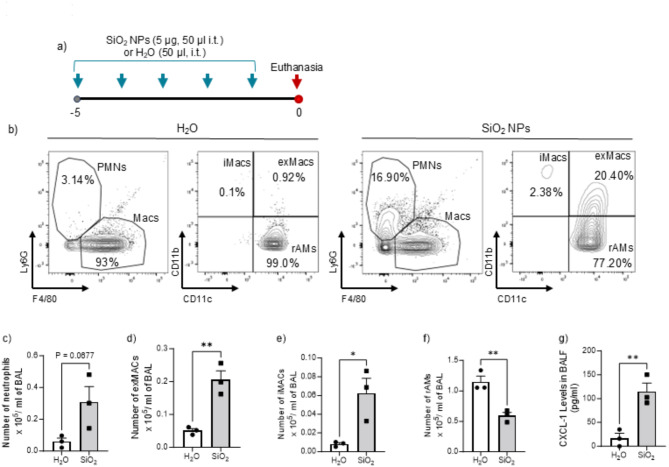
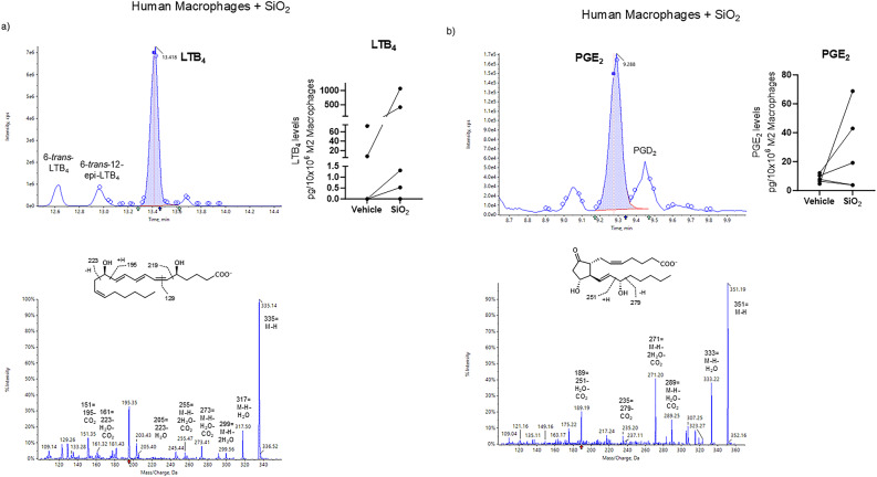
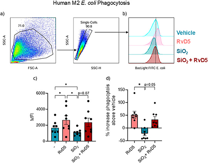
Occupational exposure to engineered nanomaterials (ENMs) is increasing in the workplace and can impact human health. Amorphous silicon dioxide nanoparticles (SiO2 NPs) are widely produced respirable ENMs used in commercial products. We have investigated their impact on lung inflammation resolution and bacterial defense. Mice exposed to SiO2 NPs, followed by bacteria, exhibited increased lung inflammation, bacterial proliferation, and lung damage compared to mice not exposed to NPs. SiO2 NPs increased human macrophage production of pro-inflammatory mediators and disrupted phagocytosis of bacteria and efferocytosis of apoptotic neutrophils - pivotal responses for host defense and inflammation resolution. A pro-resolving mediator, resolvin D5 (RvD5), restored macrophage phagocytosis of bacteria and partially controlled excess lung inflammation after SiO2 NPs. These findings demonstrate that SiO2 NPs disrupt endogenous resolution processes to give rise to heightened lung inflammation and infection. RvD5 reduced inflammation and partially restored endogenous resolution cellular processes, suggesting that RvD5 can reduce ENP disruption of resolution.

Keywords: Infection; Macrophages; Phagocytosis.; Resolution; Resolvin D5; SiO2 nanoparticles.


Conflict of interest statement

Declarations. Competing interests: The authors declare no competing interests.

Figures

Fig. 1
Fig. 1
Airway exposure to SiO2 NPs induces lung inflammation in mice. (a) C57BL/6 mice were exposed intratracheally to either SiO2 NPs (5ug/ day) or vehicle over five consecutive days. Mice were then euthanized 24 h after the last exposure and bronchoalveolar lavage (BAL) was performed. (b) Immunophenotyping of BAL with flow cytometry to evaluate (c) neutrophils, (d) exudative macrophages (CD45+ F4/80+ CD11b+ CD11c+, exMACs), (e) infiltrating macrophages (CD45+ F4/80+ CD11b+ CD11c, iMACs), and (f) resident alveolar macrophages (CD45+ F4/80+ CD11b CD11c+, rAMs). (g) BAL fluid (BALF) was assessed for CXCL-1 levels by ELISA. Data expressed as mean ± SEM, n = 3 mice in each group. Statistical significance was determined by Unpaired t-test, with *P < 0.05; **P < 0.01.
Fig. 2
Fig. 2
Exposure to SiO2 NPs promotes pro-inflammatory mediator release in human M2 macrophages. (a) LTB4 was quantified via liquid chromatography-tandem mass spectrometry (LC-MS/MS). Top panel, Multiple reaction monitoring (MRM) chromatogram of LTB4 (m/z 335 > 195) with a retention time (TR) = 13.42 min and signal to noise (S/N) ratio of 5488.1. Inset, quantitation of LTB4 in macrophages stimulated with SiO2 (10 µg/mL) or vehicle. Bottom panel, associated ESI-MS/MS spectrum with a molecular ion at m/z 335 = M-H and accompanying daughter ions at m/z 317 = M-H-H2O, m/z 299 = M-H-2H2O, m/z 273 = M-H-H2O-CO2, m/z 255 = M-H-2H2O-CO2, m/z 205 = 223-H2O, m/z 195, m/z 161 = 223-H2O-CO2, m/z 151 = 195-CO2, and m/z 129. (b) PGE2 was similarly quantified. Top panel, MRM chromatogram of PGE2 (m/z 351 > 189) with a retention time (TR) = 9.29 min and S/N ratio of 309.4. Inset, quantitation of PGE2 in macrophages stimulated with SiO2 or vehicle. Bottom panel, accompanying ESI-MS/MS spectrum with a molecular ion at m/z 351 = M-H and fragment daughter ions at m/z 333 = M-H-H2O, m/z 315 = M-H-2H2O, m/z 289 = M-H-H2O-CO2, m/z 271 = M-H-2H2O-CO2, m/z 235 = 279-CO2, and m/z 189 = 251-H2O-CO2. The retention times and fragmentation spectra of both LTB4 and PGE2 were essentially identical to those of their respective synthetic reference standards.
Fig. 3
Fig. 3
Exposure to SiO2 NPs promotes RvD5 release and impairs neutrophil efferocytosis in human macrophages. (a) RvD5 was identified via liquid chromatography-tandem mass spectrometry (LC-MS/MS). Top panel, Multiple reaction monitoring (MRM) chromatogram of RvD5 (m/z 359 > 199) with a retention time (TR) = 13.18 min and signal to noise (S/N) ratio of 455.5. Inset, quantitation of RvD5 in M2 macrophages stimulated with SiO2 (10 µg/mL) or vehicle. Bottom panel, Corresponding MS/MS spectrum with a parent ion at m/z 359 = M-H and daughter ions including m/z 341 = M-H-H2O, m/z 323 = M-H-2H2O, m/z 315 = M-H-CO2, m/z 297 = M-H-H2O-CO2, m/z 227 = 289-H2O-CO2, m/z 199 = 217-H2O, and m/z 141. These data were essentially identical to those of synthetic RvD5 reference standard. (b) To assess efferocytosis, human macrophages (Mφ) plated on a 6-well plate (2 × 106 cells/ well) were incubated with vehicle or SiO2 NPs (10–100 µg) for 15 min at 37 °C followed by the addition of CFSE-labeled apoptotic PMN (1:3 ratio; Mφ: neutrophils). Efferocytosis is expressed as the percentage above vehicle control. Data presented as mean ± SEM, n = 3 healthy human donors. Statistical significance was determined using Wilcoxon matched pairs signed rank test (a, b) and RM one-way ANOVA (c) with *p < 0.05.
Fig. 4
Fig. 4
SiO2 NP exposure disrupts human macrophage phagocytosis of E. coli. Human monocytes were isolated from healthy donors and differentiated into macrophages with 20 ng/mL of human recombinant GM-CSF for seven days. Human macrophages (Mφ), plated on a 6-well plate (2 × 106 cells/ well), were incubated with vehicle or SiO2 (10 µg) for 180 min. Macrophages were then incubated with 10 nM of RvD5, or vehicle alone (0.01% vol/vol) for 15 min at 37oC prior to the addition of BacLight Green-labeled Escherichia coli (ratio of 1:50, M2 macrophages: E. coli). Fluorescence-associated Phagocytosis was monitored using flow cytometry. (a) Representative flow cytometry gating strategy for M2 macrophages. (b) Histogram of intracellular BacLight FITC-labeled E. coli in M2 macrophages. (c) Geometric Mean Fluoresce Intensity (MFI) of phagocytosis. (d) Percentage increase in M2 phagocytosis relative. E. coli + vehicle control. Data presented as means ± SEM, n = 6 donors. Statistical significance was analyzed using one-way ANOVA, with *p < 0.05, **p < 0.01, compared to E. coli + vehicle (Panel c-d).
Fig. 5
Fig. 5
SiO2 NP exposure exacerbates severity of S. pneumoniae pneumonia in mice. (a) C57BL/6 mice were exposed intratracheally to SiO2 NPs (5ug/day) or vehicle (H2O) for five days. 24 h after the last exposure, mice were infected intranasally with a mild inoculum of Streptococcus pneumoniae (Serotype 3, 1 × 105 CFU). 48 h post-infection, mice were euthanized, and (b) BAL collected was immunophenotyped by flow cytometry to evaluate numbers of (c) neutrophils, (d) exudative macrophages (CD45+ F4/80+ CD11b+ CD11c+ - exMACs), (e) infiltrating macrophages (CD45+ F4/80+ CD11b+ CD11c - iMACs), and (f) resident alveolar macrophages (CD45+ F4/80+ CD11b CD11c+ - rAMs). (g-i) BAL fluid (BALF) was analyzed for (g) total protein content, (h) CXCL-1 levels, and (i) TNF-α levels. Bacterial counts were determined in the (j) bronchoalveolar lavage (BAL) and (k) the left lung after BAL. (l) In separate experiments, lung histology was evaluated using H&E staining. Images were captured at both 10x (bars = 100 μm) and 40x magnifications (bars = 50 μm). Arrows indicate areas of inflammatory cell infiltration and stars (*) mark damaged epithelium. Data is shown as mean ± SEM, n = 6–7 mice in each group. Statistical analysis was performed by (c-i) or unpaired t-test (j-k) Mann Whitney test, with *p < 0.05; **p < 0.01; ***p < 0.001.
Fig. 6
Fig. 6
RvD5 decreases lung inflammation and enhances S. pneumoniae clearance in mice. (a) C57BL/6 mice were exposed intratracheally to SiO2 NPs (5µg/day) or vehicle for five consecutive days. 24 h after the final exposure, mice were infected intranasally with a mild inoculum of Streptococcus pneumoniae (Serotype 3, 1 × 105 CFU). At the time of infection and 24 h later, mice were administered RvD5 (100ng/mouse, i.v.) or vehicle. 48 h after infection mice were euthanized and (b) lung histology was performed using H&E staining. Photomicrographs were taken at both 10x (scale bars = 100 μm) and 40x magnification (scale bars = 50 μm). Arrows point to areas of significant inflammatory cell infiltrates and stars (*) indicate damage to the epithelium. In a separate experiment, bacterial counts were determined in (c) bronchoalveolar lavage (BAL) and (d) the left lung after BAL. (e) BAL collected was immunophenotyped by flow cytometry to evaluate (f) neutrophils. Macrophages were further phenotyped and counts of (g) exudative macrophages (CD45 + F4/80 + CD11b + CD11c+ - exMACs), (h) infiltrating macrophages (CD45 + F4/80 + CD11b + CD11c- - iMACs), and (i) resident alveolar macrophages (CD45 + F4/80 + CD11b- CD11c+ - rAMs) were obtained. Analysis of BAL fluid (BALF) was conducted to measure the levels of (j) total protein, (k) CXCL-1, and (l) TNF- a. Data presented as mean ± SEM, n = 3–4 mice in each group pooled from 3 independent experiments. Statistical analysis was performed by one-way ANOVA, with *p < 0.05; **p < 0.01; ***p < 0.001.
Fig. 7
Fig. 7
RvD5 decreases S. pneumoniae-triggered proinflammatory cytokine secretion. C57BL/6 mice were exposed intratracheally to SiO2 NPs (5µg/day) or vehicle for five days, and 24 h after last exposure infected intranasally with a mild inoculum of Streptococcus pneumoniae (Serotype 3, 1 × 105 CFU). At the time of infection and 24 h later, mice were administered RvD5 (100ng/mouse, i.v.) or vehicle. (A) Cytokine levels from BAL harvested at 48 h after infection were evaluated by LegendPlex and summarized in a heatmap. (B-K) Analysis of BAL fluid was conducted to measure the levels of pro-inflammatory (B) IL-1a, (C) IL-1β, (D) TNF-a, (E) IFN-γ, (F) IL-6, (G) IFN-β, (H) IL-17, (I) Il-27, (J) IL-12p70, and (K) MCP-1. Data are shown as mean ± SEM, n = 3–4 mice in each group pooled from 3 independent experiments in each group. Statistical analysis was performed by one-way ANOVA, with *P < 0.05; **P < 0.01; ***P < 0.001.

References

    1. Pirela, S. V., Martin, J., Bello, D. & Demokritou, P. Nanoparticle exposures from nano-enabled toner-based printing equipment and human health: State of science and future research needs. Crit. Rev. Toxicol.47, 678–704. 10.1080/10408444.2017.1318354 (2017). - PMC - PubMed
    1. Eleftheriadou, M., Pyrgiotakis, G. & Demokritou, P. Nanotechnology to the rescue: Using nano-enabled approaches in microbiological food safety and quality. Curr. Opin. Biotechnol.44, 87–93. 10.1016/j.copbio.2016.11.012 (2017). - PMC - PubMed
    1. Health, N. I., f., O. S. & a. DHHS Publication; no. (NIOSH) 2013 – 101 (Department of Health and Human Services - Centers for Disease Control and Prevention, 2012).
    1. Nel, A., Xia, T., Madler, L. & Li, N. Toxic potential of materials at the nanolevel. Science311, 622–627. 10.1126/science.1114397 (2006). - PubMed
    1. Demokritou, P. et al. An in vivo and in vitro toxicological characterisation of realistic nanoscale CeO2 inhalation exposures. Nanotoxicology7, 1338–1350 (2013). - PMC - PubMed

MeSH terms

LinkOut - more resources